

ATF4 is necessary and sufficient for ER stress-induced upregulation of REDD1 expression
- PMID: 19114033
- PMCID: PMC2656673
- DOI: 10.1016/j.bbrc.2008.12.079
ATF4 is necessary and sufficient for ER stress-induced upregulation of REDD1 expression
Abstract
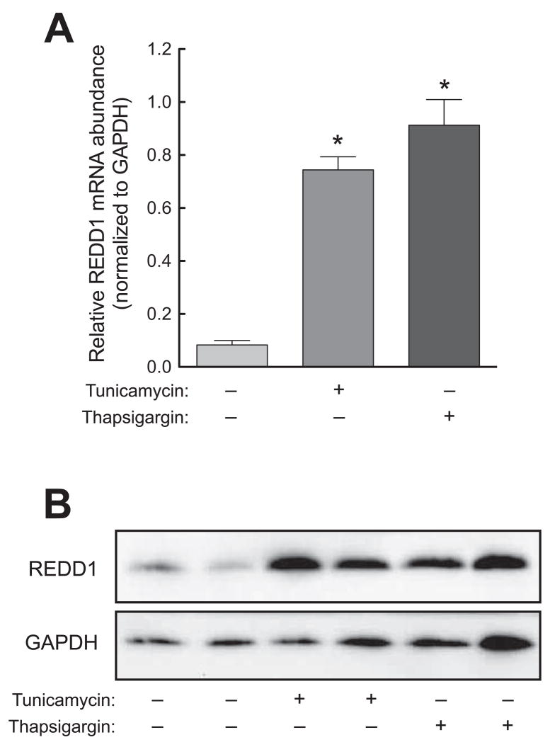
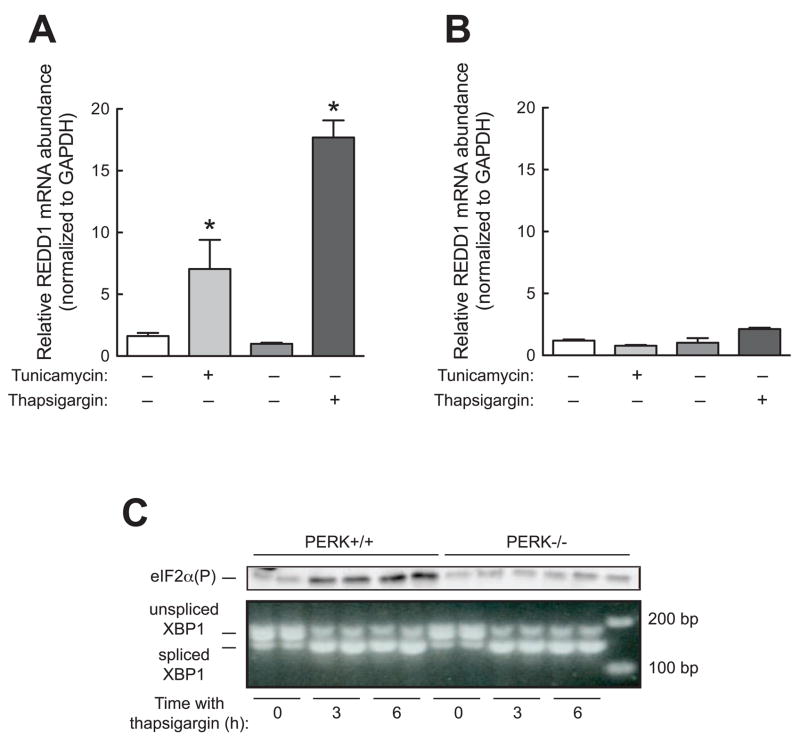
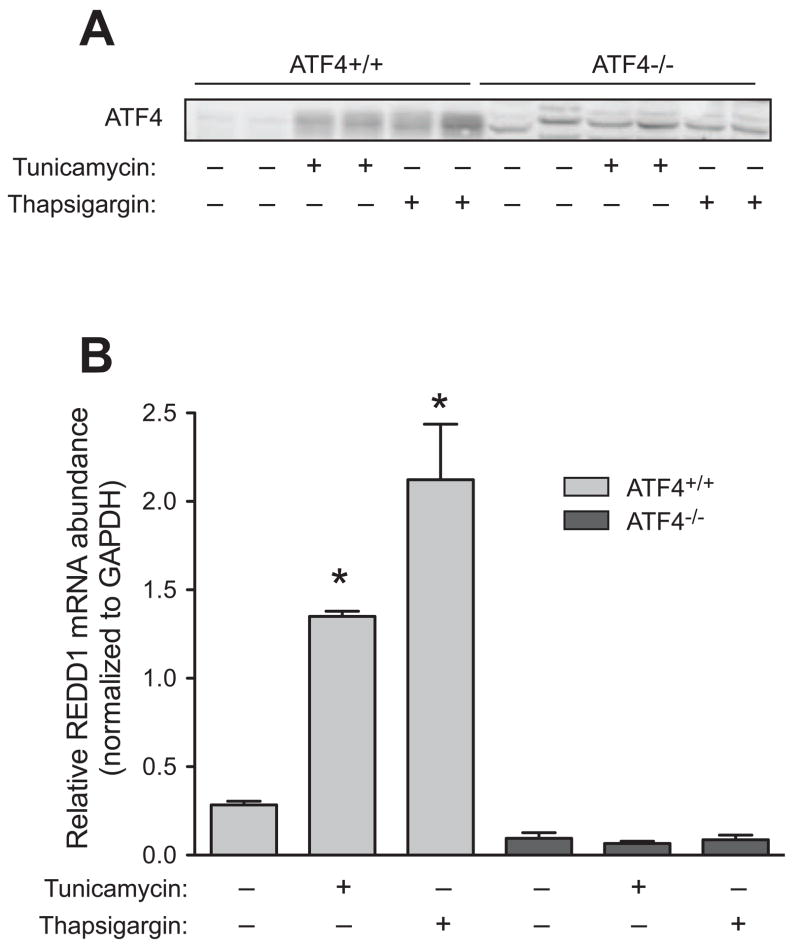
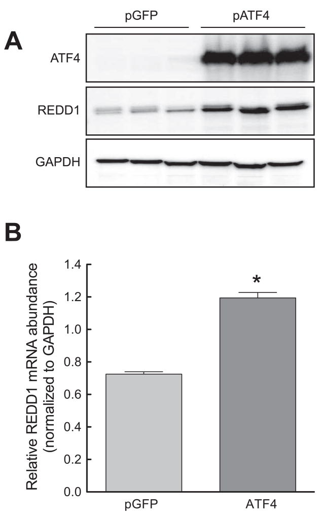
In response to a variety of cell stresses, e.g. endoplasmic reticulum (ER) stress, expression of REDD1 (regulated in development and DNA damage responses) is transcriptionally upregulated. However, the mechanism through which ER stress acts to upregulate REDD1 expression is unknown. In the present study, REDD1 expression was found to be upregulated by ER stress in several cell lines. However, in MEF cells lacking the eIF2alpha kinase PERK, ER stress failed to upregulate REDD1 expression, demonstrating that phosphorylation of eIF2alpha was necessary for the effect. Moreover, ER stress led to upregulated expression of the transcription factor ATF4, but in MEF cells lacking ATF4, REDD1 mRNA expression was not increased by ER stress. In contrast, exogenous expression of ATF4 was sufficient to induce REDD1 expression. Overall, the results suggest that REDD1 expression is upregulated during ER stress through a mechanism involving activation of PERK, phosphorylation of eIF2alpha, and increased ATF4 expression.
Figures
References
-
- Shoshani T, Faerman A, Mett I, Zelin E, Tenne T, Gorodin S, Moshel Y, Elbaz S, Budanov A, Chajut A, Kalinski H, Kamer I, Rozen A, Mor O, Keshet E, Leshkowitz D, Einat P, Skaliter R, Feinstein E. Identification of a novel Hypoxia-Inducible Factor 1-responsive gene, RTP801, involved in apoptosis. Molecular and Cellular Biology. 2002;22:2283–2293. - PMC - PubMed
-
- Ellisen LW, Ramsayer KD, Johannessen CM, Yang A, Beppu H, Minda K, Oliner JD, McKeon F, Haber DA. REDD1, a developmentally regulated transcriptional target of p63 and p53, links p63 to regulation of reactive oxygen species. Molecular Cell. 2002;10:995–1005. - PubMed
-
- Wang Z, Malone MH, Thomenius MJ, Zhong F, Xu F, Distelhorst CW. Dexamethasone-induced Gene 2 (dig2) is a novel pro-survival stress gene induced rapidly by diverse apoptotic signals. The Journal of Biological Chemistry. 2003;278:27053–27058. - PubMed
-
- Wang H, Kubica N, Ellisen LW, Jefferson LS, Kimball SR. Dexamethasone represses signaling through the mammalian target of rapamycin in muscle cells by enhancing expression of REDD1. The Journal of Biological Chemistry. 2006;281:39128–39134. - PubMed
Publication types
MeSH terms
Substances
Grants and funding
LinkOut - more resources
Full Text Sources
Other Literature Sources
