. 2009 Apr;9(2):86-95.
doi: 10.1016/j.mito.2008.12.001. Epub 2008 Dec 11.

Distinct effects of tafazzin deletion in differentiated and undifferentiated mitochondria

Affiliations

Distinct effects of tafazzin deletion in differentiated and undifferentiated mitochondria

Devrim Acehan et al. Mitochondrion. 2009 Apr.

Abstract

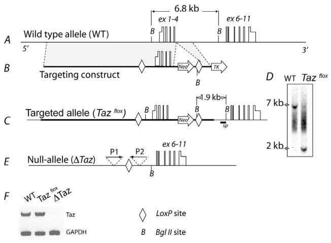
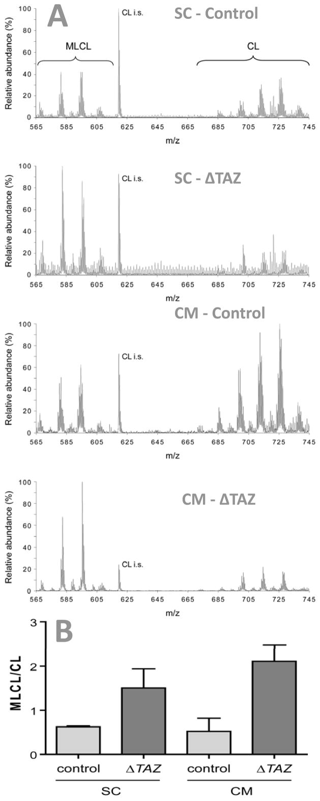
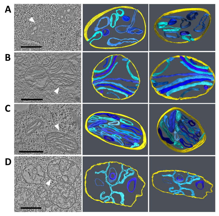
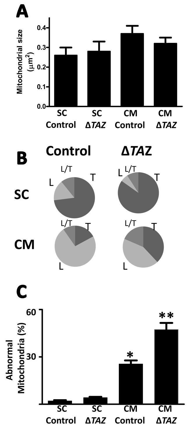
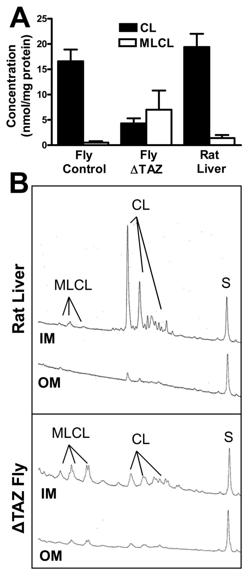
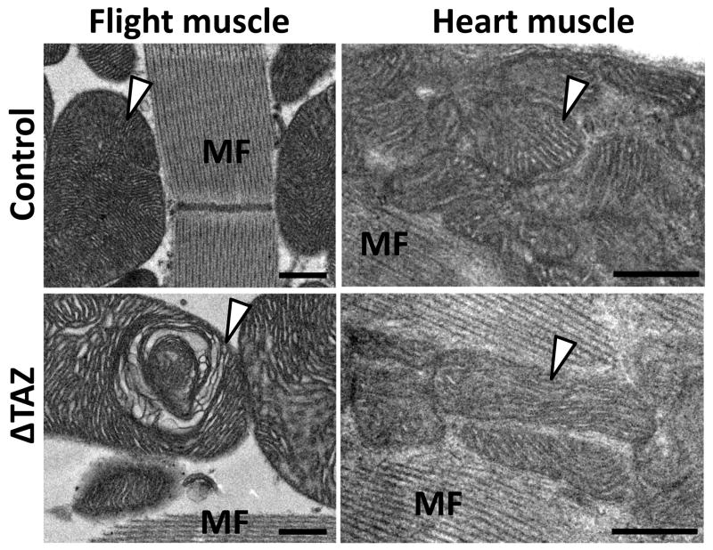
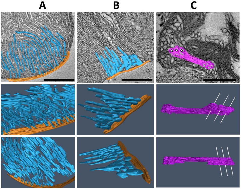
Tafazzin is a conserved mitochondrial protein that is required to maintain normal content and composition of cardiolipin. We used electron tomography to investigate the effect of tafazzin deletion on mitochondrial structure and found that cellular differentiation plays a crucial role in the manifestation of abnormalities. This conclusion was reached by comparing differentiated cardiomyocytes with embryonic stem cells from mouse and by comparing different tissues from Drosophila melanogaster. The data suggest that tafazzin deficiency affects cardiolipin in all mitochondria, but significant alterations of the ultrastructure, such as remodeling and aggregation of inner membranes, will only occur after specific differentiation.


Figures

Fig. 1
Fig. 1. Generation of tafazzin-deficient mouse embryonic stem cells
A, The normal mouse tafazzin gene is shown with exons (boxes) and untranslated regions (half-height boxes). B, The targeting vector contains the neomycin-resistant cassette (Neor) and the thymidine kinase gene (TK), which are used as positive and negative selectors, respectively. Shaded areas depict regions homologous to the sequence of the mouse tafazzin gene. C, The linearized targeting vector was inserted into embryonic stem cells by electroporation followed by selection for neomycin and ganciclovir resistance. The panel shows the correctly targeted tafazzin gene (Tazflox). D, Clones were screened for homologous recombination by Southern blot analysis with a 535 bp DNA probe (marked sp on panel C). Clones containing wild (WT) type and targeted tafazzin alleles (Tazflox) produce 6.8 kb and 1.9 kb bgl II fragments, respectively. E, The tafazzin gene was inactivated in Tazflox cells by transient expression of Cre-recombinase. Cell colonies were screened by PCR with primers P1 and P2. F, Inactivation of the tafazzin gene was verified by RT-PCR analysis of total RNA from differentiated wild type (WT) and Taz-deficient (ΔTaz) cells with mRNA-specific primers P3 and P4 (see text). Glyceroaldehydephosphate dehydrogenase (GAPDH) mRNA was amplified as a control.
Fig. 2
Fig. 2. Cardiolipin and monolysocardiolipin in mouse embryonic stem cells (SC) and differentiated cardiomyocytes (CM)
Lipid extracts of controls and tafazzin-deficient cells (ΔTAZ) were analyzed by mass spectrometry. A, Mass spectra. B, Ratio of monolysocardiolipin (MLCL) to cardiolipin (CL). Tafazzin deletion caused an increase in MLCL relative to CL and a change in the pattern of molecular species. CL i.s., internal standard (tetramyristoyl-cardiolipin).
Fig. 3
Fig. 3. Electron microscopic tomograms of mitochondria from mouse embryonic stem cells and differentiated cardiomyocytes
The left panels show a representative slice of the tomogram; the other panels show 3-D models of mitochondria in different tilt angles. White arrowheads point to the mitochondria for which models were created. Inner boundary membranes are shown in yellow and cristae membranes are shown in shades of blue. A, undifferentiated control stem cell; B, differentiated control cardiomyocyte; C, undifferentiated stem cell with tafazzin deletion; D, differentiated cardiomyocyte with tafazzin deletion. Embryonic stem cells contain mainly tubular mitochondria that are not altered by tafazzin deletion. Differentiated cardiomyocytes contain mainly lamellar mitochondria that show significant abnormalities in the absence of tafazzin. Bars: 500 nm.
Fig. 4
Fig. 4. Morphometry of mitochondria in mouse embryonic stem cells (SC) and differentiatedcardiomyocytes (CM)
Control cells and tafazzin-deficient cells (ΔTAZ) were analyzed. A, Average size of mitochondrial cross sections. B, Proportion of mitochondria with tubular (T), lamellar (L), and mixed (L/T) cristae morphology. C, Proportion of mitochondria with abnormal cristae. Embryonic stem cells contain mainly tubular mitochondria that are not altered by tafazzin deletion. Differentiated cardiomyocytes contain mainly lamellar mitochondria that show significant abnormalities in the absence of tafazzin. Data are means with s.e.m. of two experiments, in each of which we evaluated hundred mitochondrial cross sections. *Significant difference between control SC and control CM (p<0.01); **Significant difference between control CM and ΔTAZ CM (p<0.05).
Fig. 5
Fig. 5. Quantification and submitochondrial localization of cardiolipin and monolysocardiolipin
Cardiolipin (CL) and monolysocardiolipin (MLCL) were quantified in isolated mitochondria and submitochondrial membranes from Drosophila and rat liver by fluorescence-HPLC with pre-column derivatization. A, Concentration in whole mitochondria. Monolysocardiolipin is a minor component of normal mitochondria but it increases significantly in tafazzin-deficient mitochondria (ΔTAZ). B, Distribution between inner (IM) and outer (OM) membrane (membrane aliquots corresponding to 1.5 mg protein). The fluorescence yield is plotted against the retention time between 15 and 60 min. CL and MLCL were quantified by the magnitude of their peak integrals relative to the integral of the internal standard peak (S, oleoyl-tristearoyl-cardiolipin). The data show that both CL and MLCL are mainly present in the inner membrane.
Fig. 6
Fig. 6. Electron micrographs of flight muscle and heart muscle of Drosophila
The images show mitochondria (marked by arrowheads) and myofibers (MF) in flight muscle and heart from the same fly preparations. Flight muscle mitochondria are larger and have a higher cristae density than heart mitochondria. Tafazzin deficiency (ΔTAZ) causes characteristic structural abnormalities in flight muscle mitochondria but not in heart mitochondria. Bars: 500 nm.
Fig. 7
Fig. 7. Electron microscopic tomograms of mitochondria from Drosophila flight muscles
The images show 3-D models of mitochondrial compartments in different tilt angles. A, normal control; B and C, tafazzin deletion mutants (ΔTAZ). Cristae (inner membranes plus intercristae space) are shown in blue, the peripheral compartment (inner boundary membrane plus outer membrane plus intermembrane space) is shown in orange, and aggregated inner membranes are shown in purple. In the upper row, a single slice of the tomogram is presented as background. In panel C, white lines are drawn through some of the cylindrical cavities that penetrate the block of aggregated inner membranes in order to enhance the 3-D impression. ΔTAZ mitochondria show regional cristae remodeling (panel B) and hyperdense malformations consisting of stacks of aggregated inner membranes (panel C). Bars: 500 nm.

References

    1. Acehan D, Xu Y, Stokes DL, Schlame M. Comparison of lymphoblast mitochondria from normal subjects and patients with Barth syndrome using electron microscopic tomography. Lab Invest. 2007;87:40–48. - PMC - PubMed
    1. Barth PG, Scholte HR, Berden JA, Van der Klei-Van Moorsel JM, Luyt-Houwen IE, Van’t Veer-Korthof ET, Van der Harten JJ, Sobotka-Plojhar MA. An X-linked mitochondrial disease affecting cardiac muscle, skeletal muscle and neutrophil leucocytes. J Neurol Sci. 1983;62:327–355. - PubMed
    1. Barth PG, Wanders RJ, Vreken P, Janssen EA, Lam J, Baas F. X-linked cardioskeletal myopathy and neutropenia (Barth syndrome) J Inherit Metab Dis. 1999;22:555–567. - PubMed
    1. Benzer S. Behavioral mutants of Drosophila isolated by countercurrent distribution. Proc Natl Acad Sci U S A. 1967;58:1112–1119. - PMC - PubMed
    1. Brandner K, Mick DU, Frazier AE, Taylor RD, Meisinger C, Rehling P. Taz1, an outer mitochondrial membrane protein, affects stability and assembly of inner membrane protein complexes: implications for Barth Syndrome. Mol Biol Cell. 2005;16:5202–5214. - PMC - PubMed

Publication types

MeSH terms