. 2009 Mar;191(5):1369-81.
doi: 10.1128/JB.01580-08. Epub 2008 Dec 29.

Characterization of a Coxiella burnetii ftsZ mutant generated by Himar1 transposon mutagenesis

Affiliations

Characterization of a Coxiella burnetii ftsZ mutant generated by Himar1 transposon mutagenesis

Paul A Beare et al. J Bacteriol. 2009 Mar.

Abstract

Coxiella burnetii is a gram-negative obligate intracellular bacterium and the causative agent of human Q fever. The lack of methods to genetically manipulate C. burnetii significantly impedes the study of this organism. We describe here the cloning and characterization of a C. burnetii ftsZ mutant generated by mariner-based Himar1 transposon (Tn) mutagenesis. C. burnetii was coelectroporated with a plasmid encoding the Himar1 C9 transposase variant and a plasmid containing a Himar1 transposon encoding chloramphenicol acetyltransferase, mCherry fluorescent protein, and a ColE1 origin of replication. Vero cells were infected with electroporated C. burnetii and transformants scored as organisms replicating in the presence of chloramphenicol and expressing mCherry. Southern blot analysis revealed multiple transpositions in the C. burnetii genome and rescue cloning identified 30 and 5 insertions in coding and noncoding regions, respectively. Using micromanipulation, a C. burnetii clone was isolated containing a Tn insertion within the C terminus of the cell division gene ftsZ. The ftsZ mutant had a significantly lower growth rate than wild-type bacteria and frequently appeared as filamentous forms displaying incomplete cell division septa. The latter phenotype correlated with a deficiency in generating infectious foci on a per-genome basis compared to wild-type organisms. The mutant FtsZ protein was also unable to bind the essential cell division protein FtsA. This is the first description of C. burnetii harboring a defined gene mutation generated by genetic transformation.


Figures

FIG. 1.
FIG. 1.
Plasmid maps of pCR2.1-P1196-Himar1C9 and p1898-Tn. (A) In pCR2.1-P1196-Himar1C9, the promoter from CBU1169 (P1169) drives expression of the C9 variant of the Himar1 transposase. (B) In p1898-Tn, the Himar1 transposon contains CAT and mCherry genes expressed as a single transcriptional unit from P1169. Also contained within the Himar1 ITRs is a ColE1 origin of replication that allows rescue cloning of the Tn in E. coli. Outside of the transposon is the coding sequence of CBU1898.
FIG. 2.
FIG. 2.
Analysis of the ftsZ Tn insertion of C. burnetii B2c. (A) Schematic of the C. burnetii ftsZ chromosomal region in wild-type C. burnetii (NMII) bacteria and the B2c clone. The Tn is flanked by ITR elements (black arrowheads). The binding sites for PCR primers 1 (CBU0141-F), 2 (P1169-NdeI-R), and 3 (CBU0141-R) are shown. Chromosomal regions of NMII and B2c corresponding to the ftsZ probe are depicted. The locations of BsaHI restriction sites and the predicted restriction fragment sizes resulting from a chromosomal BsaHI digest are indicated below the NMII and B2c chromosomal maps. (B) PCR primers 1 and 3 amplified wild-type ftsZ (1,179 bp) from gDNA of NMII and B2, but not B2c. A 3,698-bp product indicating Tn insertion into ftsZ was amplified from B2 and B2c gDNA, but not NMII gDNA. PCR primers 2 and 3 amplified a 594-bp product from gDNA of B2 and B2c, but not NMII, confirming Tn insertion into ftsZ of B2 and B2c and also revealing the Tn orientation (A) in the chromosome. (C) Southern blot of BsaHI-digested gDNA from NMII, B2 and B2c probed for ftsZ. The ftsZ probe hybridized with a 5,540-bp band in gDNA from B2 and B2c gDNA, but not NMII. This fragment corresponds to the 2,734-bp fragment hybridized in NMII gDNA, shifted by 2,806 bp due to the insertion of the Tn into ftsZ.
FIG. 3.
FIG. 3.
Southern blot analysis of C. burnetii transformants. (A) A schematic of C. burnetii gDNA containing a Tn integration. The regions detected by the CAT and mCherry probes are demarked by solid black bars. The location of cut sites for BamHI and BsaHI and the predicted sizes of bands detected by the CAT (>1,223 bp) or mCherry (775 bp) probes are shown. The dotted vertical line indicates the border of the Tn and shows the smallest possible BamHI-BamHI or BamHI-BsaHI restriction fragment size (1,223 bp) that could be detected by the CAT probe. Double parallel lines indicate that the distance between the BamHI site within the Tn and the flanking BamHI or BsaHI site will vary depending on the insertion site of the Tn. (B) Southern blot profiles of untransformed C. burnetii (NMII) gDNA and whole genome amplified DNA from electroporation experiments 1, 2, 3, 4, 5, 6, 7a, and 7b digested with BamHI and BsaHI and probed for CAT and mCherry genes. (C) Southern blot profile of electroporation experiment 1 showing fragments detected by the CAT probe whose sizes correspond to Tn insertions within CBU1745, CBU0964, CBU1196, CBU2020, and CBU0573 recovered by rescue cloning. The 775-bp fragment detected by the mCherry probe is also indicated. DNA molecular weight marker (M) sizes are indicated.
FIG. 4.
FIG. 4.
Microscopy of B2c-infected Vero cells. (A) Vero cells were infected with B2c for 5 days and then imaged live by DIC and confocal fluorescence microscopy. A confocal Z series of B2c-infected cells was taken at 568 nm, merged, then overlaid onto the DIC image. Red fluorescent C. burnetii are apparent in infected cells. (B) Confocal fluorescence image of a Vero cell infected with B2c for 5 days, fixed with methanol, and then immunostained for the lysosomal protein CD63 (green). Bars, 5 μm.
FIG. 5.
FIG. 5.
Growth kinetics of B2c. DNA was extracted from Vero cells infected with B2c or wild-type C. burnetii (NMII) at 0, 1, 2, 3, 5, and 7 days postinfection. B2c had a slower generation time than NMII (19.8 h versus 11.7 h) during exponential phase (2 to 5 days postinfection). This correlated with a 1.63-log increase in GE over 7 days compared to a 2.51-log increase for NMII. GE between B2c and NMII were significantly different (P < 0.005) from 2 to 7 days postinfection according to the Student t test.
FIG. 6.
FIG. 6.
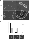
Morphological analysis of B2c. B2c and wild-type C. burnetii purified from Vero cells at 7 days postinfection were visualized by SEM. (A) Low (left) and high (right) magnifications of wild-type organisms (top) and B2c (bottom). (B) Histogram showing the percentage of B2c and wild-type C. burnetii with no division septum, one division septum, or more that one division septum (n > 250). Representative SEM images of the three scored morphologies are shown, with septae indicated by white arrowheads. B2c cells exhibited more division septae than wild-type organisms.
FIG. 7.
FIG. 7.
Domain structure of E. coli FtsZ and alignment with wild-type C. burnetii (NMII) and B2c FtsZ. (A) Diagram of the domain structure of E. coli FtsZ. The N-terminal domain (black) is involved in the formation of the Z ring which is essential for cell division. The globular C-terminal domain contains a conserved motif (gray) that is essential for binding of FtsA and ZipA. The location of the Tn insertion in B2c FtsZ is marked by a triangle. (B) CLUSTAL W alignment of E. coli FtsZ, NMII FtsZ and B2c FtsZ. The FtsA binding region of E. coli FtsZ is highlighted in gray. Tn-encoded amino acids of the B2c FtsZ protein are italicized.
FIG. 8.
FIG. 8.
FtsZ pull-down assay. (A) Production of C. burnetii Xpress-FtsZ, Xpress-FtsZ::Tn, and GST-FtsA in IVTT reactions. An aliquot of each IVTT reaction was separated by SDS-PAGE, and fusion proteins were detected by immunoblotting with anti-Xpress or anti-GST antibodies. Bands of approximately 47 and 45 kDa were detected by the anti-Xpress antibody and correspond to Xpress-tagged FtsZ and FtsZ::Tn, respectively. An ∼73-kDa band was detected with anti-GST antibody and corresponds to GST-tagged FtsA. Molecular mass marker sizes are shown. (B) Pull-down assay using GST-FtsA. An IVTT reaction containing either Xpress-FtsZ or Xpress-FtsZ::Tn was mixed with an IVTT reaction containing GST-FtsA, and then the pull-down procedure was conducted with GST-Sepharose. Pull-downs of Xpress-FtsZ, Xpress-FtsZ::Tn, or GST-FtsA IVTT reactions alone were conducted as controls. GST-FtsA bound to only wild-type C. burnetii FtsZ, as revealed by immunoblotting for the Xpress epitope tag.

References

    1. Bakin, A., J. A. Kowalak, J. A. McCloskey, and J. Ofengand. 1994. The single pseudouridine residue in Escherichia coli 16S RNA is located at position 516. Nucleic Acids Res. 223681-3684. - PMC - PubMed
    1. Baldridge, G. D., N. Burkhardt, M. J. Herron, T. J. Kurtti, and U. G. Munderloh. 2005. Analysis of fluorescent protein expression in transformants of Rickettsia monacensis, an obligate intracellular tick symbiont. Appl. Environ. Microbiol. 712095-2105. - PMC - PubMed
    1. Beare, P. A., D. Howe, D. C. Cockrell, and R. A. Heinzen. 2007. Efficient method of cloning the obligate intracellular bacterium Coxiella burnetii. Appl. Environ. Microbiol. 734048-4054. - PMC - PubMed
    1. Chen, S. Y., T. A. Hoover, H. A. Thompson, and J. C. Williams. 1990. Characterization of the origin of DNA replication of the Coxiella burnetii chromosome. Ann. N. Y. Acad. Sci. 590491-503. - PubMed
    1. Cockrell, D. C., P. A. Beare, E. R. Fischer, D. Howe, and R. A. Heinzen. 2008. A method for purifying obligate intracellular Coxiella burnetii that employs digitonin lysis of host cells. J. Microbiol. Methods 72321-325. - PMC - PubMed

Publication types

MeSH terms