. 2009 Mar;29(5):1338-53.
doi: 10.1128/MCB.01359-08. Epub 2008 Dec 29.

Ras subcellular localization defines extracellular signal-regulated kinase 1 and 2 substrate specificity through distinct utilization of scaffold proteins

Affiliations

Ras subcellular localization defines extracellular signal-regulated kinase 1 and 2 substrate specificity through distinct utilization of scaffold proteins

Berta Casar et al. Mol Cell Biol. 2009 Mar.

Abstract

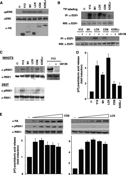
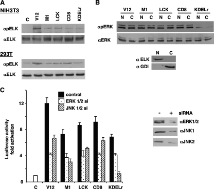
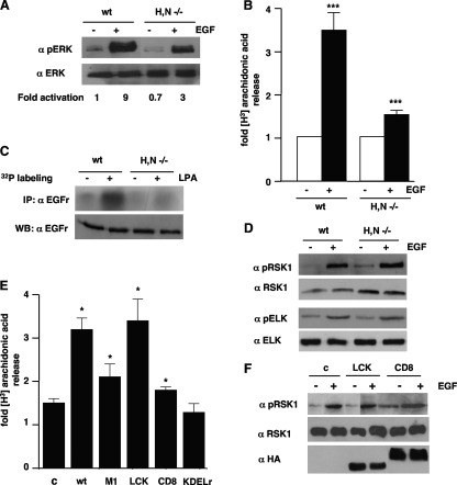
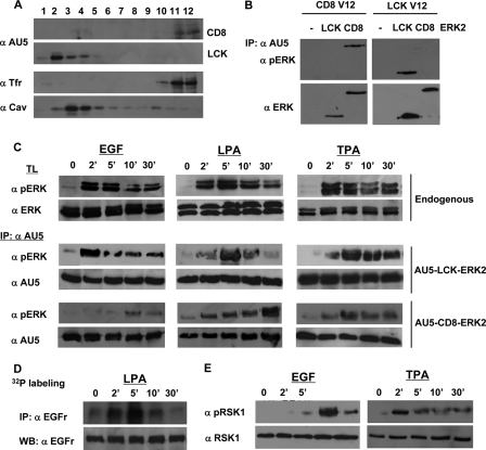
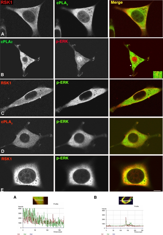
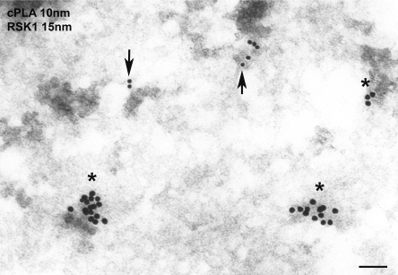
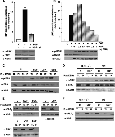
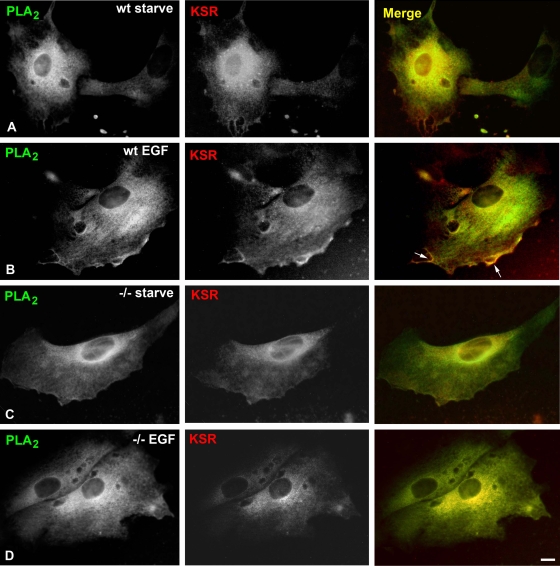
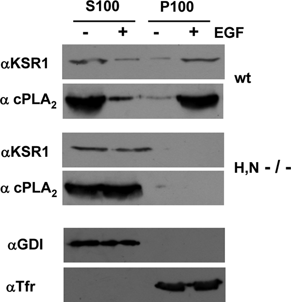
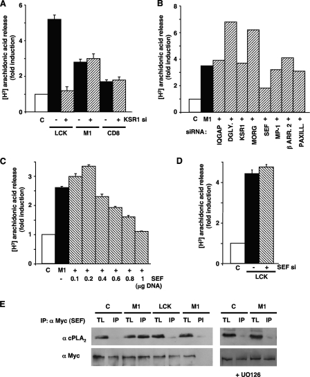
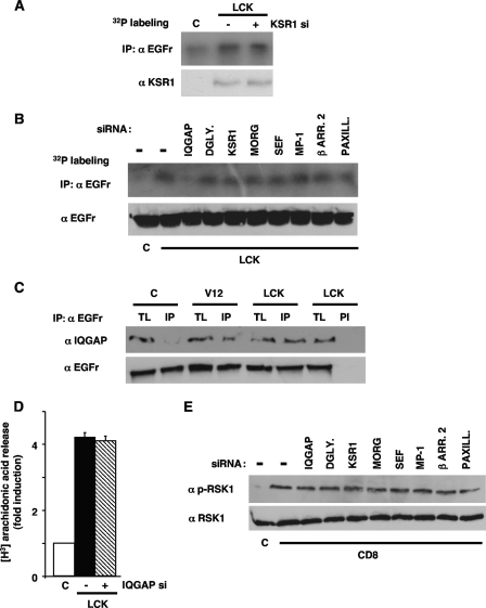
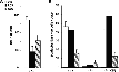
Subcellular localization influences the nature of Ras/extracellular signal-regulated kinase (ERK) signals by unknown mechanisms. Herein, we demonstrate that the microenvironment from which Ras signals emanate determines which substrates will be preferentially phosphorylated by the activated ERK1/2. We show that the phosphorylation of epidermal growth factor receptor (EGFr) and cytosolic phospholipase A(2) (cPLA(2)) is most prominent when ERK1/2 are activated from lipid rafts, whereas RSK1 is mainly activated by Ras signals from the disordered membrane. We present evidence indicating that the underlying mechanism of this substrate selectivity is governed by the participation of different scaffold proteins that distinctively couple ERK1/2, activated at defined microlocalizations, to specific substrates. As such, we show that for cPLA(2) activation, ERK1/2 activated at lipid rafts interact with KSR1, whereas ERK1/2 activated at the endoplasmic reticulum utilize Sef-1. To phosphorylate the EGFr, ERK1/2 activated at lipid rafts require the participation of IQGAP1. Furthermore, we demonstrate that scaffold usage markedly influences the biological outcome of Ras site-specific signals. These results disclose an unprecedented spatial regulation of ERK1/2 substrate specificity, dictated by the microlocalization from which Ras signals originate and by the selection of specific scaffold proteins.


Figures

FIG. 1.
FIG. 1.
Ras sublocalization defines ERK1/2 substrate specificity. (A) Phosphorylation of ERK1/2 resulting from compartmentalized Ras activation. 293T cells were transfected with H-RasV12 (0.5 μg), untargeted (V12) or specifically tethered to the ER (M1), lipid rafts (LCK), the disordered membrane (CD8), or the Golgi complex (KDELr). Phosphorylated and total ERK1/2 levels were determined in total lysates by immunoblotting. Anti-HA Western blotting (WB) reveals the expression levels of the Ras constructs. C, control. (B) Phosphorylation of EGFr resulting from compartmentalized Ras activity. Top panel, EGFr phosphorylation was determined by immunoprecipitation (IP) in 32P-labeled 293T cells transfected with the Ras constructs. Bottom panel, EGFr phosphorylation is ERK dependent. 293T cells transfected with the targeted Ras constructs and incubated with 2 μM U0126 for 2 h (+) were analyzed for EGFr phosphorylation. (C) Phosphorylation of RSK1 induced by compartmentalized Ras activation. Total and phosphorylated RSK1 levels were determined in starved NIH 3T3 and 293T cells expressing the Ras constructs. Right panel, RSK1 phosphorylation is ERK dependent. 293T cells transfected with H-RasV12 and incubated with U0126 for 2 h (+) were analyzed for RSK1 phosphorylation. (D) Regulation of cPLA2 activation by Ras sublocalization. 3H-labeled arachidonic acid release was measured in NIH 3T3 cells. (E) ERK1/2 substrate specificity is unaffected by targeted Ras expression levels. Cells were transfected with increasing concentrations (0.5 to 5 μg) of LCK- or CD8-RasV12 and RSK1 phosphorylation and cPLA2 activation were examined. (D and E) Shown are the averages ± standard errors of the means of the results from three independent experiments relative to the levels in the control cells. α, anti.
FIG. 2.
FIG. 2.
Effects of Ras sublocalization on the activation of ERK1/2 nuclear substrates. (A) Phosphorylation of Elk1 by compartmentalized Ras signals. Total and phosphorylated Elk1 levels were determined by immunoblotting in NIH 3T3 and 293T cells expressing the targeted Ras constructs. C, control. (B) Effects of compartmentalized Ras signals on ERK1/2 nucleocytoplasmic distribution. Total and phosphorylated ERK1/2 levels were examined in nuclear (N) and cytoplasmic (C) fractions from 293T cells expressing the targeted Ras constructs. Bottom panel, the purity of the fractions was ascertained by blotting against Rho-GDP dissociation inhibitor (αGDI) and Elk1. (C) Elk1 activation dependence on Ras compartmentalized signals. Left panel, Elk1 transactivation was assayed in NIH 3T3 cells transfected with GAL4-Elk1 and the targeted Ras constructs plus siRNAs against ERK1 and -2 or JNK1 and -2 (10 ng), as shown in the key. C, control. Shown are the averages ± standard errors of the means of the results from three independent experiments relative to the levels in the control cells. Right panel, diminished expression of the endogenous mitogen-activated protein kinases resulting from siRNA treatments. V12, untargeted H-RasV12; M1, H-RasV12 specifically tethered to the ER; LCK, H-RasV12 specifically tethered to lipid rafts; CD8, H-RasV12 specifically tethered to the disordered membrane; KDELr, H-RasV12 specifically tethered to the Golgi complex.
FIG. 3.
FIG. 3.
cPLA2 but not RSK1 activation is impaired in MEFs devoid of Ras at lipid rafts. (A) ERK1/2 activation in H/N-Ras−/− MEFs. Total and phosphorylated ERK1/2 levels were analyzed in wild type (wt) and H/N-Ras−/− MEFs after starvation (−) and stimulation with 10 ng/ml EGF for 5 min (+). Figures show ERK1/2 phosphorylation levels relative to those found in unstimulated, wild-type MEFs. (B) cPLA2 activation is impaired in H/N-Ras−/− MEFs. 3H-labeled arachidonic acid release was measured in wild-type and H/N-Ras−/− MEFs after stimulation (+) with EGF. Shown are the averages ± standard errors of the means of the results from three experiments relative to the levels in unstimulated cells. ***, P < 0.001, with a 95% confidence interval. (C) EGFr phosphorylation is impaired in H/N-Ras−/− MEFs. EGFr phosphorylation was assayed in wild-type and H/N-Ras−/− MEFs after stimulation with LPA (5 μM) for 5 min (+). (D) Phosphorylation of RSK1 and Elk1 is unaffected in H/N-Ras−/− MEFs. Total and phosphorylated RSK1/Elk1 levels were determined by immunoblotting in wild-type and H/N-Ras−/− MEFs after stimulation with EGF. (E) The presence of Ras at lipid rafts in H/N-Ras−/− MEFs restores PLA2 activation. 3H-labeled arachidonic acid release was measured in H/N-Ras−/− MEFs transfected with wild-type H-Ras targeted to its different microlocalizations (1 μg) after stimulation with EGF. Shown are the averages ± standard errors of the means of the results from two experiments relative to the levels detected in unstimulated cells transfected with the respective site-specific, wild-type H-Ras constructs. *, P < 0.05, with a 95% confidence interval. (F) The restoration of Ras at lipid rafts in H/N-Ras−/− MEFs does not affect RSK1 activation. Total and phosphorylated RSK1 levels were determined for H/N-Ras−/− MEFs transfected with LCK- or CD8-tethered wild-type H-Ras (1 μg) after stimulation with EGF (+). Targeted H-Ras protein expression levels were determined by anti-HA immunoblotting. α, anti; c, control.
FIG. 4.
FIG. 4.
Sublocalization dictates ERK1/2 substrate selectivity under physiological stimulation. (A) Subcellular fractionation of targeted ERK2 proteins. 293T cells expressing AU5-tagged ERK2 specifically tethered to lipid rafts (LCK) or to the disordered membrane (CD8) (1 μg) were solubilized in 0.25% Triton X-100 and partitioned in a sucrose gradient. The presence of the targeted ERK2 proteins in the different fractions was analyzed by anti-AU5 immunoblotting. Anti-caveolin-1 (α Cav) identifies the lipid raft fractions, and anti-transferrin receptor (α Tfr) identifies the disordered membrane fractions. (B) Targeted ERK2 proteins recognize signals in site-specific fashion. Cells were transfected with LCK- or CD8-AU5-tagged ERK2 in addition to LCK-RasV12 or CD8-RasV12 (1 μg each) as shown. Total and phosphorylated ERK levels were examined in anti-AU5 immunoprecipitates. (C) Time-dependent activation profiles of the targeted ERK2 proteins. 293T cells were transfected with the tethered ERK2 constructs and their kinetics of activation were compared when stimulated with EGF (10 ng/ml), LPA (5 μM), or TPA (100 nM) for the indicated times (in minutes) by monitoring total and phosphorylated ERK levels in anti-AU5 immunoprecipitates. For comparison, the phosphorylation kinetics of endogenous ERK1/2 are shown (top two panels). (D) EGFr phosphorylation correlates with the activation of ERK2 at lipid rafts. 32P-labeled 293T cells were stimulated, after starvation, with LPA for the indicated times and phosphorylation of the EGFr was analyzed. Notice that the kinetics of EGFr phosphorylation match those of LCK-ERK2 stimulated with LPA. (E) RSK1 phosphorylation correlates with the activation of ERK2 at the disordered membrane and varies accordingly depending on the stimulus. 293T cells were stimulated, after starvation, with EGF or TPA for the indicated times, and the phosphorylation of RSK1 was analyzed. Notice that the kinetics of RSK1 activation match those of CD8-ERK2 stimulated with EGF or with TPA.
FIG. 5.
FIG. 5.
RSK and cPLA2 are located in distinct subcytoplasmic microlocalizations. (A) RSK and cPLA2 do not colocalize. Growing NIH 3T3 cells were costained with anti-RSK and anti-cPLA2 antibodies and visualized by immunofluorescence and confocal microscopy. (B and C) cPLA2 but not RSK1 colocalizes with phospho-ERK (p-ERK) in NIH 3T3 cells stably expressing LCK-RasV12. Inset, detail of the sublocalization at the cytoplasm (thick arrow). (D and E) RSK1 but not cPLA2 colocalizes with phospho-ERK in NIH 3T3 cells stably expressing CD8-RasV12. To highlight the cytoplasmic signals, the cells were not permeabilized (except in panel B), and the nuclear signals are quenched. Pseudocolors were red and green and as indicated; yellow indicates a merge. Scale bar = 10 μM. Profile A, pseudocolor intensity profile, across the section shown in panel A. Notice that the colocalization of peaks is minimal. Profile B, pseudocolor intensity profile at a PM localization (thin arrow) shown in panel B.
FIG. 6.
FIG. 6.
RSK1 and cPLA2 occupy distinct cytoplasmic microdomains. Immunogold electron microscopy was performed on NIH 3T3 cells. The secondary antibodies disclosing RSK1 (asterisks) and cPLA2 (arrows) were labeled with 15-nm and 10-nm gold particles, respectively. Scale bar = 100 nm.
FIG. 7.
FIG. 7.
KSR1 couples Ras signals from lipid rafts to cPLA2 activation. (A) Depletion of KSR1 prevents cPLA2 but not RSK1 activation. 293T cells were transfected with (+) or without (-) a siRNA (10 nM) for KSR1 and stimulated with EGF (10 ng/ml, 5 min) after starvation. Endogenous cPLA2 activation was determined by [3H]arachidonic acid release. Bottom panels show total and phosphorylated RSK1 (p-RSK1), and KSR1 levels were determined by immunoblotting. (B) Biphasic activation of cPLA2 resulting from increasing KSR1 levels. Cells were transfected with increasing concentrations of FLAG-KSR1 and stimulated with EGF after starvation. Bottom panels show total and phosphorylated RSK1, and KSR1 (FLAG) levels were determined by immunoblotting. (A and B) Shown are the averages ± standard errors of the means of the results from three independent experiments relative to the levels in starved (C) cells. (C) ERK1/2-KSR1 association is enhanced by Ras signals from lipid rafts but not the disordered membrane. Endogenous ERK1/2, total and phosphorylated, present in native KSR1 immunoprecipitates was determined in cells transfected with RasV12 (0.5 μg) tethered to lipid rafts (LCK) or to the disordered membrane (CD8) or stimulated with EGF. (D) ERK1/2-KSR1 association is enhanced in wild-type but not in H/N-Ras−/− MEFs. ERK1/2, total and phosphorylated, present in KSR1 immunoprecipitates were analyzed in MEFs, starved (C) or EGF stimulated. (E) The association of KSR1 and endogenous cPLA2 is enhanced by EGF stimulation and by Ras signals from lipid rafts and is inhibited by UO126 (2 μM, 25 min prior to stimulation). Bottom panels, RSK1 does not associate with KSR1 in response to Ras signals coming from lipid rafts or to EGF stimulation. (F) The association of KSR1 and endogenous cPLA2 in response to EGF is diminished in H/N-Ras−/− MEFs compared to wild-type fibroblasts. TL, total lysate; IP, immunoprecipitations performed with a specific antibody; PI, immunoprecipitations performed with preimmune serum; α, anti.
FIG. 8.
FIG. 8.
KSR1 and cPLA2 colocalize at the PM of wild-type (wt) but not H/N-Ras−/− (−/−) MEFs. Endogenous RSK and cPLA2 were costained with anti-RSK and anti-cPLA2 antibodies and visualized by immunofluorescence and confocal microscopy. The wild-type and H/N-Ras−/− MEFs were starved (A and C) or stimulated with EGF (10 ng/ml, 5 min) (B and D). Pseudocolors were red and green as indicated; yellow indicates a merge. Scale bar = 10 μM.
FIG. 9.
FIG. 9.
KSR1 and cPLA2 localize at the PM of wild-type (wt) but not H/N-Ras−/− MEFs upon stimulation. Soluble (S100) and particulate (P100) fractions were isolated from wild-type and H/N-Ras−/− MEFs, starved (−) or stimulated (+) with EGF (10 ng/ml, 5 min). The presence of KSR1 and cPLA2 was examined by immunoblotting. The purity of the fractions was ascertained by blotting against Rho-GDP dissociation inhibitor (αGDI) and transferrin (Tfr) receptor in lysates from wild-type MEFs.
FIG. 10.
FIG. 10.
Sef couples Ras signals from the ER to cPLA2 activation. (A) Depletion of KSR1 inhibits cPLA2 activation by Ras signals from lipid rafts but not from the ER or disordered membrane. 293T cells were transfected with RasV12 (0.5 μg) tethered to lipid rafts, the ER, and the disordered membrane with (+) or without (−) a siRNA (10 nM) for KSR1. (B) Effects of depleting the levels of different scaffold proteins on cPLA2 activation by Ras signals from the ER. Cells were transfected with ER-targeted RasV12 (M1) (+) plus siRNAs (10 nM) for the shown scaffold proteins. DGLY., dystroglycan; β ARR. 2, β-arrestin 2; PAXILL., paxillin. (C) cPLA2 activation by Ras signals from the ER exhibits a biphasic response to increasing Sef concentrations. Cells were transfected with ER-targeted RasV12 (M1) (+), in addition to increasing concentrations of Myc-Sef as shown. (D) Sef does not intervene in cPLA2 activation elicited from lipid rafts. Cells were transfected with RasV12 tethered to lipid rafts with (+) or without (-) a siRNA against Sef. (A to D) cPLA2 activation was determined by [3H]arachidonic acid release after 18 h of starvation. Shown are the averages ± standard errors of the means of the results from three independent experiments relative to the levels in starved (C) cells. (E) Sef/cPLA2 association is enhanced by Ras signals from the ER but not from lipid rafts. Endogenous cPLA2, present in anti-Myc (α Myc) immunoprecipitates, was determined in cells transfected with Myc-Sef (1 μg) plus RasV12 tethered to lipid rafts (LCK) or to the ER (M1). The dependence on ERK was determined by incubation with UO126 (2 μM, 25 min prior to stimulation). TL, total lysate; IP, immunoprecipitations performed with a specific antibody; PI, immunoprecipitations performed with preimmune serum.
FIG. 11.
FIG. 11.
IQGAP1 couples Ras signals from lipid rafts to EGFr phosphorylation. (A) Depletion of KSR1 does not affect the phosphorylation of EGFr elicited by Ras signals from lipid rafts. 293T cells were transfected with lipid raft-targeted RasV12 (LCK) (0.5 μg) plus a siRNA (10 nM) (+) for KSR1. EGFr phosphorylation was determined by immunoprecipitation (IP) in 32P-labeled cells. (B) Effects of downregulating the levels of different scaffold proteins on EGFr phosphorylation induced by Ras signals from lipid rafts. Cells were transfected with lipid raft-targeted RasV12, plus siRNAs for the shown scaffold proteins. DGLY., dystroglycan; β ARR. 2, β-arrestin 2; PAXILL., paxillin. (C) EGFr/IQGAP1 association is enhanced by Ras signals from lipid rafts. Endogenous IQGAP1, present in anti-EGFr immunoprecipitates, was measured in cells transfected with RasV12, untargeted or tethered to lipid rafts (LCK). TL, total lysate; IP, immunoprecipitations performed with a specific antibody; PI, immunoprecipitations performed with preimmune serum. (D) IQGAP1 does not intervene in cPLA2 activation elicited from lipid rafts. Cells were transfected with RasV12 tethered to lipid rafts with (+) or without (−) a siRNA against IQGAP1. cPLA2 activation was determined by [3H]arachidonic acid release after starvation. (E) RSK1 phosphorylation by Ras signals from the disordered membrane is unaffected by the depletion of scaffold proteins. Cells were transfected with disordered membrane-targeted RasV12 (CD8), plus siRNAs (10 ng) for the shown scaffold proteins. Total and phosphorylated RSK1 levels were detected by immunoblotting. α, anti.
FIG. 12.
FIG. 12.
KSR1 affects the ability of site-specific Ras signals to induce senescence. (A) Transforming potential of Ras site-specific signals. MEFs were transfected with vectors encoding H-RasV12 constructs as shown in the inset key (1 μg each) in addition to c-Myc (1 μg). +/+, wild-type MEFs. (B) Site-specific Ras signals require KSR1 to induce senescence. MEFs, wild-type (+/+) or deficient for KSR1 (−/−), were transfected with vectors encoding the H-RasV12 constructs as shown in the inset key in panel A (1 μg each). Where indicated, ectopic KSR1 (1 μg) was also transfected. After 3 weeks, the cells were stained and either the number of foci or the number of senescent cells was scored. Shown are the averages ± standard errors of the means of the results from three independent experiments. +ve, positive.

Similar articles

Cited by

References

    1. Agudo-Ibanez, L., F. Nunez, F. Calvo, I. M. Berenjeno, X. R. Bustelo, and P. Crespo. 2007. Transcriptomal profiling of site-specific Ras signals. Cell. Signal. 192264-2276. - PMC - PubMed
    1. Ait-Mamar, B., M. Cailleret, C. Rucker-Martin, A. Bouabdallah, G. Candiani, C. Adamy, P. Duvaldestin, F. Pecker, N. Defer, and C. Pavoine. 2005. The cytosolic phospholipase A2 pathway, a safeguard of beta2-adrenergic cardiac effects in rat. J. Biol. Chem. 28018881-18890. - PubMed
    1. Ajenjo, N., E. Canon, I. Sanchez-Perez, D. Matallanas, J. Leon, R. Perona, and P. Crespo. 2004. Subcellular localization determines the protective effects of activated ERK2 against distinct apoptogenic stimuli in myeloid leukemia cells. J. Biol. Chem. 27932813-32823. - PubMed
    1. Arozarena, I., D. S. Aaronson, D. Matallanas, V. Sanz, N. Ajenjo, S. P. Tenbaum, H. Teramoto, T. Ighishi, J. C. Zabala, J. S. Gutkind, and P. Crespo. 2000. The Rho family GTPase Cdc42 regulates the activation of Ras/MAP kinase by the exchange factor Ras-GRF. J. Biol. Chem. 27526441-26448. - PubMed
    1. Arozarena, I., D. Matallanas, M. T. Berciano, V. Sanz-Moreno, F. Calvo, M. T. Munoz, G. Egea, M. Lafarga, and P. Crespo. 2004. Activation of H-Ras in the endoplasmic reticulum by the RasGRF family guanine nucleotide exchange factors. Mol. Cell. Biol. 241516-1530. - PMC - PubMed

Publication types

MeSH terms