. 2009 Jan;133(1):79-91.
doi: 10.1085/jgp.200810105.

The skeletal L-type Ca(2+) current is a major contributor to excitation-coupled Ca(2+) entry

Affiliations

The skeletal L-type Ca(2+) current is a major contributor to excitation-coupled Ca(2+) entry

Roger A Bannister et al. J Gen Physiol. 2009 Jan.

Abstract

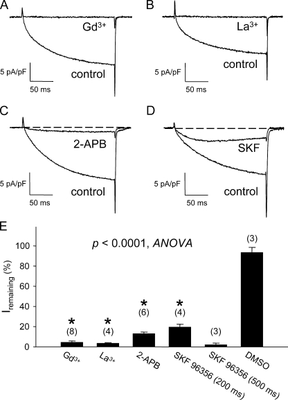
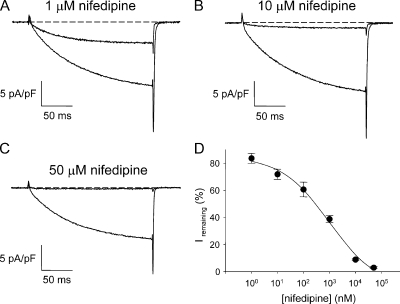
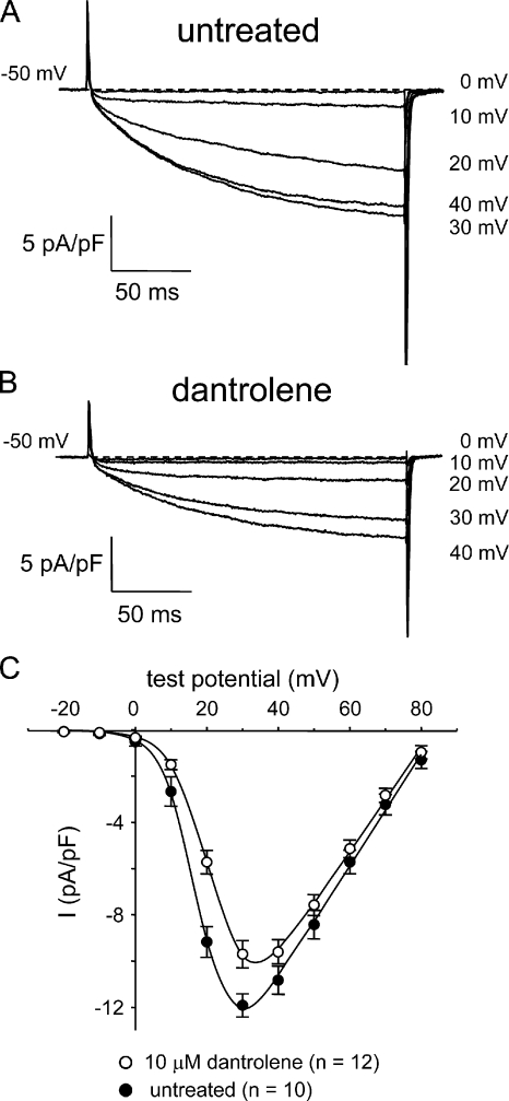
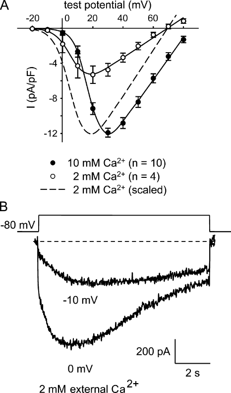
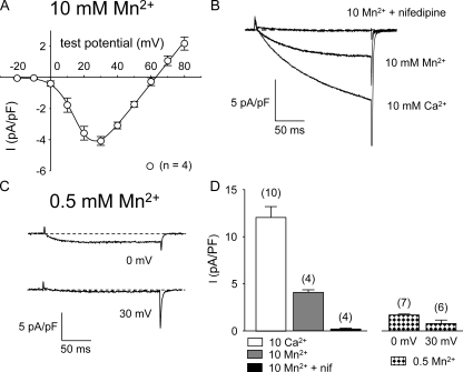
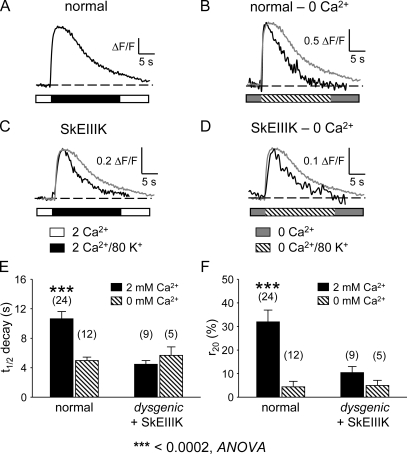
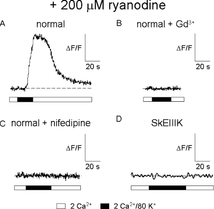
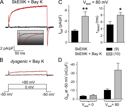
The term excitation-coupled Ca(2+) entry (ECCE) designates the entry of extracellular Ca(2+) into skeletal muscle cells, which occurs in response to prolonged depolarization or pulse trains and depends on the presence of both the 1,4-dihydropyridine receptor (DHPR) in the plasma membrane and the type 1 ryanodine receptor in the sarcoplasmic reticulum (SR) membrane. The ECCE pathway is blocked by pharmacological agents that also block store-operated Ca(2+) entry, is inhibited by dantrolene, is relatively insensitive to the DHP antagonist nifedipine (1 microM), and is permeable to Mn(2+). Here, we have examined the effects of these agents on the L-type Ca(2+) current conducted via the DHPR. We found that the nonspecific cation channel antagonists (2-APB, SKF 96356, La(3+), and Gd(3+)) and dantrolene all inhibited the L-type Ca(2+) current. In addition, complete (>97%) block of the L-type current required concentrations of nifedipine >10 microM. Like ECCE, the L-type Ca(2+) channel displays permeability to Mn(2+) in the absence of external Ca(2+) and produces a Ca(2+) current that persists during prolonged ( approximately 10-second) depolarization. This current appears to contribute to the Ca(2+) transient observed during prolonged KCl depolarization of intact myotubes because (1) the transients in normal myotubes decayed more rapidly in the absence of external Ca(2+); (2) the transients in dysgenic myotubes expressing SkEIIIK (a DHPR alpha(1S) pore mutant thought to conduct only monovalent cations) had a time course like that of normal myotubes in Ca(2+)-free solution and were unaffected by Ca(2+) removal; and (3) after block of SR Ca(2+) release by 200 microM ryanodine, normal myotubes still displayed a large Ca(2+) transient, whereas no transient was detectable in SkEIIIK-expressing dysgenic myotubes. Collectively, these results indicate that the skeletal muscle L-type channel is a major contributor to the Ca(2+) entry attributed to ECCE.


Figures

Figure 1.
Figure 1.
Skeletal muscle L-type current is blocked by nonspecific cation channel antagonists. Acute block of L-type currents evoked by step depolarizations to +30 mV by 100 μM Gd3+ (A), 100 μM La3+ (B), 100 μM 2-APB (C), and 20 μM SKF 96356 (D). The test pulse was applied after a prepulse protocol (see Materials and methods) (Adams et al., 1990). Results of these experiments and application of DMSO vehicle (0.01%) control experiments are summarized in E. For trivalent cations, maximal block was generally achieved in <30 s after gravity perfusion of the 30-mm dish, whereas longer perfusion times were required for maximal block by 2-APB (1–2 min) and SKF 96356 (2–3 min). The asterisk indicates a significant difference for current (measured at the end of a 200-ms test pulse) remaining in myotubes exposed to blockers relative to DMSO-treated control cells (P < 0.0001, ANOVA). Although not indicated, block by SKF 96356 measured at the end of a 500-ms test pulse was also significantly (P < 0.00008, t test) different than DMSO controls. Throughout, the error bars represent ± SEM.
Figure 2.
Figure 2.
Block of skeletal L-type current by nifedipine. (A) Incomplete block of L-type current by 1 μM nifedipine. (B) Incomplete block of L-type current by 10 μM nifedipine. (C) Complete block of L-type current by 50 μM nifedipine. Currents evoked by step depolarizations to +30 mV from −50 mV after a prepulse protocol (see Materials and methods). (D) Dose–response relationship for the effects of nifedipine, which were measured ∼1 min after addition. Note that the minimal apparent block (at 1 nM) was ∼80%, most likely as a consequence of current rundown. Each symbol represents data collected from 2–10 cells.
Figure 3.
Figure 3.
Dantrolene inhibits skeletal L-type Ca2+ currents. Recordings of L-type currents elicited by 200-ms depolarizations from −50 mV to the indicated test potentials are shown for an untreated normal myotube (A) or a normal myotube exposed to 10 μM dantrolene for >10 min at ∼25°C (B). (C) Comparison of I-V relationships for untreated (•; n = 10) and dantrolene-treated myotubes (○; n = 12). Currents were evoked at 0.1 Hz in 10-mV increments after a prepulse protocol (see Materials and methods). Current amplitudes were normalized by linear cell capacitance (pA/pF). The smooth curves are plotted according to Eq. 1, with best-fit parameters presented in Table I.
Figure 4.
Figure 4.
In 2 mM of external Ca2+, L-type currents are activated by weak depolarizations and inactivate very slowly. (A) Comparison of I-V relationships for normal myotubes in 10 mM of external Ca2+ (•; n = 10) or 2 mM of external Ca2+ (○; n = 4) elicited at 0.1 Hz by test potentials ranging from −20 through +80 mV in 10-mV increments after a prepulse protocol. The smooth curves are plotted according to Eq. 1. The best-fit parameters for each plot are presented in Table I. (B) Recordings of L-type currents in 2 mM of external Ca2+ elicited by 9,800-ms depolarizations from −80 mV to the indicated test potentials.
Figure 5.
Figure 5.
The skeletal muscle L-type channel conducts Mn2+. (A) I-V relationship for normal myotubes in 10 mM of external Mn2+ (○; n = 4) for currents elicited at 0.1 Hz by test potentials ranging from −20 through +80 mV in 10-mV increments after a prepulse protocol. The smooth curves are plotted according to Eq. 1. The best-fit parameters for each plot are presented in Table I. (B) Representative currents recorded at +30 mV from a normal myotube in 10 mM Ca2+ (bottom trace), in 10 mM Mn2+ (middle trace), and in 10 mM Mn2+ after acute application of 10 μM nifedipine (top trace). (C) L-type current evoked by steps from −50 to 0 mV (top) and to +30 mV (bottom) in 0.5 mM Mn2+. (D) Average peak currents in 10 mM of divalent cation at a test potential of +30 mV (left) and for 0.5 mM Mn2+ at the indicated test potentials (right).
Figure 6.
Figure 6.
Ca2+ influx via the L-type channel accounts for the bulk of Ca2+ influx in response to depolarization. Representative Ca2+ transients are shown for a normal myotube in 2 mM of bath Ca2+ (A), a normal myotube in nominal 0 mM of bath Ca2+ (B), a dysgenic myotube expressing SkEIIIK in 2 mM Ca2+ (C), and a dysgenic myotube expressing SkEIIIK in 0 mM Ca2+ (D). Summary of t1/2 decay (E) and r20 (F) values for normal myotubes and dysgenic myotubes expressing SkEIIIK in the presence (solid bars) and absence (hatched bars) of 2 mM Ca2+. In B–D, a scaled version of the transient shown in A has been included for comparison of the rates of transient decay (gray traces). Transients were elicited locally by 20-s applications of 80 mM KCl via micropipette placed close to the myotube of interest. See Materials and methods for definitions of t1/2 decay and r20. Asterisks indicate a significant difference (P < 0.0005, ANOVA).
Figure 7.
Figure 7.
SkEIIIK supports minimal Ca2+ entry during long global depolarizations. Representative Ca2+ transients evoked by global perfusion of 80 mM KCl Ringer's solution for normal myotubes (A), normal myotubes exposed to 100 μM Gd3+ (B), normal myotubes exposed to 50 μM nifedipine (C), and dysgenic myotubes expressing SkEIIIK (D). In each case, myotubes were exposed to 200 μM ryanodine for >1 h at 37°C before experiments to block the contribution of RYR1 to the Ca2+ transient. In experiments with SkEIIIK, a slower sampling rate was used to minimize any bleaching of the Fluo-3 dye. Expression of SkEIIIK was confirmed by electrically evoked (100 V, 5 ms) contractions (17 of 20 myotubes tested) before ryanodine treatment (see Papadopoulos et al., 2004).
Figure 8.
Figure 8.
Potentiated SkEIIIK conducts inward Ca2+ current. Representative currents are shown for a ±Bay K 8644–treated (5 μM) dysgenic myotube expressing SkEIIIK (A) and a ±Bay K 8644–treated, uninjected dysgenic myotube (B). The inset illustrates the tail currents shown in A on an expanded time base. (C) Summary of amplitudes (left) and deactivation time constants (right) of SkEIIIK tail currents recorded in the absence (n = 9) and presence (n = 10) of ±Bay K 8644. (D) Total charge moved obtained by integration of tail currents recorded in the absence (n = 9) and presence (n = 10) of ±Bay K 8644 from dysgenic myotubes expressing SkEIIIK. Asterisks indicate significant differences between untreated and ±Bay K 8644–treated cells (*, P < 0.05; **, P < 0.01, t test).

References

    1. Adams, B.A., and K.G. Beam. 1989. A novel calcium current in dysgenic skeletal muscle. J. Gen. Physiol. 94:429–444. - PMC - PubMed
    1. Adams, B.A., T. Tanabe, A. Mikami, S. Numa, and K.G. Beam. 1990. Intramembrane charge movement restored in dysgenic skeletal muscle by injection of dihydropyridine receptor cDNAs. Nature. 346:569–572. - PubMed
    1. Armstrong, C.M., F.M. Bezanilla, and P. Horowicz. 1972. Twitches in the presence of ethylene glycol bis(-aminoethyl ether)-N,N′-tetraacetic acid. Biochim. Biophys. Acta. 267:605–608. - PubMed
    1. Avila, G., and R.T. Dirksen. 2000. Functional impact of the ryanodine receptor on the skeletal muscle L-type Ca2+ channel. J. Gen. Physiol. 115:467–480. - PMC - PubMed
    1. Beam, K.G., and C. Franzini-Armstrong. 1997. Functional and structural approaches to the study of excitation-contraction coupling. Methods Cell Biol. 52:283–306. - PubMed

Publication types