. 2009 Jan 6;106(1):238-43.
doi: 10.1073/pnas.0811466106. Epub 2008 Dec 29.

A regulatory interplay between miR-27a and Runx1 during megakaryopoiesis

Affiliations

A regulatory interplay between miR-27a and Runx1 during megakaryopoiesis

Oren Ben-Ami et al. Proc Natl Acad Sci U S A. .

Abstract

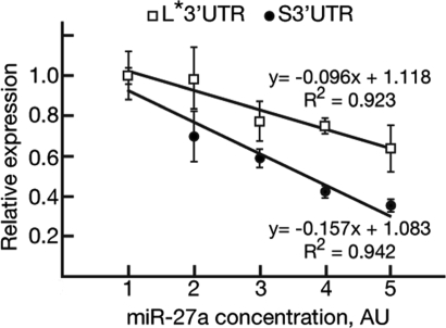
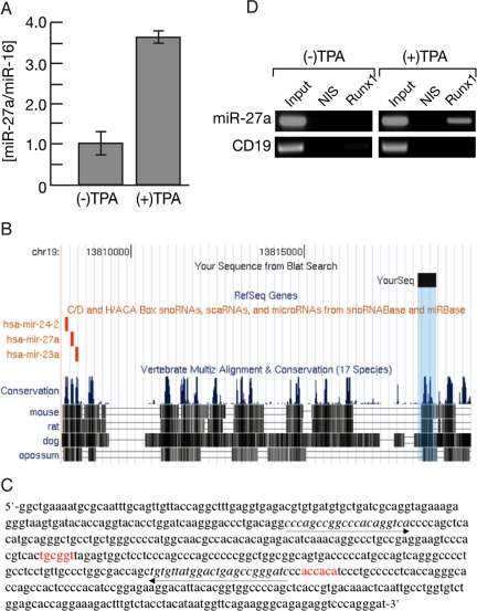
The transcription factor Runx1 is a key regulator of definitive hematopoiesis in the embryo and the adult. Lineage-specific expression of Runx1 involves transcription and post-transcription control through usage of alternative promoters and diverse 3'UTR isoforms, respectively. We identified and mapped microRNA (miR) binding sites on Runx1 3'UTR and show that miR-27a, miR-9, miR-18a, miR-30c, and miR-199a* bind and post-transcriptionally attenuate expression of Runx1. miR-27a impacts on both the shortest (0.15 kb) and longest (3.8 kb) 3'UTRs and, along with additional miRs, might contribute to translation attenuation of Runx1 mRNA in the myeloid cell line 416B. Whereas levels of Runx1 mRNA in 416B and the B cell line 70Z were similar, the protein levels were not. Large amounts of Runx1 protein were found in 70Z cells, whereas only minute amounts of Runx1 protein were made in 416B cells and overexpression of Runx1 in 416B induced terminal differentiation associated with megakaryocytic markers. Induction of megakaryocytic differentiation in K562 cells by 12-o-tetradecanoylphorbol-13-acetate markedly increased miR-27a expression, concomitantly with binding of Runx1 to miR-27a regulatory region. The data indicate that miR-27a plays a regulatory role in megakaryocytic differentiation by attenuating Runx1 expression, and that, during megakaryopoiesis, Runx1 and miR-27a are engaged in a feedback loop involving positive regulation of miR-27a expression by Runx1.


Conflict of interest statement

The authors declare no conflict of interest.

Figures

Fig. 1.
Fig. 1.
Post-transcriptional attenuation of Runx1 in 416B cells. (A) qRT-PCR analysis of Runx1 mRNA in 416B and 70Z cells. HPRT1 was used as an internal control. (B) Western blot analysis of nuclear lysates of 416B and 70Z cells reacted with anti-Runx1 polyclonal antibody. LaminB1 was used as protein loading control. (C) Northern blot analysis of miRs expressed in HEK293 (lane 2) and 416B (lane 4) cells. 5S rRNA bands were used as reference for RNA loading.
Fig. 2.
Fig. 2.
MiR-27a inhibits Runx1 3′UTR-dependent expression. (A) Inhibition of luciferase 3′UTR reporter. Relative expression of luciferase activity in HEK293 cells co-transfected with pRL-S3′UTR, pRL-Mut1-S3′UTR, pRL-Mut2-S3′UTR, or pRL-Mut(1 + 2)-S3′UTR and miR-27a (0.1 nM). Cells co-transfected with scrambled miR oligo (miR-NC 0.1 nM) served as controls. Relative to miR-NC, the effect of miR-27a was significant [P = 0.007, P = 0.002, P = 0.01, and P = 0.02 for S3′UTR, Mut1, Mut2, and Mut(1 + 2), respectively]. Data shown are representative of at least two independent experiments done in triplicate. Error bars indicate SD. (B) Antago-miR27a diminished miR-27a activity. Relative expression of luciferase activity in HEK293 cells co-transfected with miR-27a, pRL-S3′UTR, and increasing amounts of antago-miR-27a. Cells co-transfected with scrambled antago-miR-NC served as controls. Data shown are representative of at least two independent experiments done in triplicate. Error bars indicate SD. Firefly luciferase was used as an internal control in A and B. (C) Inhibition of Runx1 cDNA expression by miR-27a. Western blot analysis of nuclear lysates of HEK293 cells co-transfected with pcRunx1, pEGFP-3xNLS, and miR-27a and either miR-NC or miR-9 as controls. miR-9 was used as a negative control along with miR-NC, because pcRunx1 contained the S3′UTR lacking miR-9 binding site. EGFP-3xNLS was used to monitor transfection efficiency and protein loading. Control of non-transfected cells is also indicated (Non). (D) Inhibition of endogenous Runx1 mRNA translation by miR-27a. 70Z cells were transfected with miR-27a, miR-9, and miR-NC and endogenous Runx1 mRNA and protein were monitored by qRT-PCR (Lower) and Western blots (Upper). HPRT1 was used as qRT-PCR internal control and Emerin as a control for protein loading. Ratios of Runx1/Emerin were derived by densitometry analysis using ImageJ (55). Note that miR-27a mediates stronger effect on the protein compared with miR-9. This may be related to the proximal positioning and higher “quality” of miR-27a binding sites within the 3′UTR of Runx1 (Figs. S2 and S3). Neither miR-9 nor miR-27a affected the level of Runx1 mRNA, but mediated translation inhibition leading to an approximate two- to threefold decrease in the steady-state level of Runx1 protein.
Fig. 3.
Fig. 3.
The size of the 3′UTR affects the ability of miR-27a to attenuate Runx1 expression. HEK293 cells were co-transfected with pRL-S3′UTR or pFF-L*3′UTR and increasing amounts of miR-27a or miR-NC (transfection control). Numbers at the x axis [arbitrary units (AU) 1–5] correspond to increasing concentrations of miR-27a as follows: 0 nM, 0.1 nM, 0.3 nM, 1 nM, and 3 nM. The effect of miR-27a on reporter activity was calculated relative to the corresponding miR-NC transfected cells (defined as 1.0). Reporter expression was normalized to internal control luciferase activity. The slope values (−0.096 and −0.157 for L*3′UTR and S3′UTR, respectively) indicate that the S3′UTR-dependent translation is more sensitive than L*3′UTR-dependent translation to inhibition by miR-27a. Of note, a square r of ≈0.93 indicates the close relationships between the values comprising the slopes. Data shown are representative of at least two independent experiments done in triplicate. Error bars indicate SD.
Fig. 4.
Fig. 4.
Regulation of miR-27a expression by Runx1 during megakaryocytic differentiation. (A) qRT-PCR analysis of miR-27a levels in K562 cells untreated or treated with TPA (30 nM, 96 h). miR-16 served as an endogenous control. (B) Conserved regions of the ≈0.5 kb genomic element (denoted by the “YourSeq” black rectangle) identified by an anti-Runx1-ChIP-Seq of CMK cells. This putative regulatory element is located on chr19: 13,818,269–13,818,800 of the March 2006 Human Genome Assembly [http://genome.ucsc.edu/ (56)], ≈10 kb upstream of the miR-27a locus. (C) DNA sequence of the ≈0.5 kb genomic element (denoted by the “YourSeq” black rectangle in B). RUNX binding sites (shown in red) are located at nucleotide positions 238 and 361. The proximal Runx site is conserved down to Opossum. (D) ChIP-PCR assay demonstrates TPA-induced binding of Runx1 to miR-27a regulatory element. ChIP was performed on K562 cells incubated with or without TPA (30 nM, 8 h). Primers used for PCR (italicized and underlined by arrows in C) spanned the more proximal RUNX binding site on DNA derived from ChIP before (input) and after IP with the Runx1 antibody (Runx1) or with control non-immune serum (NIS). PCR of CD19 promoter region lacking RUNX binding sites served as an internal negative control.

References

    1. Cameron ER, Neil JC. The Runx genes: lineage-specific oncogenes and tumor suppressors. Oncogene. 2004;23:4308–4314. - PubMed
    1. De Brujin MF, Speck NA. Core-binding factors in hematopoiesis and immune function. Oncogene. 2004;23:4238–4248. - PubMed
    1. Levanon D, Groner Y. Structure and regulated expression of mammalian RUNX genes. Oncogene. 2004;23:4211–4219. - PubMed
    1. Ghozi MC, Bernstein Y, Negreanu V, Levanon D, Groner Y. Expression of the human acute myeloid leukemia gene AML1 is regulated by two promoter regions. Proc Natl Acad Sci USA. 1996;93:1935–1940. - PMC - PubMed
    1. Pozner A, et al. Transcription-coupled translation control of AML1/RUNX1 is mediated by cap- and internal ribosome entry site dependent mechanisms. Mol Cell Biol. 2000;20:2297–2307. - PMC - PubMed

Publication types

LinkOut - more resources