. 2009 Jan 6;106(1):145-50.
doi: 10.1073/pnas.0808657106. Epub 2008 Dec 29.

Force generation by a dynamic Z-ring in Escherichia coli cell division

Affiliations

Force generation by a dynamic Z-ring in Escherichia coli cell division

Jun F Allard et al. Proc Natl Acad Sci U S A. .

Abstract

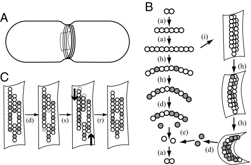
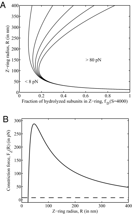
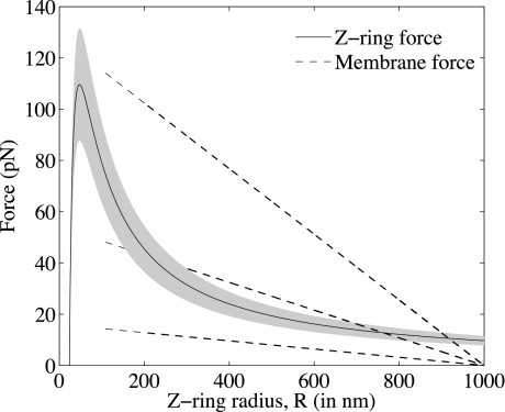
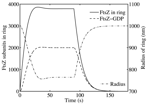
FtsZ, a bacterial homologue of tubulin, plays a central role in bacterial cell division. It is the first of many proteins recruited to the division site to form the Z-ring, a dynamic structure that recycles on the time scale of seconds and is required for division to proceed. FtsZ has been recently shown to form rings inside tubular liposomes and to constrict the liposome membrane without the presence of other proteins, particularly molecular motors that appear to be absent from the bacterial proteome. Here, we propose a mathematical model for the dynamic turnover of the Z-ring and for its ability to generate a constriction force. Force generation is assumed to derive from GTP hydrolysis, which is known to induce curvature in FtsZ filaments. We find that this transition to a curved state is capable of generating a sufficient force to drive cell-wall invagination in vivo and can also explain the constriction seen in the in vitro liposome experiments. Our observations resolve the question of how FtsZ might accomplish cell division despite the highly dynamic nature of the Z-ring and the lack of molecular motors.


Conflict of interest statement

The authors declare no conflict of interest.

Figures

Fig. 1.
Fig. 1.
A schematic illustration of FtsZ kinetics and mechanics. (A) A rod-shaped bacterial cell (E. coli) shown with an invagination in the cell wall and a Z-ring composed of a collection of filaments. (B) From top left: FtsZ-GTP assembles into straight filaments [denoted (a)]; filaments incorporate into the Z-ring (i); subunits hydrolyze their GTP to GDP [white-to-gray transition, denoted (h)] and upon doing so, adopt a highly curved conformation; those in the Z-ring exert a force on the membrane and cell wall; GDP-bound subunits dissociate from the filament tips (d), exchange GDP for GTP (e) and continue to cycle. (C) Four filaments within the Z-ring. From left to right: Hydrolyzed subunits at the tips of filaments disassemble (d); under the influence of the curvature-induced constriction force (thick arrows), weak lateral bonds are broken, filaments slide past each other (s) and reconnect (r).
Fig. 2.
Fig. 2.
Predicted Z-ring forces. (A) The inward radial force, represented as a contour plot, varies with the fraction of subunits that are hydrolyzed, fD, and with the radius of the ring, R, from Eq. 3, for S = 4,000. In this plot, fD and R are treated as parameters. (B) Predicted constriction force generated for different radii, R, calculated by using the full model with the kinetic Eqs. 1 and 2 in quasi-steady state. For radii >25 nm, the force (solid curve) is always >8 pN (dashed line), the required constriction force predicted by Lan et al. (32).
Fig. 3.
Fig. 3.
Plots of the membrane force (left-hand side of Eq. 4, dashed lines) and Z-ring force (right-hand side of Eq. 4, solid line) with S set to its steady-state value as a function of R for kin = 2.1 · 10−4 s−1 μM−1 nm−1 and FM = 16, 54, and 128 pN, which correspond to liposomes 2, 3, and 4 bilayers thick. Notice that for FM corresponding to 2 bilayers, there is no solution.
Fig. 4.
Fig. 4.
Constriction of the Z-ring in a liposome. For kin = 2.1 · 10−4 s−1 μM−1 nm−1 and FM = 54 pN, the ring constricts from 1,000 nm to 765 nm with a half-time of 6.4 s. Once the ring is in steady state, at 90 s, GTP is artificially depleted by imposing ZT = 0. The ring dissolves and the radius expands to its original 1,000 nm with a half-time of 8.8 s.

References

    1. Vaughan S, Wickstead B, Gull K, Addinall S. Molecular evolution of FtsZ protein sequences encoded within the genomes of archaea, bacteria, and eukaryota. J Mol Evol. 2004;58:19–29. - PubMed
    1. Miyagishima S. Origin and evolution of the chloroplast division machinery. J Plant Res. 2005;118:295–306. - PubMed
    1. Bi E, Lutkenhaus J. FtsZ ring structure associated with division in Escherichia coli. Nature. 1991;354:161–164. - PubMed
    1. Lutkenhaus J. FtsZ ring in bacterial cytokinesis. Mol Microbiol. 1993;9:403–409. - PubMed
    1. Ma X, Ehrhardt D, Margolin W. Colocalization of cell division proteins FtsZ and FtsA to cytoskeletal structures in living Escherichia coli cells by using green fluorescent protein. Proc Natl Acad Sci USA. 1996;93:12998–13003. - PMC - PubMed

Publication types

MeSH terms

LinkOut - more resources