. 2009 Jan 16;206(1):249-58.
doi: 10.1084/jem.20080129. Epub 2008 Dec 29.

Chemerin expression marks early psoriatic skin lesions and correlates with plasmacytoid dendritic cell recruitment

Affiliations

Chemerin expression marks early psoriatic skin lesions and correlates with plasmacytoid dendritic cell recruitment

Cristina Albanesi et al. J Exp Med. .

Abstract

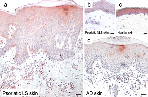
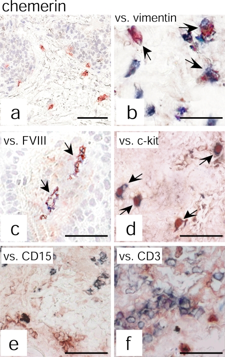
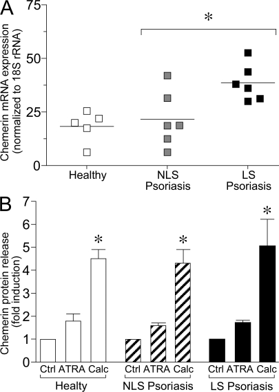
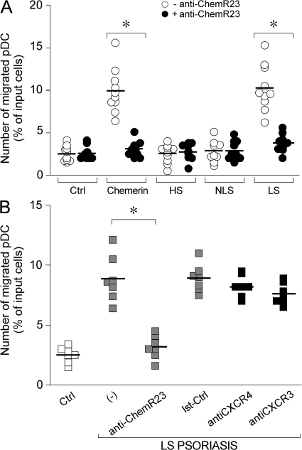
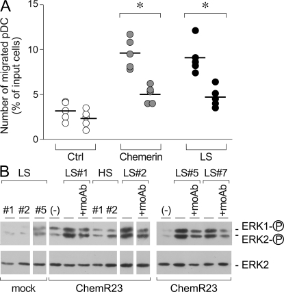
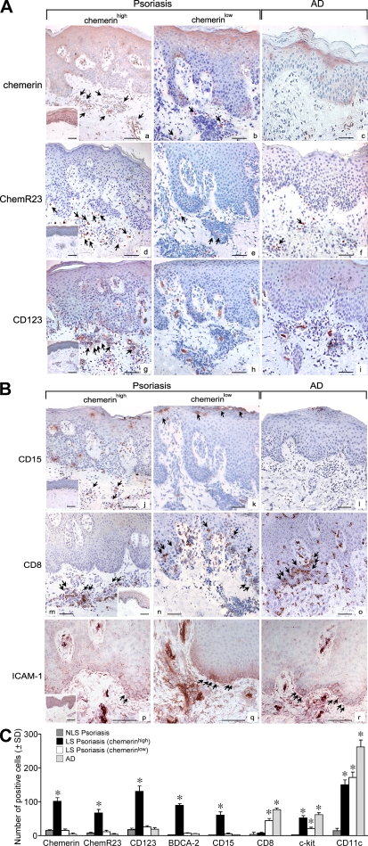
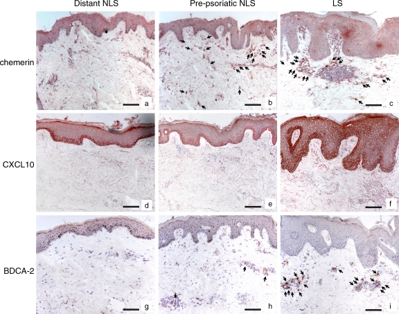
Psoriasis is a type I interferon-driven T cell-mediated disease characterized by the recruitment of plasmacytoid dendritic cells (pDC) into the skin. The molecules involved in pDC accumulation in psoriasis lesions are unknown. Chemerin is the only inflammatory chemotactic factor that is directly active on human blood pDC in vitro. The aim of this study was to evaluate the role of the chemerin/ChemR23 axis in the recruitment of pDC in psoriasis skin. Prepsoriatic skin adjacent to active lesions and early lesions were characterized by a strong expression of chemerin in the dermis and by the presence of CD15(+) neutrophils and CD123(+)/BDCA-2(+)/ChemR23(+) pDC. Conversely, skin from chronic plaques showed low chemerin expression, segregation of neutrophils to epidermal microabscesses, and few pDC in the dermis. Chemerin expression was localized mainly in fibroblasts, mast cells, and endothelial cells. Fibroblasts cultured from skin of psoriatic lesions expressed higher levels of chemerin messenger RNA and protein than fibroblasts from uninvolved psoriatic skin or healthy donors and promoted pDC migration in vitro in a chemerin-dependent manner. Therefore, chemerin expression specifically marks the early phases of evolving skin psoriatic lesions and is temporally strictly associated with pDC. These results support a role for the chemerin/ChemR23 axis in the early phases of psoriasis development.


Figures

Figure 1.
Figure 1.
Chemerin expression in healthy and diseased skin. Immunohistochemistry was performed with an anti-chemerin mAb (red) on the following: a, LS (12 different subjects); b, nonlesional (NLS; 10 different subjects); c, normal skin from healthy donors (6 different subjects); and d, AD LS skin (5 different subjects). Bars, 20 μm. Chemerin was highly expressed in the dermis of psoriatic lesions but not in NLS psoriatic or healthy skin. Few chemerin+ cells were present in the dermis of AD LS skin. In addition, chemerin immunoreactivity was intense throughout the epidermis of healthy and NLS psoriatic skin, whereas in LS it was weak and confined to the suprabasal layer keratinocytes. The figure is representative of the results on skin biopsies obtained from all the different donors indicated.
Figure 2.
Figure 2.
Chemerin expression colocalizes with vimentin+, FVIII+, and c-kit+ cells. Chemerin expression was evaluated by double immunohistochemistry in psoriatic plaque lesions. Anti-chemerin mAb (red) immunoreactivity was detected in dermal cells having fibroblast-like morphology (a). Double-staining analysis revealed that chemerin colocalizes with vimentin (b), FVIII (c), and c-kit (d) but not with CD15 (e) or CD3 (f) staining (all blue). The figure shows the staining of one biopsy that is representative of eight different patients evaluated. Arrows indicate cells that are double positive for chemerin and for vimentin (b), FVIII (c), or c-kit (d). Bars, 20 μm.
Figure 3.
Figure 3.
Selective expression of chemerin by fibroblasts isolated of psoriatic LS skin. (A) Chemerin expression was determined in fibroblast primary cultures prepared from skin cells isolated from healthy donors (five donors) as well as NLS and LS skin of the same psoriatic patients (six donors). Levels of mRNA expression were determined after normalization with 18S ribosomal RNA values. Horizontal lines indicate mean values for each experimental group. (B) Chemerin protein expression was evaluated by ELISA on supernatants from fibroblast cultures derived from NLS and LS psoriatic patients (10 donors) and healthy subject (10 donors) skin cells. Cells were stimulated or not with 10 μM retinoic acid (ATRA) or 1 μM calcitriol (Calc) for 24 h. Data are represented as fold of induction, with basal chemerin release being 321 ± 31, 436 ± 84, and 702 ± 110 pg/ml for healthy, NLS, and LS psoriatic cells, respectively. Error bars are SD of mean values of 10 different donors per group. *, P < 0.05.
Figure 4.
Figure 4.
Psoriatic fibroblasts induced pDC migration in vitro. Supernatants from cultured fibroblasts were tested for their ability to induce blood-purified pDC migration in Transwell experiments. (A) pDC transmigration was assessed using supernatants collected from healthy (HS; 1:9 dilution), NLS (1:9 dilution), and LS (1:81 dilution) psoriasis fibroblast cultures or 100 pM of recombinant chemerin. Anti-ChemR23 antibody was used at 10 μg/ml. (B) Migration of pDC to LS psoriasis supernatants was completely inhibited in the presence of a blocking anti-ChemR23 mAb (IgG2b) but not by an isotype-matched mAb (Ist-Ctrl; anti-CCR6 mAb) or blocking anti CXCR4 and CXCR3 mAbs. Squares represents the results obtained from normal subjects (white), psoriasis patients (gray), and psoriasis patients treated with control mAbs (black). *, P < 0.01 versus respective control group (i.e., without blocking mAb). Results are expressed as single values obtained using supernatants generated from individual cultures obtained from 10 and 7 different donors investigated (A and B, respectively). Horizontal lines indicate mean values for each experimental group.
Figure 5.
Figure 5.
LS skin fibroblasts from psoriatic lesions release bioactive chemerin. (A) Blood-purified pDC were preincubated with 300 pM of recombinant human chemerin at 37°C. Cells were then washed and tested in Transwell assay for their ability to transmigrate an endothelial cell monolayer in response to medium (Ctrl), 100 pM chemerin, or LS supernatant (1:81 dilution). Data were obtained with supernatants obtained from five different donors. *, P < 0.05. Horizontal lines indicate mean values for each experimental group. (B) H460 cells transfected with the empty vector (mock) or ChemR23 were stimulated with fibroblast-derived supernatant from LS skin (1:81 dilution) or healthy donors (HS; 1:9 dilution). Anti-ChemR23 mAb was used at 10 μg/ml. Western blots were performed as detailed in Materials and methods.
Figure 6.
Figure 6.
Infiltration of pDC in psoriatic LS skin correlates with the expression of chemerin and the presence of neutrophils in the dermis. (A and B) Immunohistochemistry was performed using sections from AD lesions (n = 5) and from psoriatic skin showing a chemerinhigh (n = 8) and chemerinlow (n = 12) phenotype. Bars, 20 μm. The insets represent stainings of uninvolved psoriatic skin. Arrows point to positive cells. (C) Positive cells were enumerated in the dermis of psoriatic uninvolved skin (NLS) in chemerinhigh, chemerinlow, and AD skin sections (n = 3). Slides were analyzed blind by two observers and positive cells were counted in 10 adjacent fields at a magnification of 200×. Results are expressed as the mean number of positive cells ±SD. *, P < 0.05 versus NLS skin.
Figure 7.
Figure 7.
Chemerin is highly expressed in prepsoriatic skin where BDCA-2+ pDC start to be accumulated. Immunohistochemistry was performed using sections from early psoriatic plaques at sites of peripheral lesions (LS) together with the adjacent prepsoriatic NLS skin and biopsies from normal skin appearing 3 cm distant from the evolving plaques of the same patients (five total patients examined). Arrows indicate cells that are positive for chemerin (b and c) or BDCA-2 (h and i). Bars, 20 μm.

References

    1. Colonna, M., G. Trinchieri, and Y.J. Liu. 2004. Plasmacytoid dendritic cells in immunity. Nat. Immunol. 5:1219–1226. - PubMed
    1. Liu, Y.J. 2005. IPC: professional type 1 interferon-producing cells and plasmacytoid dendritic cell precursors. Annu. Rev. Immunol. 23:275–306. - PubMed
    1. Colonna, M., and M. Cella. 2007. Crosspresentation: plasmacytoid dendritic cells are in the business. Immunity. 27:419–421. - PubMed
    1. Sapoznikov, A., J.A. Fischer, T. Zaft, R. Krauthgamer, A. Dzionek, and S. Jung. 2007. Organ-dependent in vivo priming of naive CD4+, but not CD8+, T cells by plasmacytoid dendritic cells. J. Exp. Med. 204:1923–1933. - PMC - PubMed
    1. Cella, M., F. Facchetti, A. Lanzavecchia, and M. Colonna. 2000. Plasmacytoid dendritic cells activated by influenza virus and CD40L drive a potent TH1 polarization. Nat. Immunol. 1:305–310. - PubMed

Publication types

MeSH terms