. 2009 Jan 28;23(3):319-28.
doi: 10.1097/QAD.0b013e328321b778.

HIV-1 infection of human penile explant tissue and protection by candidate microbicides

Affiliations

HIV-1 infection of human penile explant tissue and protection by candidate microbicides

Lucia Fischetti et al. AIDS. .

Abstract

Objective: Factors governing events between exposure of male genital mucosa surfaces and the establishment of infection are poorly understood. Furthermore, little is known about the safety and efficacy of microbicides on male genital mucosa.

Design: Here we present a novel penile tissue explant model to characterize the mechanisms of HIV-1 infection of male genital tissue and evaluate candidate microbicides.

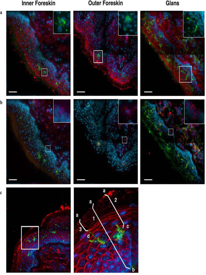
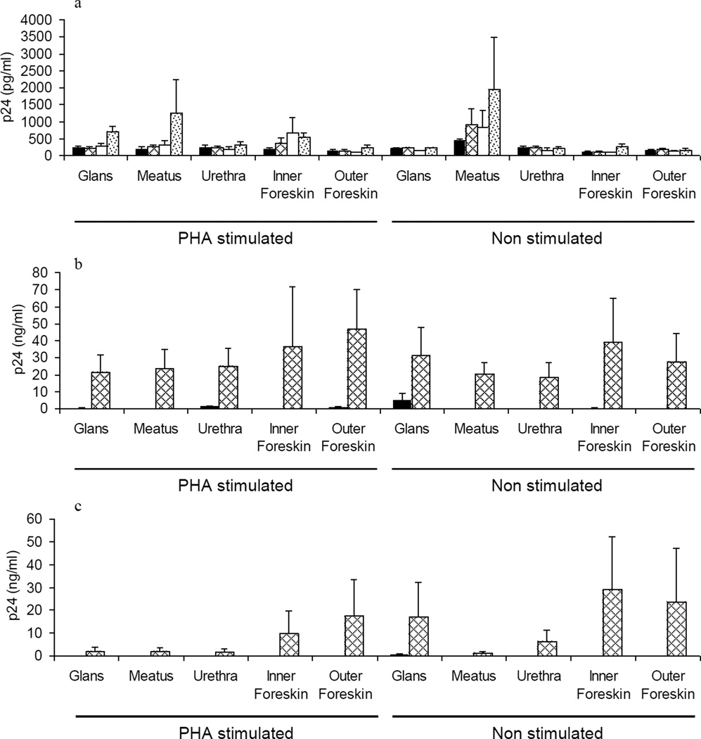
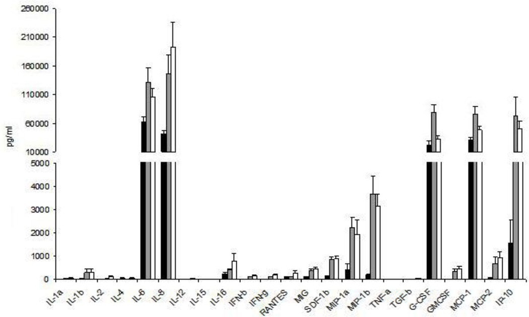
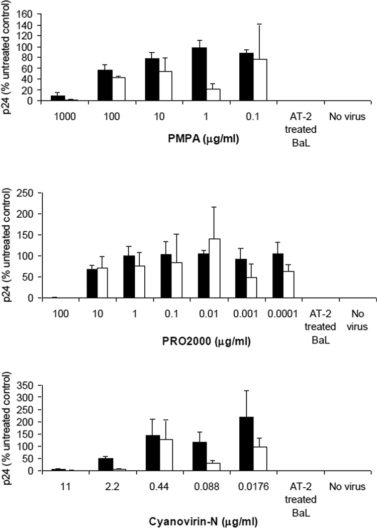
Methods: Mucosal explant culture conditions were determined for glans, urethra and foreskin obtained from gender reassignment and circumcision. Density and distribution of CD4 and CD1a cells were visualized by microscopy. In vitro HIV-1 infection was determined by measuring p24 release, whereas microbicide biocompatibility and efficacy were assessed by measurement of tissue viability, cytokine expression and p24 production.

Results: Cultured glans and foreskin showed comparable epithelial thickness but some differences in CD4 and CD1a cell density. All tissue sites examined (foreskin, glans, meatus, urethra) were equally susceptible to R5 HIV-1 infection, which was productively disseminated by migratory cells emigrating from tissue. In contrast, X4 HIV-1 failed to infect mucosal tissue and dissemination by migratory cells was less efficient. The three candidate microbicides poly(methyl 2-propionamidoacrylate), PRO 2000 and Cyanovirin-N, showed good tissue compatibility and efficient prevention of HIV-1 infection, causing only minor changes in tissue cytokine profile.

Conclusion: The described model provides a useful model to study the determinants of HIV-1 infection of male genital tissue and is likely to be an important tool for the future development of microbicide candidates and concepts.


Figures

Figure 1
Figure 1. Identification of LCs and CD4+ cells in epithelial tissue and illustration of measurements taken to characterize cell localization
Tissue specimens were collected from healthy donors and cultured for up to 1 week. These samples were frozen in OCT, then sectioned and stained with fluorescent antibodies to identify LCs and CD4+ cells. Images are representative of inner foreskin, outer foreskin and glans tissue specimens that were kept for up to 3 days in culture. The same image is shown in both (a) and (b), with different staining patterns. (a) LCs are identified through staining for CD1a (green) and nuclei (blue). Tissue integrity is displayed through staining with fluorescent wheat germ agglutinin (WGA, red). The boxed area, enlarged in the upper right corner, shows LCs (green) and nuclei (blue). (b) CD4+ cells are identified through positive staining for CD4 (red). LCs (green) and nuclei (blue) can also be seen, as before. The boxed area is enlarged in the upper right corner and shows characteristic CD4+ cells (red) and nuclei (blue). Scale bars, 40µm. (c). LCs (green) can be observed in this inner foreskin specimen, which has also been stained with WGA (red) and for nuclei (blue). The boxed area is enlarged at right to show the LC in greater detail. For each cell studied, we measured (1) epithelial thickness, (2) the distance of the cell body to the surface, and, in the case of LCs, (3) the proximity of the closest LC projection to the surface. (1) Epithelial thickness was calculated from the tissue surface (a) to below the epithelial basement membrane (b). (2, 3) The proximity of cell bodies and LC projections to surface were determined by measuring the distance between the tissue surface (a) and either the cell body (c) or LC projection (d).
Figure 2
Figure 2. Density and distribution of LCs and CD4+ cells in genital epithelium
Tissue samples at the initiation of culture (2h, black bars) and 3 days in culture (grey bars) were stained to identify LCs and CD4+ cells and compared. (a) Average thickness of epithelium. (b) Average distance of LC cell bodies to tissue surface. (c) Average distance of LC projections to tissue surface. (d) Average number of LCs found in epithelium per 100µm2 imaged. (e) Average number of CD4+ T cells detected in each tissue type per 100µm2 imaged (stroma + epithelium). (f) Average number of CD4+ T cells found within the surface epithelium per 100µm2 imaged. (g) Average distance of CD4+ cells, found within surface epithelium, to tissue surface. Error bars denote standard error of the mean. P values on top of grey bars indicate significance between 2 hours and 3 days in culture. P values spanning black bars indicate significance between different tissue sites after 2 hours in culture. P values < 0.05 marked with *, <0.01 marked with **, <0.001 marked with ***.
Figure 3
Figure 3. Replication of HIVBaL and HIVLAV in penile tissue explants and dissemination of the infection by migratory cells
Figure 3a shows the time course of HIVBaL replication in tissue explants at days 4 (black bars), 7 (grey net bars), 9 (white bars) and 11 (grey dotted bars) days post infection. Figure 3b and 3c show dissemination of HIVBaL (Figure 3b) and HIVLAV (Figure 3c) infection by migratory cells isolated from different tissue explants, folowing 4 (black bars) and 7 (grey net bars) day co-culture with indicator T cells. Data represent the mean p24 ± standard error of the mean of 3 separate donors; each condition was tested in triplicate. P24 levels for uninfected controls and in the samples exposed to AT-2 treated virus were undetectable (data not shown).
Figure 4
Figure 4. Cytokine production by male genital tissue
Cytokine expression was quantified in glans (black bars), inner foreskin (grey bars) and outer foreskin (white bars) after culturing tissue explants for 24 hours in medium alone. Data represent the mean ± standard error of 9 separate donors, where each donor was tested in triplicate.
Figure 5
Figure 5. Inhibition of HIV infection and viral dissemination by candidate microbicides
Penile tissue explants were exposed to compound (PMPA (Figure 5a), PRO 2000 (Figure 5b) and Cyanovirin-N (Figure 5c)) just prior to exposure to HIV-1BaL in the presence of compound for 2 hours. Virus and compound were removed and explants cultured overnight. Explants were then separated from any cells that had migrated from the tissue and cultured separately for 10 days (black bars). Viral replication was determined by measurement of p24 release into culture supernatant and is expressed as % of the untreated control. Viral dissemination by migratory cells (white bars) was determined by p24 release following 4 day co-culture with indicator T cells. Data represent the mean ± standard error of 3 separate donors, where each donor was tested in triplicate.

References

    1. WHO. AIDS epidemic update. 2007. 2007
    1. European Study Group on Heterosexual Transmission of HIV. Comparison of female to male and male to female transmission of HIV in 563 stable couples. BMJ. 1992;304:809–813. - PMC - PubMed
    1. Hugonnet S, Mosha F, Todd J, Mugeye K, Klokke A, Ndeki L, et al. Incidence of HIV infection in stable sexual partnerships: a retrospective cohort study of 1802 couples in Mwanza Region, Tanzania. J Acquir Immune Defic Syndr. 2002;30:73–80. - PubMed
    1. Nicolosi A, Correa Leite ML, Musicco M, Arici C, Gavazzeni G, Lazzarin A. The efficiency of male-to-female and female-to-male sexual transmission of the human immunodeficiency virus: a study of 730 stable couples. Italian Study Group on HIV Heterosexual Transmission. Epidemiology. 1994;5:570–575. - PubMed
    1. Padian NS, Shiboski SC, Jewell NP. Female-to-male transmission of human immunodeficiency virus. JAMA. 1991;266:1664–1667. - PubMed

Publication types

MeSH terms