. 2008;3(12):e4070.
doi: 10.1371/journal.pone.0004070. Epub 2008 Dec 30.

Akt regulates drug-induced cell death through Bcl-w downregulation

Affiliations

Akt regulates drug-induced cell death through Bcl-w downregulation

Michela Garofalo et al. PLoS One. 2008.

Retraction in

Expression of concern in

Abstract

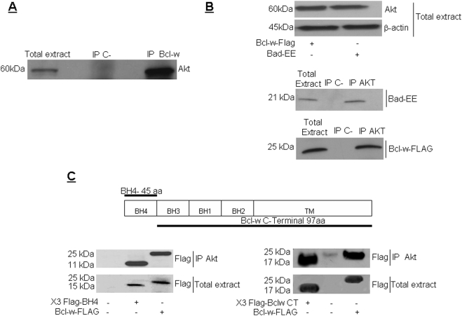
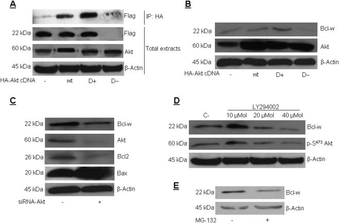
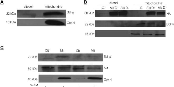
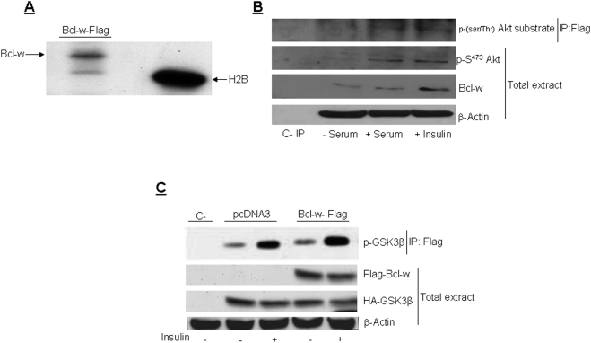
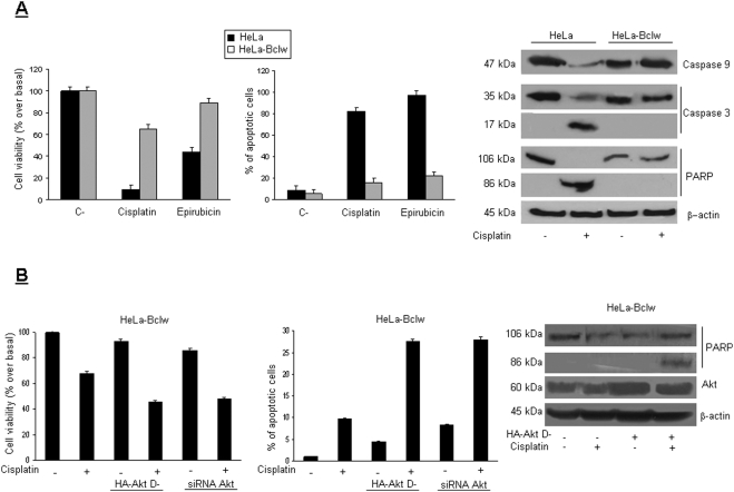
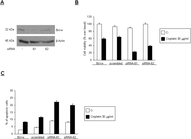
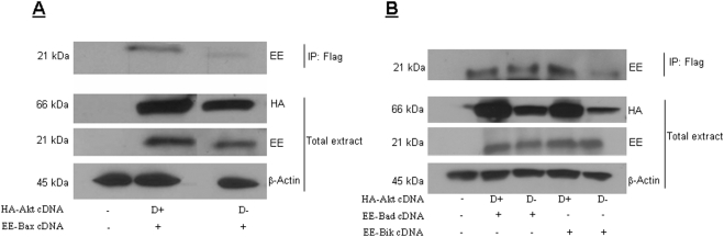
Akt is a serine threonine kinase with a major role in transducing survival signals and regulating proteins involved in apoptosis. To find new interactors of Akt involved in cell survival, we performed a two-hybrid screening in yeast using human full-length Akt c-DNA as bait and a murine c-DNA library as prey. Among the 80 clones obtained, two were identified as Bcl-w. Bcl-w is a member of the Bcl-2 family that is essential for the regulation of cellular survival, and that is up-regulated in different human tumors, such as gastric and colorectal carcinomas. Direct interaction of Bcl-w with Akt was confirmed by immunoprecipitation assays. Subsequently, we addressed the function of this interaction: by interfering with the activity or amount of Akt, we have demonstrated that Akt modulates the amount of Bcl-w protein. We have found that inhibition of Akt activity may promote apoptosis through the downregulation of Bcl-w protein and the consequential reduction in interaction of Bcl-w with pro-apoptotic members of the Bcl-2 family. Our data provide evidence that Bcl-w is a new member of the Akt pathway and that Akt may induce anti-apoptotic signals at least in part through the regulation of the amount and activity of Bcl-w.


Conflict of interest statement

Competing Interests: The authors have declared that no competing interests exist.

Figures

Figure 1
Figure 1. Akt interacts with Bcl-w.
(A) Co-immunoprecipitation of endogenous Akt with Bcl-w. Wt HeLa cells were lysed and 1 mg of protein extract immunoprecipitated using an anti-Bcl-w antibody. Immunoprecipitates and total lysates (50 µg) were separated on 12%SDS polyacrilamide gel and blotted with an anti-Akt antibody. As negative control, proteins were incubated with beads without antibody (B) Co-immunoprecipitation of transfected Akt with FLAG-Bcl-w or EE-BAD. HEK-293 cells were tansfected with 2 µg of HA-Akt and 2 µg of FLAG-Bcl-w or EE-BAD cDNAs, as indicated. After 48 hr, cells were lysed, and 1 mg of protein extract was immunoprecipitated using an anti-HA antibody. Immunoprecipitates were subsequently blotted with anti-HA, anti-Flag or anti-EE antibodies, as indicated. (C) HEK-293 cells were transfected with 2 µg of either wt-Bcl-w cDNA or the deletion mutants, Bcl-w/BH4 or Bcl-w/CT, as indicated. Protein extracts were immunoprecipitated using an anti-Akt antibody. Immunoprecipitates and total lysates were resolved on 12%SDS-PAGE and transferred to Hybond-C nitrocellulose. Membranes were incubated with an anti-FLAG antibody. Both deletion mutants, Bclw/BH4 and Bclw/CT, immunoprecipitated with Akt.
Figure 2
Figure 2. Akt activity regulates Bcl-w expression.
(A) HeLa cells were transfected with 2 µg of HA-Akt wt, Akt D+, or HA-Akt D− cDNA and 2 µg Flag-Bcl-w for 48 hrs. Protein extracts were immunoprecipitated with an anti-HA monoclonal antibody. Immunoprecipitates were resolved on 12% SDS-PAGE and transferred to Hybond-C nitrocellulose. Membranes were incubated with anti-Flag antibody (0.2 µg/ml). 50 µg of total sample extracts were also analyzed by western blot using the indicated antibodies. Loading control was obtained using anti-β actin antibody. (B) HeLa cells were transfected with 4 µg of HA-Akt wt, HA-Akt D+, or HA-Akt D− cDNA for 48 hrs. Protein extracts were blotted with anti-Bcl-w antibody in order to detect endogenous levels of Bcl-w. Loading control was obtained with anti-β actin antibody. (C) Cells were transfected with 100 nM of siAkt-RNA for 48 hrs. Cellular proteins were solubilized and analyzed by western blot using the indicated antibodies. (D) HeLa cells were treated with 10, 20 or 40 µM of LY294002 for 24 hrs. Protein extracts were analyzed by western blot using the indicated antibodies. Loading control was obtained using anti-β actin antibody. (E) Bcl-w HeLa cells were treated with 10 µM of MG-132 for 8 hrs. 40 µg of protein extracts were analyzed by western blot with anti-Bcl-w antibodies. Loading control was obtained using anti-β actin antibody.
Figure 3
Figure 3. Akt controls Bcl-w localization.
(A) HeLa cells were subjected to fractionated separation of mitochondrial/cytosolic proteins using a mitochondria/cytosol fractionation kit (Biovision). Protein extracts were loaded onto 15% SDS polyacrilamide gel, and analyzed by western blot by anti-Bcl-w antibody. As a control of the mitochondrial fraction, an anti-cox4 antibody was used. (B) HeLa cells were transfected with 2 µg of HA-Akt WT, D+, or D− for 48 hrs. Cells were subjected to mitochondria/cytosol separation as above. Protein extracts were analyzed by western blot using anti-Bcl-w, anti-Akt, or anti-cox4 antibodies. (C) Cells were transfected with 100 nM of siAkt-RNA for 48 hrs. Cytosol and mitochondria were isolated as described in the methods and analyzed by western blot using the indicated antibodies.
Figure 4
Figure 4. Akt phosphorylates Bcl-w in vitro and in vivo.
(A) HeLa cells were transfected with 2 µg of DNA of Flag Bcl-w, solubilized, and 1 mg of protein extract was immunoprecipitated with an anti-M2 Flag antibody. Immunoprecipitates were incubated with recombinant constitutive active Akt (rAkt), and in vitro kinase assay was conducted as described in the methods. Samples were loaded onto 2.5% SDS-PAGE and analyzed by autoradiography. As positive control we used Histone2B (H2B). (B) HeLa Bcl-w stable expressing clones were serum starved for 18 hrs and then stimulated with 100 nM insulin or with 20% serum for 15 min as indicated. Cells were solubilized and immunoprecipitated with an anti-M2 Flag antibody. Immunoprecipitates were loaded onto SDS-PAGE and blotted with an anti-phospho Akt substrate antibody that recognizes all the phosphorylated Akt substrates. Total extracts were analyzed by western blot using the indicated antibodies. (C) HeLa cells were transfected with 2 µg of pcDNA3 empty vector or 2 µg of HA-GSK3β, and 2 µg of Flag-Bcl-w for 48 hrs. Cells were stimulated with 100 nM insulin for 15 min, solubilized, immunoprecipitated using an anti-HA antibody, and analyzed by western blot using an anti-phospho-Gsk3 antibody. Total extracts were analyzed by western blot using the indicated antibodies. Bcl-w overexpression does not affect Akt activity.
Figure 5
Figure 5. Akt regulates the anti-apoptotic function of Bcl-w.
(A) HeLa control cells and HeLa cells stably expressing Flag-Bcl-w were plated in 96 well plates in triplicate and treated with 30 µg/ml of cisplatin or 10 µg/ml of epirubicin for 24 hr. Apoptosis was analyzed by Cell Vitality assay, by propidium iodide staining and FACS analysis, or by western blot for caspase cascade activation with anti-caspase-3, -9, and PARP antibodies. Loading control was obtained with anti β-actin. (B) HeLa-Flag Bcl-w cells were transfected with 4 µg of HA-Akt D− cDNA or with 100 nM of siAkt-RNA for 48 hrs and then treated with 30 µg/ml of cisplatin for 24 hr. Cell death was then analyzed as described above. Total lysates were analyzed by western blot using an anti-PARP antibody. Loading control was obtained with an anti-β-actin antibody. Inactivation of Akt activity resulted in a reduction in the protective effect of Bcl-w on cell death.
Figure 6
Figure 6. Effects of Bcl-w si RNA on cell death.
(A) Cells were transfected with 150 nM of siBcl-w-RNAs for 72 hrs. Total lysates were analyzed by western blot using anti-Bcl-w antibodies. Loading control was obtained with an anti-β-actin antibody. (B, C) Cells were transfected with 150 nM of siBcl-w-RNAs for 48 hrs. Then, the cells were splitted into 96 wells and then treated with 30 µg/ml of cisplatin for 24 hr. Cell death was then analyzed with MTT (B) or propidium iodide staining and FACS analysis (C). Bcl-w down-regulation induces an increase of cell death.
Figure 7
Figure 7. (A) Akt activity regulates Bcl-w interaction with Bcl-2 family members.
Flag-Bcl-w/HeLa cells were transfected with 2 µg of either HA-Akt D+ or HA-Akt D− cDNA, and 2 µg of EE-Bax cDNA for 48 hr. Cells were harvested and 1 mg of total lysate immunoprecipitated using an anti-Flag antibody. The immunoprecipitates were then blotted with an anti-EE antibody. Total protein was normalized using anti-EE, -HA or -β-actin antibodies, as indicated. (B) Flag Bcl-w/HeLa cells were transfected with 2 µg of HA-Akt D+ or HA-Akt D− cDNA, and 2 µg of either EE-Bad or EE-Bik cDNA, as indicated, for 48 hr. Cells were harvested and 1 mg of total lysate immunoprecipitated using anti-Flag antibody. The immunoprecipitates were then blotted with an anti-EE antibody. Total protein was normalized using anti-EE, -HA or -β-actin antibodies, as indicated. Inactivation of Akt induced a reduction of Bcl-w interaction with the pro-apoptotic Bcl-2 members.

References

    1. Manning BD, Cantley LC. AKT/PKB signaling: navigating downstream. Cell. 2007;129:1261–1274. - PMC - PubMed
    1. Song G, Ouyang G, Bao S. The activation of Akt/PKB signaling pathway and cell survival. J Cell Mol Med. 2005;9:59–71. - PMC - PubMed
    1. Bellacosa A, Franke TF, Gonzalez-Portal ME, Datta K, Taguchi T, et al. Structure, expression and chromosomal mapping of c-akt: relationship to v-akt and its implications. Oncogene. 1993;8:745–754. - PubMed
    1. Datta SR, Brunet A, Greenberg ME. Cellular survival: a play in three Akts. Genes Dev. 1999;13:2905–2927. - PubMed
    1. Yoeli-Lerner M, Toker A. Akt/PKB signaling in cancer: a function in cell motility and invasion. Cell Cycle. 2006;5:603–605. - PubMed

Publication types

Substances