. 2009 Mar;20(5):1555-64.
doi: 10.1091/mbc.e08-11-1094. Epub 2008 Dec 30.

The subcellular distribution of an RNA quality control protein, the Ro autoantigen, is regulated by noncoding Y RNA binding

Affiliations

The subcellular distribution of an RNA quality control protein, the Ro autoantigen, is regulated by noncoding Y RNA binding

Soyeong Sim et al. Mol Biol Cell. 2009 Mar.

Abstract

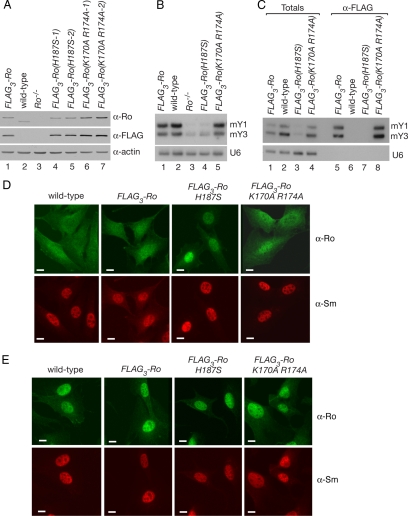
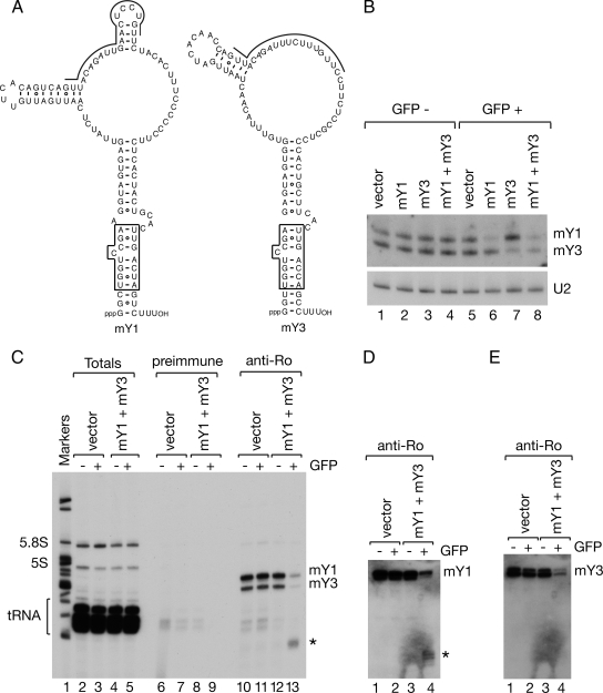
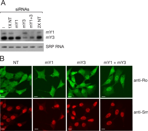
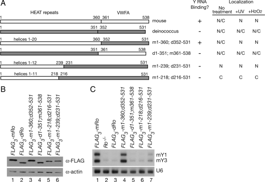
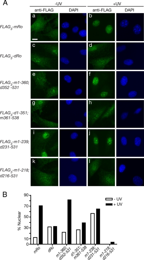
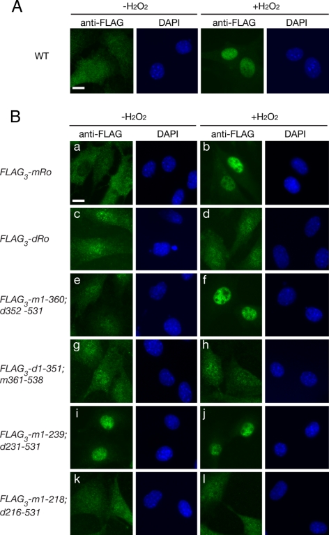
The Ro autoantigen is a ring-shaped RNA-binding protein that binds misfolded RNAs in nuclei and is proposed to function in quality control. In the cytoplasm, Ro binds noncoding RNAs, called Y RNAs, that inhibit access of Ro to other RNAs. Ro also assists survival of mammalian cells and at least one bacterium after UV irradiation. In mammals, Ro undergoes dramatic localization changes after UV irradiation, changing from mostly cytoplasmic to predominantly nuclear. Here, we report that a second role of Y RNAs is to regulate the subcellular distribution of Ro. A mutant Ro protein that does not bind Y RNAs accumulates in nuclei. Ro also localizes to nuclei when Y RNAs are depleted. By assaying chimeric proteins in which portions of mouse Ro were replaced with bacterial Ro sequences, we show that nuclear accumulation of Ro after irradiation requires sequences that overlap the Y RNA binding site. Ro also accumulates in nuclei after oxidative stress, and similar sequences are required. Together, these data reveal that Ro contains a signal for nuclear accumulation that is masked by a bound Y RNA and suggest that Y RNA binding may be modulated during cell stress.


Figures

Figure 1.
Figure 1.
A mutant Ro protein that does not bind Y RNAs accumulates in nuclei. (A) Lysates from wild-type (lane 2), Ro−/− (lane 3) and Ro−/− cell lines stably expressing either FLAG3-Ro (lane 1) or FLAG3-Ro containing the indicated mutations (lanes 4–7) were subjected to Western blotting with anti-Ro (top) and anti-FLAG antibodies (middle). To control for loading, the blot was reprobed to detect actin (bottom). (B) RNA extracted from the cell lines was subjected to Northern blotting to detect the two mouse Y RNAs, mY1 and mY3. The blot was reprobed to detect U6 snRNA (bottom). (C) Lysates from the cell lines were subjected to immunoprecipitation with anti-FLAG antibodies. RNAs from the immunoprecipitates (lanes 5–8) and equivalent amounts of the total extracts (lanes 1–4) were subjected to Northern blotting to detect mY1 and mY3. As a control, the blot was reprobed to detect the U6 snRNA. (D and E) The indicated cell lines were subjected to immunofluorescence with anti-Ro (top) and anti-Sm antibodies (bottom). Cells were either unirradiated (D) or irradiated with 10 J/m2 UVC and allowed to recover for 24 h (E) before staining. Bars, 10 μm.
Figure 2.
Figure 2.
Y RNAs are efficient targets of the RNA interference pathway. (A) Proposed secondary structures of mY1 and mY3 RNAs are shown. The boxed region is a conserved helix that is critical for Ro recognition (Green et al., 1998). The sequences targeted by the shRNAs are indicated by lines. (B) Mouse astrocytes were transfected with plasmids expressing shRNAs against mY1, mY3, or both RNAs and sorted for GFP expression. Vector-transfected cells were used as a negative control. RNA extracted from GFP-negative cells (lanes 1–4) and GFP-positive cells (lanes 5–8) was subjected to Northern blotting to detect mY1 and mY3 RNA. As a loading control, the membrane was reprobed for the U2 small nuclear RNA (bottom). (C) Extracts from GFP-positive cells were subjected to immunoprecipitation with preimmune (lanes 6–9) or anti-Ro antibodies (lanes 10–13). RNAs in immunoprecipitates (lanes 6–13), and a small fraction of the total lysates (lanes 2–5) were labeled at the 3′ end with [32P]pCp. Lane 1, molecular size markers. Asterisk, a fragment of Y RNAs. (D and E) Unlabeled RNAs from the immunoprecipitates shown in C were subjected to Northern blotting to detect the 5′ half of the conserved helix of mY1 (D) and mY3 (E). Asterisk, a fragment corresponding to the 5′ end of mY1.
Figure 3.
Figure 3.
Ro accumulates in nuclei after siRNA-mediated knockdown of Y RNAs. (A) siRNAs against the indicated Y RNAs were transfected into mouse astrocytes. After 2 d, the levels of Y RNAs were analyzed by Northern blotting. As a control, the blot was reprobed to detect SRP RNA. NT, nontarget control siRNAs. (B) At 2 d after transfection, the cells were subjected to immunofluorescence with anti-Ro (top) and anti-Sm antibodies (bottom). Bars, 10 μm.
Figure 4.
Figure 4.
The HEAT repeat domain of mouse Ro is required for Y RNA binding. (A) Summary of the constructs assayed for Y RNA binding and nuclear accumulation after stress. Mouse sequences are shown in white, whereas deinococcal sequences are shaded. Numbers correspond to the amino acids of each Ro protein present within the construct. Because the mouse HEAT repeat domain of mouse Ro contains several insertions that are not present in the bacterial domain, chimeras containing mouse HEAT repeats contain slightly more amino acids than those containing bacterial HEAT repeats. Each construct also contains three copies of the FLAG epitope at the N terminus. (B) After transfection and selection of stable cell lines, Ro protein expression was examined by Western blotting by using an anti-FLAG antibody. To control for loading, the blot was reprobed to detect actin. (C) RNA extracted from the indicated cell lines was subjected to Northern analysis to detect mY1 and mY3 RNAs. As a control, the blot was reprobed to detect U6 snRNA.
Figure 5.
Figure 5.
Sequences for nuclear accumulation reside in the mouse HEAT repeat domain. (A) Ro−/− fibroblasts stably expressing either FLAG-tagged mouse Ro (a and b), D. radiodurans Ro (c and d), or the indicated chimeric proteins (e–l) were assayed for their location in unirradiated cells (a, c, e, g, i, and k) and 24 h after irradiation with 10 J/m2 UVC (b, d, f, h, j, and l). Left, immunofluorescence with anti-FLAG antibodies. Right, nuclei were visualized by staining with DAPI. Bar, 10 μm. (B) Histogram showing the percentage of cells in each cell line that exhibited predominantly nuclear staining before and after UV irradiation. For each measurement, at least 100 cells were counted. For FLAG3-m1-218; d216-531, no unirradiated cells with predominantly nuclear staining were detected.
Figure 6.
Figure 6.
Alignment of Ro proteins. Sequence alignment of Ro proteins from Mus musculus, X. laevis, Danio rerio, Caenorhabditis elegans, Chlamydomonas reinhardtii, and D. radiodurans. Residues that are identical or similar (L = V = I = M, F = Y = W, S = T, E = D, R = K = H) are shaded. α-Helices (gray bars) are assigned based on the X. laevis structure (Stein et al., 2005). Asterisks indicate amino acids shown by mutagenesis to contribute to Y RNA binding (Stein et al., 2005; Fuchs et al., 2006).
Figure 7.
Figure 7.
Nuclear accumulation of Ro during oxidative stress is regulated by Y RNA binding. (A) Wild-type mouse fibroblasts were subjected to immunofluorescence with anti-Ro antibodies before (left) and after incubation with 50 μM hydrogen peroxide for 3 h (right). Nuclei were detected by staining with DAPI. Bar, 10 μm. (B) Ro−/− fibroblasts expressing either FLAG-tagged mouse Ro (a and b), D. radiodurans Ro (c and d), or chimeric proteins (e–l) were assayed for their location in untreated cells (a, c, e, g, i, and k) and after treatment with 50 μM H2O2 for 3 h (b, d, f, h, j, and l). Left, immunofluorescence with anti-FLAG antibodies. Right, nuclei were visualized by staining with DAPI. Bar, 10 μm.

References

    1. Bouffard P., Barbar E., Briere F., Boire G. Interaction cloning and characterization of RoBPI, a novel protein binding to human Ro ribonucleoproteins. RNA. 2000;6:66–78. - PMC - PubMed
    1. Chen X., Quinn A. M., Wolin S. L. Ro ribonucleoproteins contribute to the resistance of Deinococcus radiodurans to ultraviolet irradiation. Genes Dev. 2000;14:777–782. - PMC - PubMed
    1. Chen X., Smith J. D., Shi H., Yang D. D., Flavell R. A., Wolin S. L. The Ro autoantigen binds misfolded U2 small nuclear RNAs and assists mammalian cell survival after UV irradiation. Curr. Biol. 2003;13:2206–2211. - PubMed
    1. Chen X., Wolin S. L. The Ro 60 kDa autoantigen: insights into cellular function and role in autoimmunity. J. Mol. Med. 2004;82:232–239. - PubMed
    1. Chen X., Wurtmann E. J., Van Batavia J., Zybailov B., Washburn M. P., Wolin S. L. An orthologue of the Ro autoantigen functions in 23S rRNA in D. radiodurans. Genes Dev. 2007;21:1328–1339. - PMC - PubMed

Publication types