. 2009 Mar;20(5):1592-604.
doi: 10.1091/mbc.e08-08-0819. Epub 2008 Dec 30.

Genome-wide analysis of AP-3-dependent protein transport in yeast

Affiliations

Genome-wide analysis of AP-3-dependent protein transport in yeast

Vikram C Anand et al. Mol Biol Cell. 2009 Mar.

Abstract

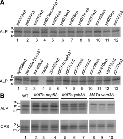
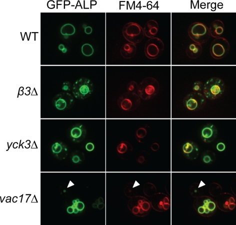
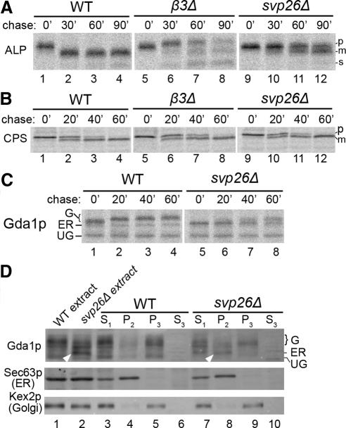
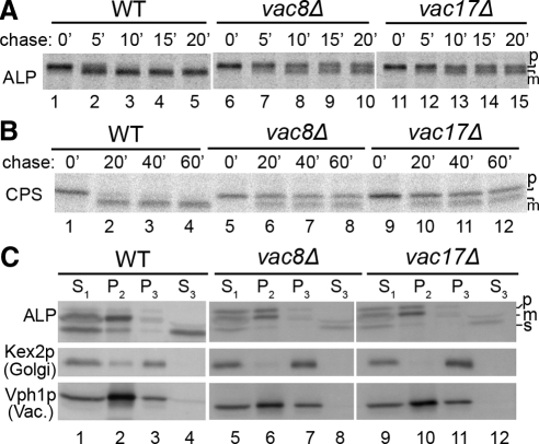
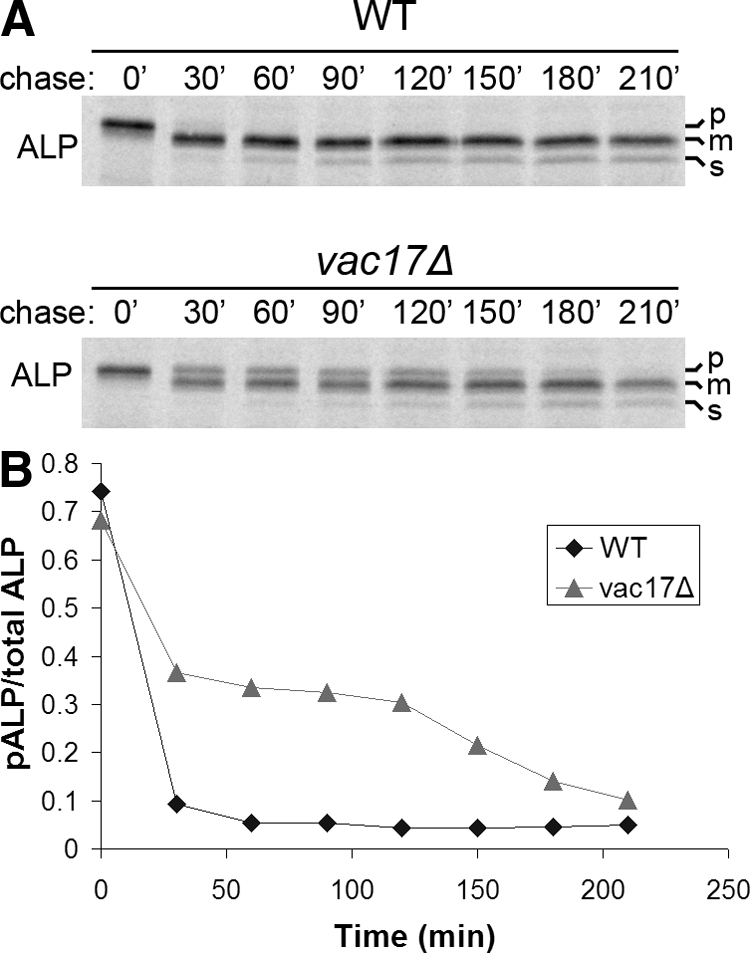
The evolutionarily conserved adaptor protein-3 (AP-3) complex mediates cargo-selective transport to lysosomes and lysosome-related organelles. To identify proteins that function in AP-3-mediated transport, we performed a genome-wide screen in Saccharomyces cerevisiae for defects in the vacuolar maturation of alkaline phosphatase (ALP), a cargo of the AP-3 pathway. Forty-nine gene deletion strains were identified that accumulated precursor ALP, many with established defects in vacuolar protein transport. Maturation of a vacuolar membrane protein delivered via a separate, clathrin-dependent pathway, was affected in all strains except those with deletions of YCK3, encoding a vacuolar type I casein kinase; SVP26, encoding an endoplasmic reticulum (ER) export receptor for ALP; and AP-3 subunit genes. Subcellular fractionation and fluorescence microscopy revealed ALP transport defects in yck3Delta cells. Characterization of svp26Delta cells revealed a role for Svp26p in ER export of only a subset of type II membrane proteins. Finally, ALP maturation kinetics in vac8Delta and vac17Delta cells suggests that vacuole inheritance is important for rapid generation of proteolytically active vacuolar compartments in daughter cells. We propose that the cargo-selective nature of the AP-3 pathway in yeast is achieved by AP-3 and Yck3p functioning in concert with machinery shared by other vacuolar transport pathways.


Figures

Figure 1.
Figure 1.
Example of results from analysis of ALP maturation in strains from the MATa deletion library. (A) Indicated strains were grown in YPD, extracts were prepared, and ALP maturation was assessed by SDS-PAGE and immunoblotting. Strains scored as positive for pALP accumulation are designated with an asterisk. The positions of precursor (p), mature (m), and soluble (s) forms of ALP are indicated. The abundance of the sALP form can vary between experiments depending on growth state and other undetermined factors. (B) Example of results from analysis of ALP and CPS maturation in multiple samples of each strain scored as positive in the genome-wide screen. Three individual colonies from each of the indicated gene deletion strains and WT cells were independently cultured, cell extracts were prepared, and ALP and CPS were detected by immunoblotting. Forms of ALP are indicated as in A. The position of precursor (p) and mature (m) forms of CPS are indicated. The forked line indicates the intermediate band position where pCPS and mCPS forms comigrate. Examples of strains scored in Table 1 as no defect (yck3Δ), moderate defect (pep8Δ), and severe defect (vam3Δ) are shown.
Figure 2.
Figure 2.
Specific defect in ALP transport in yck3Δ cells. (A) Pulse-chase immunoprecipitation of ALP. Cells from WT, apl6Δ-AP-3 β subunit gene deletion strain (β3Δ), and yck3Δ strains were metabolically labeled with [35S]methionine for 7 min, and labeling was quenched with nonradioactive amino acids. Samples of cells were removed at the indicated times after initiation of the chase. Cells were lysed, ALP was immunoprecipitated, and the immunoprecipitates were subjected to SDS-PAGE. Gels were exposed to a phosphor screen and digitally imaged. (B) Pulse-chase immunoprecipitation of CPS. Pulse-chase analysis of CPS was performed as described in A, except that samples were removed at 0, 10, 20, and 40 min after addition of the chase. (C) Subcellular fractionation of yck3Δ cells. Cell lysates from wild-type (WT), apl6Δ (β3Δ), and yck3Δ strains were fractionated by differential centrifugation to generate low-speed (300 × g for 5 min) supernatant (S1), medium-speed (10,000 × g for 15 min) supernatant and pellet (S2, P2), and high-speed (200,000 × g for 17 min) supernatant and pellet (S3, P3) fractions. Fractions were analyzed by SDS-PAGE and immunoblotted for ALP, the Golgi protein Kex2p, and the vacuole membrane (Vac) protein Vph1p. (D) ALP maturation in sorting defective Yck3p mutants. Cells from the MATa yck3Δ strain harboring empty vector, or plasmids containing wild-type YCK3 or two mutant yck3 genes (yck3-1, yck3-2) that lack a YDSI tyrosine-based AP-3 pathway sorting sequence (Sun et al., 2004). Cells were cultured in selective media, cell extracts were prepared and subjected to immunoblotting for ALP.
Figure 3.
Figure 3.
Mislocalization of GFP-ALP in yck3Δ but not vac17Δ cells. WT, β3Δ, yck3Δ, and vac17Δ cells harboring a plasmid encoding GFP-ALP were grown to early-logarithmic phase in minimal media, collected by centrifugation, and stained with FM4-64 for 20 min. The cells were then washed and allowed to internalize the dye for 60 min. Cells were imaged by confocal microscopy to visualize GFP-ALP and FM4-64 localization. Arrowhead indicates a vacuole probably generated de novo.
Figure 4.
Figure 4.
Selective defects in type II membrane protein transport in svp26Δ cells. (A) Pulse-chase immunoprecipitation of ALP. Wild-type (WT), apl6Δ (β3Δ), and svp26Δ cells were analyzed by pulse-chase immunoprecipitation of ALP as described in the legend to Figure 2A, except that chase samples were removed at 0, 30, 60, and 90 min. (B) Pulse-chase immunoprecipitation of CPS. Pulse-chase immunoprecipitation of CPS was performed as described in the legend to Figure 2B, except that samples were removed at 0, 20, 40, and 60 min after addition of the chase. (C) Pulse-chase immunoprecipitation of Gda1p. Pulse-chase immunoprecipitation of Gda1p from the indicated strains was performed as described in the legend to Figure 2A, except that samples were removed at 0, 20, 40, and 60 min after addition of the chase. UG, unglycosylated form of Gda1p (does not diminish in size after treatment with endoglycosidase H [data not shown]); ER, endoplasmic core glycosylated form of Gda1p; G, Golgi glycosylated forms of Gda1p (Vowels and Payne, 1998). (D) Subcellular fractionation of svp26Δ cells. Subcellular fractionation was performed as described in the legend to Figure 3. White arrowheads indicate the accumulated, glycosylated form of Gda1p not present in WT extract. Endoglycosidase H treatment reduces all forms of Gda1p (G, Golgi form; ER, endoplasmic reticulum form; UG, unglycosylated form) in both WT and svp26Δ strains to the UG form (data not shown). Fractions were immunoblotted for Gda1p, the ER protein Sec63p, and Kex2p.
Figure 5.
Figure 5.
ALP and CPS maturation is delayed in vac8Δ and vac17Δ cells. (A) Pulse-chase immunoprecipitation of ALP. Pulse-chase immunoprecipitation of ALP from the indicated strains was performed as described in the legend to Figure 2A except that chase samples were removed at 0, 5, 10, 15, and 20 min. (B) Pulse-chase immunoprecipitation of CPS. Pulse-chase immunoprecipitation of CPS was performed as described in the legend to Figure 4B. (C) Subcellular fractionation of vac8Δ and vac17Δ cells. Subcellular fractionation vac8Δ and vac17Δ cells and immunoblotting analysis was performed as described in the legend to Figure 2C.
Figure 6.
Figure 6.
Accumulated pALP in vac17Δ cells matures after 120 min delay. (A) Pulse-chase immunoprecipitation of ALP from WT and vac17Δ cells was performed as described in the legend to Figure 2A except that chase samples were removed at 0, 30, 60, 90, 120, 150, 180, and 210 min. (B) Bands in A were quantified as described in Materials and Methods, and the ratio of pALP to total ALP was calculated.
Figure 7.
Figure 7.
Model for Yck3p function. Vps41p phosphorylation by Yck3p targets Vps41p for function at the Golgi in AP-3–dependent vesicle formation. Unphosphorylated Vps41p functions as a component of HOPS in vesicle docking/fusion at the vacuole membrane. See text for details. Phosphorylated Vps41p may function at the Golgi as part of the HOPS complex although it is pictured acting independently. The site of Vps41p dephosphorylation and release from AP-3 vesicles is unknown.

Similar articles

Cited by

References

    1. Avaro S., Belgareh-Touze N., Sibella-Arguelles C., Volland C., Haguenauer- Tsapis R. Mutants defective in secretory/vacuolar pathways in the EUROFAN collection of yeast disruptants. Yeast. 2002;19:351–371. - PubMed
    1. Berger A. C., Salazar G., Styers M. L., Newell-Litwa K. A., Werner E., Maue R. A., Corbett A. H., Faundez V. The subcellular localization of the Niemann-Pick Type C proteins depends on the adaptor complex AP-3. J. Cell Sci. 2007;120:3640–3652. - PubMed
    1. Bonangelino C. J., Catlett N. L., Weisman L. S. Vac7p, a novel vacuolar protein, is required for normal vacuole inheritance and morphology. Mol. Cell. Biol. 1997;17:6847–6858. - PMC - PubMed
    1. Bonangelino C. J., Chavez E. M., Bonifacino J. S. Genomic screen for vacuolar protein sorting genes in Saccharomyces cerevisiae. Mol. Biol. Cell. 2002;13:2486–2501. - PMC - PubMed
    1. Bonifacino J. S. The GGA proteins: adaptors on the move. Nat. Rev. Mol. Cell Biol. 2004;5:23–32. - PubMed

Publication types

MeSH terms