. 2009 May;150(5):2419-28.
doi: 10.1210/en.2008-1385. Epub 2008 Dec 30.

Characterization of relaxin receptor (RXFP1) desensitization and internalization in primary human decidual cells and RXFP1-transfected HEK293 cells

Affiliations

Characterization of relaxin receptor (RXFP1) desensitization and internalization in primary human decidual cells and RXFP1-transfected HEK293 cells

András Kern et al. Endocrinology. 2009 May.

Abstract

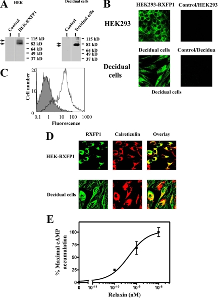
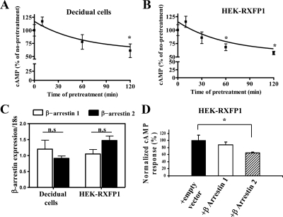
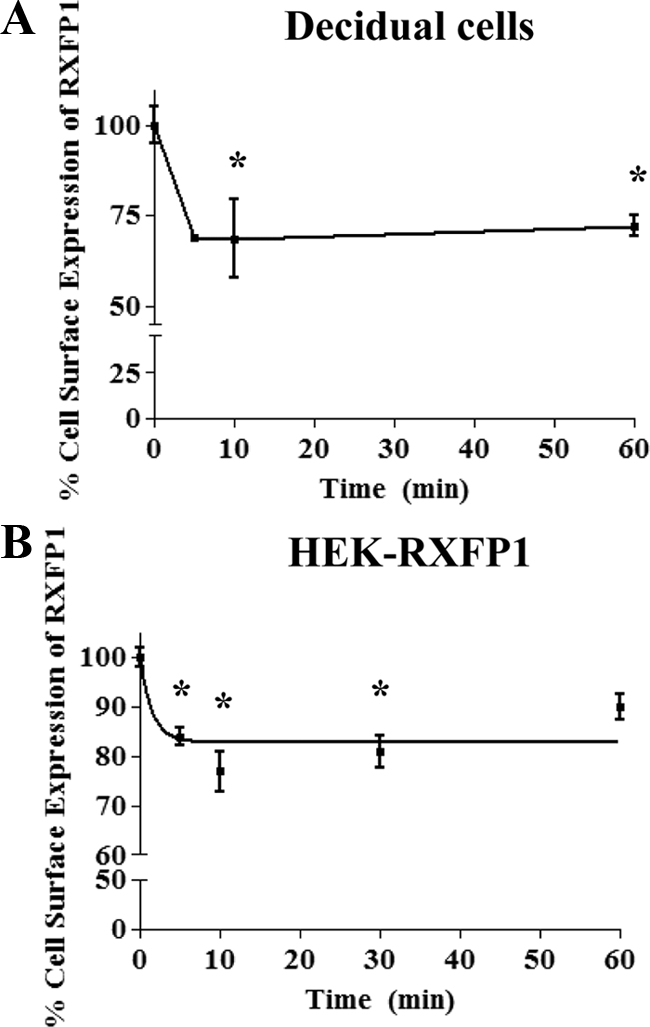
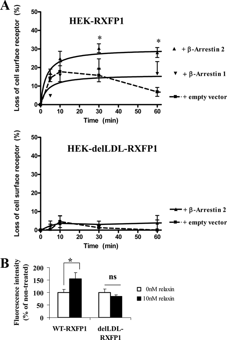
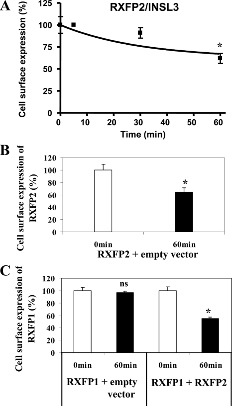
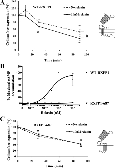
We report here the desensitization and internalization of the relaxin receptor (RXFP1) after agonist activation in both primary human decidual cells and HEK293 cells stably transfected with RXFP1. The importance of beta-arrestin 2 in these processes has also been demonstrated. Thus, in HEK-RXFP1 cells the desensitization of RXFP1 was significantly increased when beta-arrestin 2 was overexpressed. After relaxin activation, beta-arrestin 2 was translocated to the cell membrane and RXFP1 underwent rapid internalization. We have previously shown that RXFP1 forms dimers/oligomers during its biosynthesis and trafficking to the plasma membrane, we now show that internalization of RXFP1 occurs through this dimerization/oligomerization. In nonagonist stimulated cells, it is known that the majority of the RXFP1 is located intracellularly and was confirmed in the cells used here. Constitutive internalization of RXFP1 could account for this and indeed, slow but robust constitutive internalization, which was increased after agonist stimulation was demonstrated. A carboxyl-terminal deleted RXFP1 variant had a similar level of constitutive agonist-independent internalization as the wild-type RXFP1 but lost sensitivity to agonist stimulation. This demonstrated the importance of the carboxyl terminus in agonist-stimulated receptor internalization. These data suggest that the autocrine/paracrine actions of relaxin in the decidua are under additional controls at the level of expression of its receptor on the surface of its target cells.


Figures

Figure 1
Figure 1
A, RXFP1 protein expression in HEK-RXFP1 and primary decidual cells. Membrane preparations from HEK-RXFP1 cells, decidual cells, and untransfected HEK293 cells (control) were analyzed by Western blotting. RXFP1 expression was detected in HEK-RXFP1 and decidual cells, indicated by arrows, and not in untransfected HEK293 cells (control). B, Immunocytochemistry of RXFP1 in HEK-RXFP1 cells and decidual cells. Nonpermeabilized cells were stained with a monoclonal antibody to RXFP1 followed by a mouse AlexaFluor 488-conjugated secondary antibody, showing membrane localization of RXFP1. No staining was detected in untransfected HEK293 cells (control). C, FACS analysis of isolated decidual cells. Cell surface expression of RXFP1 was measured by FACS in nonpermeabilized decidual cells. The fluorescence of the control cells (no primary antibody to RXFP1) is shown by the solid histogram compared with the cell population stained with primary antibody to RXFP1 and mouse AlexaFluor 488 secondary antibody in the open histogram. D, The subcellular localization of RXFP1 in HEK-RXFP1 (upper row) and decidual cells (bottom row). Cells were fixed, permeabilized and double labeled for confocal microscopy. The ER resident proteins were labeled with antibody to calreticulin (middle panels), and RXFP1 proteins were labeled in parallel using a monoclonal antibody to RXFP1 (left panels). Secondary antibodies were AlexaFluor 488 or AlexaFluor 546. The overlay (right panels) shows colocalization of RXFP1 with the ER marker calreticulin. E, Functional characterization of RXFP1 in isolated decidual cells. Relaxin caused a dose-dependent stimulation of cAMP production from decidual cells. cAMP accumulation is expressed as percentage of the maximal relaxin response. The results shown are mean of ± sem of four independent decidual cell isolation experiments performed in duplicate.
Figure 2
Figure 2
Desensitization of RXFP1 and the effect of β-arrestins. Isolated decidual cells (A) or HEK-RXFP1 (B) were exposed to 0.1 nm relaxin for the times indicated. Cells were rapidly washed and restimulated with 0.1 nm relaxin. The intracellular cAMP levels were measured and its accumulation expressed as percentage of the maximal relaxin response. Pretreatment with relaxin caused a time-dependent attenuation of cAMP levels after the second treatment with relaxin. The results shown are mean of ± sem of three independent experiments performed in duplicate. *, P < 0.05 compared with 0 min. C, qRT-PCR analysis of mRNA expression of β-arrestins 1 and 2 in decidual cells and HEK-RXFP1 cells. Total RNA was obtained from decidual cells isolated from different patients (n = 4) and HEK-RXFP1cells (n = 4). After RT qRT-PCR was performed using Applied Biosystems primers for β-arrestins 1 and 2. β-Arrestin expression was normalized to the expression of 18S in each sample and shown as relative gene expression ± sem. Both arrestins were expressed in both cell types, and there were no significant (ns) differences in their levels of expression. D, Effects of β-arrestin 1 and 2 on desensitization of RXFP1 in HEK-RXFP1 cells. HEK-RXFP1 cells were cotransfected with either β-arrestin 1 or 2 or cotransfected with an empty vector (control). These cells were pretreated with 0.1 nm relaxin for 60 min, quickly washed, and restimulated with 0.1 nm relaxin. The cAMP responses (percent) were normalized to the control cells. Overexpression of β-arrestin 2 significantly increased attenuation of the RXFP1 response after relaxin treatment. Data are means ± sem of three independent experiments performed in duplicate. *, P < 0.05 compared with controls (HEK-RXFP1 transfected with an empty vector).
Figure 3
Figure 3
Internalization of RXFP1 after relaxin treatment. A, Relaxin induced cell surface expression of RXFP1 measured by FACS analysis in decidual cells. Decidual cells were treated with 10 nm relaxin for different times (5–60 min) and FACS performed. Fluorescence was normalized to the basal RXFP1 cell surface expression at 0 min (untreated). Relaxin treatment resulted in the rapid loss of RXFP1 cell surface expression by 5 min, remaining constant for up to 60 min. *, P < 0.05 compared with unstimulated cells. B, Relaxin induced cell surface expression of RXFP1 measured by cell surface ELISA in HEK-RXFP1 cells. Cells were treated with 10 nm relaxin for different times (5–60 min) and the cell surface ELISA performed. Data were normalized to the basal RXFP1 cell surface expression for untreated cells at 0 min. Relaxin treatment caused the rapid loss of RXFP1 cell surface expression by 5 min. Data are means ± sem of three independent experiments performed in duplicate. *, P < 0.05 compared with unstimulated cells.
Figure 4
Figure 4
A. Effects of β-arrestins 1 and 2 on the internalization of RXFP1 and delLDL-RXFP1 in stably transfected HEK293 cells. HEK-RXFP1 cells (upper panel) or HEK-delLDL-RXFP1 cells (lower panel) were transfected with either of the β-arrestins or an empty vector. Cells were stimulated with 10 nm relaxin for the time indicated (5–60 min) and the cell surface expression of RXFP1 measured by ELISA. Overexpression of β-arrestin 2 caused the significantly increased loss of cell surface expression in HEK-RXFP1 cells, whereas overexpression of β-arrestin 1 had no significant effect (upper panel). There was no internalization of the delLDL-RXFP1 variant with or without cotransfection with β-arrestin 2 and treatment with relaxin up to 60 min. Data are means ± sem of three independent experiments performed in duplicate and expressed as the percentage loss of cell surface expression compared with unstimulated cells (0 min). *, P < 0.05 compared with cells transfected with an empty vector. B, Translocation of β-arrestin 2-YFP to the membrane after relaxin treatment. HEK-RXFP1 cells and HEK-delLDL-RXFP1 cells were cotransfected with β-arrestin 2-YFP and stimulated with 10 nm relaxin for 10 min. Membranes were isolated and β-arrestin 2-YFP monitored by fluorescence measurement of YFP. A significant increase in the fluorescence of the membrane preparation from the relaxin-treated HEK-RXFP1 cells but no significant (ns) increase in the delLDL-RXFP1 variant after relaxin treatment. β-Arrestin 2 therefore has an important role in RXFP1 internalization. Data are means ± sem of three independent experiments performed in duplicate and expressed as percentage of unstimulated cells (0 min, 100%). *, P < 0.05 compared with unstimulated cells.
Figure 5
Figure 5
RXFP1 internalization occurs through dimerization. A, INSL3 induced loss of cell surface expression of RXFP2 measured by cell surface ELISA in RXFP2 transiently transfected HEK293 cells. Cells were treated with 10 nm INSL3 for the times shown (5–60 min) and the cell surface ELISA performed. *, P < 0.05 compared with unstimulated cells. B and C, RXFP1 internalization was caused through RXFP1/RXFP2 heterodimerization. HEK293 cells transiently cotransfected with RXFP2 and an empty vector were treated with 10 nm INSL3 for 60 min and the cell surface ELISA performed (B). HEK293 cells transiently cotransfected with either RXFP1 and an empty vector or with RXFP1 and RXFP2, were treated with 10 nm INSL3 for 60 min, and the cell surface ELISA performed (C). Cell surface expression was normalized to the basal RXFP2 (A and B) or RXFP1 (C) cell surface expression for the untreated cells at 0 min. Cell surface expression of RXFP1 was significantly decreased in the cells cotransfected with RXFP1 and RXFP2, confirming that dimerization had occurred. Data are means ± sem of three independent experiments performed in duplicate. *, P < 0.05 compared with unstimulated cells. ns, Not significant.
Figure 6
Figure 6
Constitutive internalization of RXFP1. A, HEK-RXFP1 cells were preincubated with an HA antibody, washed, and incubated at 37 C for the times indicated, in the absence (♦) or presence (▪) of 10 nm relaxin. Receptors remained at the cell surface after incubation at 37 C either with or without relaxin were expressed as percentage of receptors at the cell surface at 0 min. Relaxin significantly enhanced the constitutive internalization of RXFP1. Data are means ± sem of three independent experiments performed in duplicate. *, P < 0.05 compared with 0 min; #, P < 0.05 compared with nonstimulated cells. WT-RXFP1, Wild-type RXFP1 . B, Functional characterization of variant RXFP1-687 in HEK293 cells. HEK293 cells were transiently transfected with either wild-type RXFP1 or variant RXFP1-687 with the carboxyl terminus deleted. Relaxin caused a dose-dependent stimulation of cAMP production in cells transfected with WT-RXFP1 (▪), whereas it had no effect on the cells transfected with RXFP1-687 (▾). This showed the importance of the carboxyl terminus of RXFP1 in signal transduction. Accumulation of cAMP was expressed as percentage of the maximal relaxin response of the WT-RXFP1. Data are mean ± sem of two independent experiments performed in duplicate. C, Constitutive internalization of RXFP1-687 in HEK293 cells. HEK293 cells transiently transfected with RXFP1-687 were preincubated with an HA antibody, washed, and warmed to 37 C for the times indicated in the absence (♦) or presence (▪) of 10 nm relaxin. Receptors remained at the cell surface after warming to 37 C in either the absence or presence of relaxin and were expressed as a percentage of receptors at the cell surface at 0 min. The RXFP1-687 variant underwent constitutive internalization to the similar extent as WT-RXFP1. However, there was no significant change in the presence of relaxin, showing that the carboxyl terminus of RXFP1 is important in agonist-induced internalization of RXFP1. Data are means ± sem of three independent experiments performed in duplicate. *, P < 0.05 compared with 0 min.

References

    1. James R, Niall H, Kwok S, Bryant-Greenwood GD 1977 Primary structure of porcine relaxin: homology with insulin and related growth factors. Nature 267:544–546 - PubMed
    1. Sherwood OD 2004 Relaxin’s physiological roles and other diverse actions. Endocr Rev 25:205–234 - PubMed
    1. Hombach-Klonisch S, Bialek J, Trojanowicz B, Weber E, Holzhausen HJ, Silvertown JD, Summerlee AJ, Dralle H, Hoang-Vu C, Klonisch T 2006 Relaxin enhances the oncogenic potential of human thyroid carcinoma cells. Am J Pathol 169:617–632 - PMC - PubMed
    1. Samuel CS, Hewitson TD 2006 Relaxin in cardiovascular and renal disease. Kidney Int 69:1498–1502 - PubMed
    1. Mookerjee I, Solly NR, Royce SG, Tregear GW, Samuel CS, ML. T 2006 Endogenous relaxin regulates collagen deposition in an animal model of allergic airway disease. Endocrinology 147:754–761 - PubMed

Publication types

MeSH terms