. 2009 Feb;174(2):380-9.
doi: 10.2353/ajpath.2009.080565. Epub 2008 Dec 30.

Activin-like kinase 5 (ALK5) mediates abnormal proliferation of vascular smooth muscle cells from patients with familial pulmonary arterial hypertension and is involved in the progression of experimental pulmonary arterial hypertension induced by monocrotaline

Affiliations

Activin-like kinase 5 (ALK5) mediates abnormal proliferation of vascular smooth muscle cells from patients with familial pulmonary arterial hypertension and is involved in the progression of experimental pulmonary arterial hypertension induced by monocrotaline

Matthew Thomas et al. Am J Pathol. 2009 Feb.

Abstract

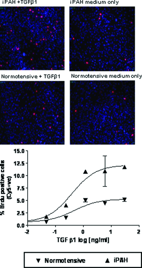
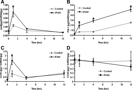
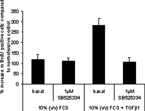
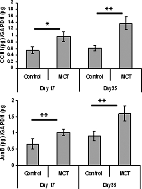
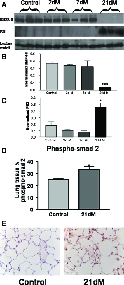
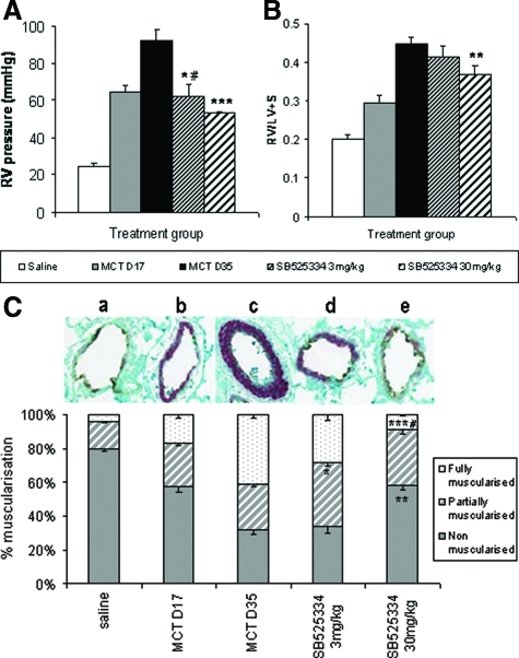
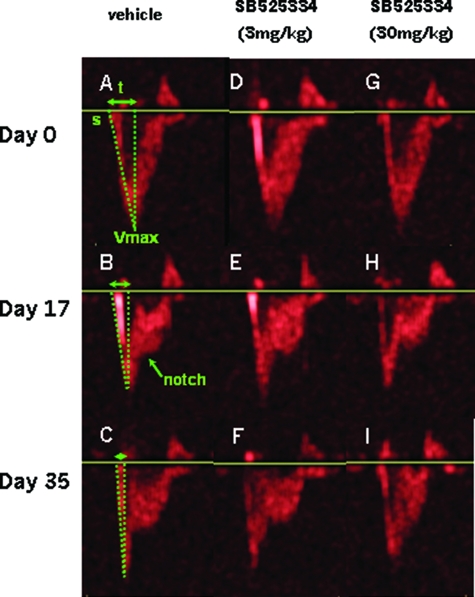
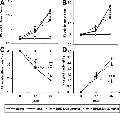
Mutations in the gene for the transforming growth factor (TGF)-beta superfamily receptor, bone morphogenetic protein receptor II, underlie heritable forms of pulmonary arterial hypertension (PAH). Aberrant signaling via TGF-beta receptor I/activin receptor-like kinase 5 may be important for both the development and progression of PAH. We investigated the therapeutic potential of a well-characterized and potent activin receptor-like kinase 5 inhibitor, SB525334 [6-(2-tert-butyl-5-{6-methyl-pyridin-2-yl}-1H-imidazol-4-yl)-quinoxaline] for the treatment of PAH. In this study, we demonstrate that pulmonary artery smooth muscle cells from patients with familial forms of idiopathic PAH exhibit heightened sensitivity to TGF-beta1 in vitro, which can be attenuated after the administration of SB525334. We further demonstrate that SB525334 significantly reverses pulmonary arterial pressure and inhibits right ventricular hypertrophy in a rat model of PAH. Immunohistochemical studies confirmed a significant reduction in pulmonary arteriole muscularization induced by monocrotaline (used experimentally to induce PAH) after treatment of rats with SB525334. Collectively, these data are consistent with a role for the activin receptor-like kinase 5 in the progression of idiopathic PAH and imply that strategies to inhibit activin receptor-like kinase 5 signaling may have therapeutic benefit.


Figures

Figure 1
Figure 1
PASMCs derived from iPAH or normotensive patients were plated at equal cell densities in 96-well plates. Cells were serum-starved for 48 hours before treatment with 0.625 ng/ml of TGF-β1. Proliferation was measured by BrdU incorporation after 6 days. The percent of positive cells was calculated as the number of BrdU-positive cells/total number of cells determined by Hoechst staining. Original magnifications, ×100.
Figure 2
Figure 2
Enhanced transcriptional responses to TGF-β1 on iPAH PASMCs. iPAH and control PASMCs were cultured to confluence, serum-starved, and treated for 0, 1, 4, and 12 hours in the presence or absence of TGF-β1 (2 ng/ml). Cells were cultured from four individuals with iPAH and three control individuals. Total RNA was harvested, reverse-transcribed into cDNA, and subjected to real-time PCR analysis performed in duplicate with primers recognizing JunB (A), PAI-1 (B), CCN1 (C), and CCN3 (D). Ratios of target genes/GAPDH RNA levels ± SD plotted. *P < 0.05 (Student’s t-test) compared with respective control time point.
Figure 3
Figure 3
PASMCs derived from iPAH patients were plated at equal cell densities in 96-well plates. Cells were starved for 48 hours before treatment with 0.625 ng/ml of TGF-β1 in growth media containing 10% (v/v) fetal calf serum. One μmol/L of SB525334 was added 15 minutes before the addition of TGF-β1. Proliferation was measured by BrdU incorporation after 6 days. The percentage of cells that were BrdU-positive was calculated and normalized to the average BrdU incorporation in untreated normotensive cells.
Figure 4
Figure 4
MCT-treated rat lungs exhibit enhanced transcription of TGF-β-regulated genes. Total RNA from rat lungs were harvested 17 days after MCT treatment (n = 10) or vehicle alone (n = 5) and 35 days after MCT treatment (n = 10) or vehicle alone (n = 5). The expression of JunB and CCN1 was determined by real-time PCR analysis. CCN1 (top) and JunB (bottom) expression were determined and the ratios of target genes/GAPDH RNA levels ± SEM plotted. *P < 0.05, **P < 0.01 (Student’s t-test) compared with respective control time point.
Figure 5
Figure 5
A: Changes in the expression of bone morphogenetic protein (BMPR-II) and phosphorylated Smad 3 (PS3) in the lungs of MCT-treated rats was assessed by immunoblotting (dM indicates days of MCT treatment). B and C: Quantification of immunoblotting for BMPR-II (B) and PS3 (C). The data shown are the mean density of the appropriate band ± SEM. *P < 0.05, ***P < 0.001 (Student’s t-test) compared with the respective control time point. D: Quantification of the expression of phosphorylated Smad 2 in the lung by immunohistochemistry. The data plotted are the mean area ± SEM of positive stained tissue expressed as a percentage of the total parenchymal area. *P < 0.05 (Student’s t-test) compared with control lung tissue area. E: Representative pictures of immunohistochemistry in lung tissue for phospho-Smad 2, brown indicates phospho-Smad 2 expression. Original magnifications, ×40.
Figure 6
Figure 6
RV systolic pressure levels (A) and Fulton index measures (RV/LV + S weight ratio) (B) in rats exposed to MCT or saline-negative control. Analysis was performed in rats at the point of established PAH pathology (day 17, gray bars), after which animals were orally treated with either vehicle (black bars), 3 mg/kg (fine hatched bars), or 30 mg/kg (thick hatched bars) SB525334 until day 35. Values are means ± SEM. C: Arteriole remodeling in rats after MCT exposure. Inflated lung sections (n = 10 per group) were stained with vWL and anti-α-smooth muscle actin, and then 200 small vessels (<100 μmol/L) per section were analyzed by an investigator unaware of the source of tissue. Each vessel was assigned as either nonmuscularized (no α-smooth muscle actin staining), partially muscularized, or fully muscularized (thick unbroken wall of smooth muscle), and then the percentage distribution of each calculated per group. A representative picture of the predominantly altered vessel phenotype is provided above each group (ae). Values are the means ± SEM, #P < 0.05 for MCT day 17 versus vehicle-treated control. *P < 0.01, **P < 0.001, ***P < 0.001 for rct day 35 versus vehicle-treated control (Student’s t-test).
Figure 7
Figure 7
Pulsed wave Doppler profiles of blood flow through the pulmonary artery outflow tract. The start of flow (s) reaches a maximum velocity (Vmax) within a given time (t), known as the pulmonary artery acceleration time, which increases in accordance with pulmonary artery pressure (A–C). The smooth flow deceleration of a normal RV circulation can also change with hypertension to show a characteristic midsystolic notch (B). Day 0 profiles (A, D, and G) are of normal animals (Veh), whose hypertensive state is seen at day 17 after MCT exposure, and immediately before commencement of compound dosing (B, E, and H). The resultant stabilization, or reversal of hypertensive pathologies, is shown at day 35 (C, F, and I) in the same animals shown at day 17.
Figure 8
Figure 8
Echocardiographic measurement of pulmonary hypertensive parameters in animals serially investigated after MCT exposure at day 0, the establishment of reliable pathologies at day 17, and the dosing of either vehicle, or SB525334 from days 17 to 35 (noted above). RV wall thickness during systole (A) and diastole (B), pulmonary artery acceleration time (C) and degree of midsystolic notch (D) are shown. Vehicle versus MCT-exposed; vehicle-treated groups are represented by open and closed circles, respectively. Treated groups are shown by dashed lines at 3 mg/kg (closed triangle) and 30 mg/kg (closed square). Values are means ± SEM. *P < 0.01 versus day 35 vehicle-treated control (one-way analysis of variance and Kruskal-Wallis test).

References

    1. Chan SY, Loscalzo JJ. Pathogenic mechanisms of pulmonary arterial hypertension. J Mol Cell Cardiol. 2008;44:14–30. - PMC - PubMed
    1. Lane KB, Machado RD, Pauciulo MW, Thomson JR, Phillips JA, III, Loyd JE, Nichols WC, Trembath RC. Heterozygous germline mutations in BMPR2, encoding a TGF-beta receptor, cause familial primary pulmonary hypertension. The International PPH Consortium. Nat Genet. 2000;26:81–84. - PubMed
    1. Morrell NW, Yang X, Upton PD, Jourdan KB, Morgan N, Sheares KK, Trembath RC. Altered growth responses of pulmonary artery smooth muscle cells from patients with primary pulmonary hypertension to transforming growth factor-beta(1) and bone morphogenetic proteins. Circulation. 2001;104:790–795. - PubMed
    1. Richter A, Yeager ME, Zaiman A, Cool CD, Voelkel NF, Tuder RM. Impaired transforming growth factor-beta signalling in idiopathic pulmonary arterial hypertension. Am J Respir Crit Care Med. 2004;170:1340–1348. - PubMed
    1. Jachec W, Foremny A, Domal-Kwiatkowska D, Smolik S, Tomasik A, Mazurek U, Wodniecki J. Expression of TGF-beta1 and its receptor genes (TbetaR I, TbetaR II, and TbetaR III-betaglycan) in peripheral blood leucocytes in patients with idiopathic pulmonary arterial hypertension and Eisenmenger’s syndrome. Int J Mol Med. 2008;21:99–107. - PubMed

Publication types

MeSH terms

Substances