. 2009 Jan;174(1):101-14.
doi: 10.2353/ajpath.2009.080599. Epub 2008 Dec 30.

Collagen fragmentation promotes oxidative stress and elevates matrix metalloproteinase-1 in fibroblasts in aged human skin

Affiliations

Collagen fragmentation promotes oxidative stress and elevates matrix metalloproteinase-1 in fibroblasts in aged human skin

Gary J Fisher et al. Am J Pathol. 2009 Jan.

Abstract

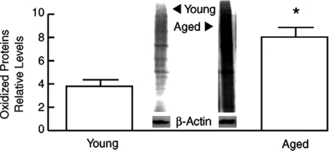
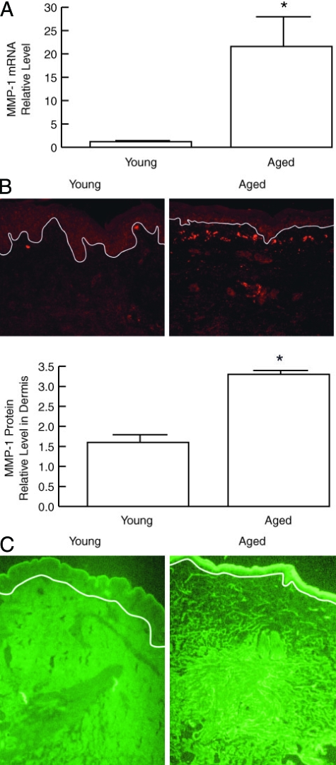
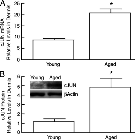
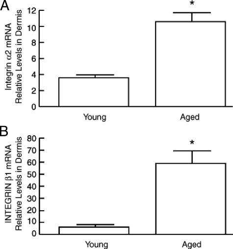
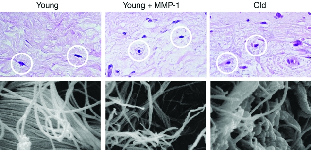
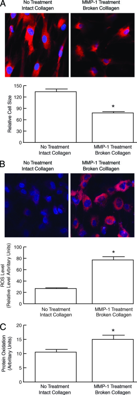
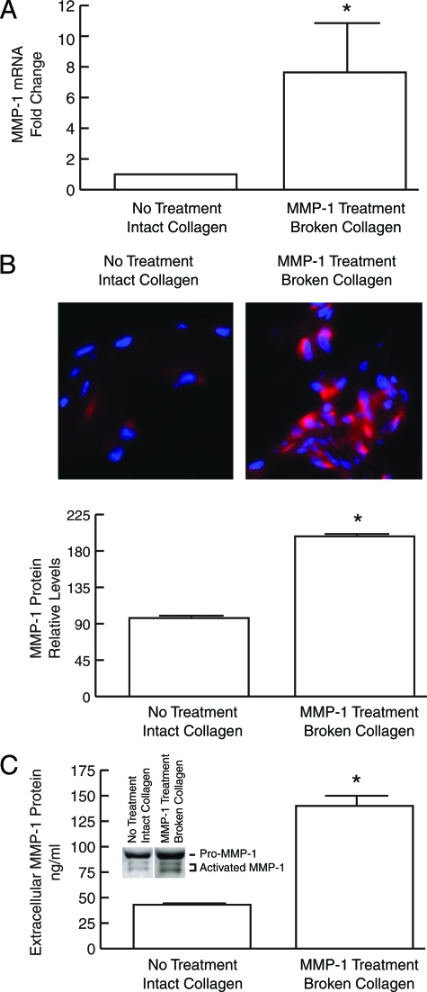
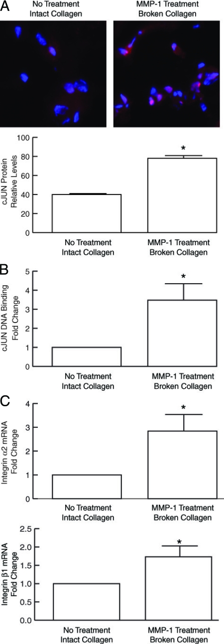
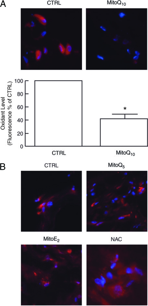
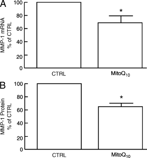
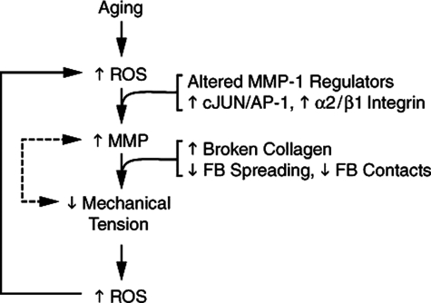
Aged human skin is fragile because of fragmentation and loss of type I collagen fibrils, which confer strength and resiliency. We report here that dermal fibroblasts express increased levels of collagen-degrading matrix metalloproteinases-1 (MMP-1) in aged (>80 years old) compared with young (21 to 30 years old) human skin in vivo. Transcription factor AP-1 and alpha2beta1 integrin, which are key regulators of MMP-1 expression, are also elevated in fibroblasts in aged human skin in vivo. MMP-1 treatment of young skin in organ culture causes fragmentation of collagen fibrils and reduces fibroblast stretch, consistent with reduced mechanical tension, as observed in aged human skin. Limited fragmentation of three-dimensional collagen lattices with exogenous MMP-1 also reduces fibroblast stretch and mechanical tension. Furthermore, fibroblasts cultured in fragmented collagen lattices express elevated levels of MMP-1, AP-1, and alpha2beta1 integrin. Importantly, culture in fragmented collagen raises intracellular oxidant levels and treatment with antioxidant MitoQ(10) significantly reduces MMP-1 expression. These data identify positive feedback regulation that couples age-dependent MMP-1-catalyzed collagen fragmentation and oxidative stress. We propose that this self perpetuating cycle promotes human skin aging. These data extend the current understanding of the oxidative theory of aging beyond a cellular-centric view to include extracellular matrix and the critical role that connective tissue microenvironment plays in the biology of aging.


Figures

Figure 1
Figure 1
Increased protein oxidation in aged human dermis in vivo. Dermis was obtained by dissection of full thickness human skin, from young (21 to 30 years old) and aged (>80 years old) individuals. Protein oxidation was determined by Western analysis of carbonyl residues. Results are means ± SEM. n = 7, *P < 0.05. Inset shows representative immunoblot.
Figure 2
Figure 2
MMP-1 mRNA, protein, and activity are increased in the dermis of aged human skin in vivo. A: MMP-1 mRNA levels in dermal fibroblasts in young (21 to 30 years old) and aged (>80 years old) human skin. Fibroblasts (150 cells) were obtained by LCM, total RNA was isolated, mRNA was amplified, and MMP-1 mRNA was quantified in 5 ng of amplified mRNA by real-time RT-PCR. Results are means ± SEM of MMP-1 mRNA normalized to 36B4 (housekeeping gene internal control) mRNA. n = 6, *P < 0.01. B: Quantitation of MMP-1 protein immunostaining in young and aged human skin. Figures show representative immunostaining. White line indicates dermal-epidermal boundary. n = 4, *P < 0.02. C: MMP-1 activity in young and aged human skin in vivo. MMP-1 activity was detected by in situ zymography, using FITC-labeled type I collagen as substrate (green fluorescence). MMP-1-catalyzed collagen cleavage causes loss of green fluorescence, resulting in darkened areas, which are most noticeable in the upper dermis of aged skin. Images are representative of three experiments.
Figure 3
Figure 3
c-Jun mRNA and protein are elevated in aged human dermis in vivo. A: c-Jun mRNA levels in young (21 to 30 years old) and aged (>80 years old) human dermis were analyzed by real-time RT-PCR. Results are means ± SEM of c-Jun mRNA normalized to 36B4 (internal control) mRNA. n = 6, *P < 0.0001. B: c-Jun protein levels in young and aged human dermis was quantified by Western analysis. Inset shows representative immunoblot. Results are means ± SEM. n = 6, *P < 0.05.
Figure 4
Figure 4
α2 and β1 integrins are elevated in aged human dermis in vivo. α2 integrin (A) and β1 integrin (B) mRNA levels were determined in young (21 to 30 years old) and aged (>80 years old) human dermis by real-time RT-PCR. Results are means ± SEM of integrin α2 and β1 mRNA normalized to 36B4 mRNA (internal control). n = 3 to 6, *P < 0.05.
Figure 5
Figure 5
MMP-1 treatment of young human skin in organ culture causes collagen fragmentation similar to that observed in aged human skin in vivo. Samples of young skin were treated with vehicle (left, top and bottom) or MMP-1 (middle, top and bottom). Top panels display paraffin-embedded sections stained with H&E. MMP-1 treatment causes fibroblasts (circled in white) to appear less stretched and more rounded. Bottom panels display scanning electron micrographs of dermal collagen, showing fragmentation of collagen fibrils in MMP-1-treated versus vehicle-treated young skin. Alterations of fibroblast morphology and collagen fragmentation observed in young skin after MMP-1 treatment resemble those observed in aged human skin (right, top and bottom).
Figure 6
Figure 6
Dermal fibroblasts cultured in MMP-1-fragmented three-dimensional collagen lattices have reduced spreading and increased levels of oxidants. Human skin dermal fibroblasts were cultured in intact or MMP-1-fragmented collagen lattices. A: Fibroblast morphology was assessed by incubation of cultures with CellTracker fluorescent dye for 1 hour. Fibroblasts were imaged by confocal microscopy. Reddish fluorescence delineates cell cytoplasm; blue fluorescence delineates nuclei. Results are means ± SEM. n = 5, *P < 0.05. Images are representative of three experiments. B: Oxidant levels were assessed by incubation of cultures with RedoxSensor Red CC-1 for 1 hour. Cells were imaged by confocal microscopy. Reddish fluorescence indicates relative oxidant levels; blue fluorescence delineates nuclei. Relative red fluorescence levels were quantified by ImageJ (NIH). Results are means ± SEM. n = 3, *P < 0.05. Top panels show representative images. C: Cellular lysates were analyzed for protein oxidation (carbonyls) by Western blot. Immunoreactive oxidized proteins were visualized by chemifluorescence, and quantified by ImageQuant software. Results are means + SEM of signal intensity in the entire lane. n = 3, *P < 0.05.
Figure 7
Figure 7
MMP-1 is elevated in dermal fibroblasts cultured in MMP-1-fragmented three-dimensional collagen lattices. Human dermal fibroblasts were cultured in intact or MMP-1-fragmented, collagen lattices. A: Total RNA was extracted and MMP-1 mRNA levels were determined by real-time RT-PCR. Results are means ± SEM of MMP-1 mRNA normalized to 36B4 (internal control) mRNA. n = 4, *P < 0.05. B: MMP-1 protein expression in fibroblasts was determined by laser confocal immunofluorescence microscopy, and quantified by image analysis. Top panels show representative images. Cell nuclei are stained blue, and MMP-1 protein is stained red. Results are means ± SEM. n = 3, *P < 0.05. C: Secreted MMP-1 protein was quantified by enzyme-linked immunosorbent assay. Inset shows representative Western blot demonstrating increased levels of both full-length MMP-1 and cleaved activated MMP-1 secreted by fibroblasts in fragmented collagen lattices. n = 4, *P < 0.05.
Figure 8
Figure 8
c-Jun protein and DNA-binding are elevated in dermal fibroblasts cultured in MMP-1-fragmented three-dimensional collagen lattices. Human dermal fibroblasts were cultured in intact or MMP-1-fragmented lattices. A: c-Jun protein levels were determined by laser confocal immunofluorescence microscopy and quantified by image analysis. Top Panels show representative images. Cell nuclei are stained blue, and c-Jun protein is stained red. Results are means ± SEM. n = 3, *P < 0.05. B: Nuclear extracts were prepared and c-Jun DNA binding was determined using the TransAM AP-1 kit (Active Motif). Results are means ± SEM. n = 3, *P < 0.05. C: α2 and β1 integrins are elevated in dermal fibroblasts cultured in MMP-1-fragmented three-dimensional collagen lattices. Human dermal fibroblasts were cultured in intact and MMP-1-fragmented three-dimensional collagen lattices. Total RNA was extracted. α2 and β1 integrin mRNA levels were analyzed by real-time RT-PCR (10 ng total RNA). Results are mean ± SEM of α2 or β1 integrin mRNA normalized to 36B4 (internal control) mRNA. n = 3, *P < 0.03.
Figure 9
Figure 9
Antioxidant MitoQ10 reduces oxidant levels in human dermal fibroblasts cultured in MMP-1-fragmented collagen lattices. A: Fibroblast cultures were treated with vehicle (CTRL) or MitoQ10 (1 nmol/L) for 72 hours, followed by incubation with RedoxSensor Red for 1 hour. Lattices were embedded in OCT, sectioned, and imaged by fluorescence microscopy. Top panels show representative images. Nuclei are stained blue; oxidized RedoxSensor Red is stained red. Bottom panel is quantitative image analyses of RedoxSensor Red fluorescence. n = 5, *P < 0.01. B: MitoQ10 analogues, MitoQ5 and MitoE2, and N-acetyl cysteine (NAC) do not reduce oxidant levels in dermal fibroblasts cultured in MMP-1-fragmented collagen lattices. Fibroblast cultures were treated with vehicle (CTRL), MitoQ5 (100 nmol/L), MitoE2 (100 nmol/L), or NAC (10 mmol/L) for 72 hours, and analyzed for RedoxSensor Red fluorescence as described in A. Results are representative images from three experiments.
Figure 10
Figure 10
Antioxidant MitoQ10 reduces MMP-1 expression in human dermal fibroblasts cultured in collagen lattices. Fibroblast cultures were treated with vehicle (CTRL) or MitoQ10 (1 nmol/L) for 72 hours. A: Fibroblasts were harvested and MMP-1 mRNA levels were determined by real-time RT-PCR. B: MMP-1 protein levels were determined by enzyme-linked immunosorbent assay. Results are means + SEM of five experiments. *P < 0.05.
Figure 11
Figure 11
Proposed model for self-perpetuating, MMP-1-mediated age-dependent collagen degradation in human skin connective tissue. MMP-1 induction in fibroblasts in aged dermis in vivo is mediated by coordinate alteration of MMP-1 regulatory pathways involving the transcription factor AP-1 and α2β1 integrin. MMP-1 breaks down collagen fibrils, thereby weakening the structural integrity of the extracellular matrix. The weakened extracellular matrix provides less resistance to mechanical forces exerted on it by dermal fibroblasts, thereby reducing mechanical tension in the fibroblasts. This reduced mechanical tension in the fibroblasts results in increased intracellular levels of oxidants, which in turn, through AP-1 and α2β1 integrin-dependent mechanisms, stimulate expression of MMP-1. Reduced mechanical tension may also stimulate MMP-1 expression through oxidant-independent pathways (dashed line). The positive feedback (or vicious cycle) nature of this model of age-dependent skin collagen connective tissue fragmentation is consistent with the biology of aging, which is epitomized by continual reduction of homeostatic control.

Similar articles

Cited by

References

    1. Uitto J, Pulkkinen L, Chu M-L. Collagen. Fitzpatrick TB, Eisen AZ, Wolff K, Freedberg IM, Austen KF, editors. New York: McGraw-Hill,; Dermatology in General Medicine. 2003:pp 165–179.
    1. Smith J, Davidson E, Sams W, Clark R. Alterations in human dermal connective tissue with age and chronic sun damage. J Invest Dermatol. 1962;39:347–350. - PubMed
    1. Uitto J. Biology of dermal cells and extracellular matrix. Fitzpatrick T, Eisen A, Wolff K, Freedberg I, Austen K, editors. New York: McGraw-Hill,; Dermatology in General Medicine. 1993:pp 299–314.
    1. Warren R, Gartstein V, Kligman A, Montagna W, Allendorf R, Ridder G. Age, sunlight, and facial skin: a histologic and quantitative study. J Am Acad Dermatol. 1991;25:751–760. - PubMed
    1. West M. The cellular and molecular biology of skin aging. Arch Dermatol. 1994;130:87–95. - PubMed

Publication types

MeSH terms