Review
. 2009 Feb;7(2):110-9.
doi: 10.1038/nrmicro2053. Epub 2008 Dec 31.

Enteric infection meets intestinal function: how bacterial pathogens cause diarrhoea

Affiliations
Review

Enteric infection meets intestinal function: how bacterial pathogens cause diarrhoea

V K Viswanathan et al. Nat Rev Microbiol. 2009 Feb.

Abstract

Infectious diarrhoea is a significant contributor to morbidity and mortality worldwide. In bacterium-induced diarrhoea, rapid loss of fluids and electrolytes results from inhibition of the normal absorptive function of the intestine as well as the activation of secretory processes. Advances in the past 10 years in the fields of gastrointestinal physiology, innate immunity and enteric bacterial virulence mechanisms highlight the multifactorial nature of infectious diarrhoea. This review explores the various mechanisms that contribute to loss of fluids and electrolytes following bacterial infections, and attempts to link these events to specific virulence factors and toxins.


Figures

Figure 1
Figure 1. The 7 metre-long human intestine absorbs nutrients and forms a barrier to luminal contents
The absorptive surface of the intestine is maximized by the presence of undulating ridges of the intestinal submucosa, organization of the mucosa into villi and crypts, and the elaboration of finger-like microvilli on epithelial cells. A single layer of polarized columnar epithelial cells rests on a basement membrane, below which is the supporting thin layer of vascular connective tissue known as the lamina propria, and a layer of smooth muscle. The apical part of the lateral membrane of intestinal epithelial cells contains the tight junctions, which serve as a regulatable barrier between the luminal and serosal sides of the epithelial monolayer (barrier function). In addition, by restricting the free movement of apical and basolateral components of epithelial cell membranes (fence function), tight junctions maintain polarity and thereby aid the directional movement of water, electrolytes and nutrients. JAM-A, junction adhesion molecule A.
Figure 2
Figure 2. Mechanisms by which enteric pathogens cause diarrhoea
These mechanisms interfere with the normal absorptive and secretory functions of the intestinal epithelial monolayer. a | Increased Cl secretion through the cystic fibrosis transmembrane receptor (CFTR; as by cholera toxin) or decreased Cl absorption owing to inhibition of downregulated in adenoma (DRA). b | Decreased Na+ absorption owing to inhibition of Na+/H+ exchanger 3 (NHE3). c | Downregulation of the Na+/glucose cotransporter (SGLT1). d | Direct inhibition of water channels or aquaporins (AQPs). e | Disruption of epithelial barrier function by promoting myosin light chain (MLC) phosphorylation (possibly by stimulating tumour necrosis factor-α (TNFα) production), leading to contraction of the peri-junctional actomyosin ring and opening of the tight junctions. f,g | Direct alteration of tight junction (TJ) protein localization leading to disruption of barrier and fence functions and pro-inflammatory signalling through engagement of Toll-like receptors (TLRs) by bacterial ligands, eventually leading to neutrophil transmigration into the lumen, and release of 5′-AMP and its conversion to adenosine, which subsequently causes cyclic AMP (cAMP)-dependent Cl secretion. MLCK, MLC kinase; NF-κB; nuclear factor-κB; PMN, polymorphonuclear leukocyte.
Figure 3
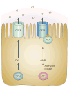
Figure 3. Cl secretion by intestinal epithelial cells is stimulated through the activation of cyclic AMP or Ca2+
A number of bacterial toxins activate these signaling molecules in different ways. Thermostable direct haemolysin (TDH) from Vibrio parahaemolyticus activates Ca2+, whereas cholera toxin (CT) from Vibrio cholerae increases the cAMP concentration by ADP-ribosylating adenylate cyclase. Activation of Ca2+ leads to Cl secretion by the calcium-activated chloride channels (CLCAs). Cholera toxin activates cAMP through ADP-ribosylation of adenylate cyclase. cAMP then activates protein kinase A (PKA), which phosphorylates the cystic fibrosis transmembrane receptor (CFTR) leading to Cl secretion.
Figure 4
Figure 4. Enteropathogenic Escherichia coli (EPEC) alter apical Na+, Cl and glucose absorption
a | Surface expression of the Na+/glucose cotransporter SGLT1 is decreased by two distinct mechanisms, both of which depend on the injection of effector proteins. The first mechanism is rapid and causes the movement of apical SGLT1 into intracellular vesicles. The second, and slower, pathway results from a loss of microvilli and hence SGLT1 on host cells, a process that is dependent on two secreted effector proteins, Map and EspF. b | The primary routes of apical Na+ uptake are the Na+/H+ exchangers (NHEs). EPEC differentially regulate the two apical NHE isoforms, increasing the activity of NHE2 through the activation of phospholipase C (PLC), which leads to activation of Ca2+ and protein kinase Cs (PKCs), whereas NHE3 activity is decreased through an effect of EspF. c | In addition to changes in Na+ uptake, apical Cl absorption is also altered. The injected effector proteins EspG and EspG2 disrupt microtubules, leading to internalization of the apical Cl–OH exchanger downregulated in adenoma (DRA). In EPEC-infected cells DRA is displaced from the cell surface into subapical membrane vesicles. This blocks one route of apical Cl absorption, which has been previously implicated in congenital Cl diarrhoea.

References

    1. Cheng AC, McDonald JR, Thielman NM. Infectious diarrhea in developed and developing countries. J Clin Gastroenterol. 2005;39:757–773. - PubMed
    1. Kosek M, Bern C, Guerrant RL. The global burden of diarrhoeal disease, as estimated from studies published between 1992 and 2000. Bull World Health Organ. 2003;81:197–204. - PMC - PubMed
    1. Petri WA, Jr, et al. Enteric infections, diarrhea, and their impact on function and development. J Clin Invest. 2008;118:1277–1290. This recent review on diarrhoea emphasizes epidemiological aspects, host susceptibility, vaccine development and oral rehydration therapy. - PMC - PubMed
    1. Merrell DS, et al. Host-induced epidemic spread of the cholera bacterium. Nature. 2002;417:642–645. - PMC - PubMed
    1. Butler SM, et al. Cholera stool bacteria repress chemotaxis to increase infectivity. Mol Microbiol. 2006;60:417–426. - PMC - PubMed

Publication types

Substances