

Spent culture medium from virulent Borrelia burgdorferi increases permeability of individually perfused microvessels of rat mesentery
- PMID: 19116656
- PMCID: PMC2605548
- DOI: 10.1371/journal.pone.0004101
Spent culture medium from virulent Borrelia burgdorferi increases permeability of individually perfused microvessels of rat mesentery
Abstract
Background: Lyme disease is a common vector-borne disease caused by the spirochete Borrelia burgdorferi (Bb), which manifests as systemic and targeted tissue inflammation. Both in vitro and in vivo studies have shown that Bb-induced inflammation is primarily host-mediated, via cytokine or chemokine production that promotes leukocyte adhesion/migration. Whether Bb produces mediators that can directly alter the vascular permeability in vivo has not been investigated. The objective of the present study was to investigate if Bb produces a mediator(s) that can directly activate endothelial cells resulting in increases in permeability in intact microvessels in the absence of blood cells.
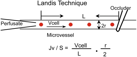
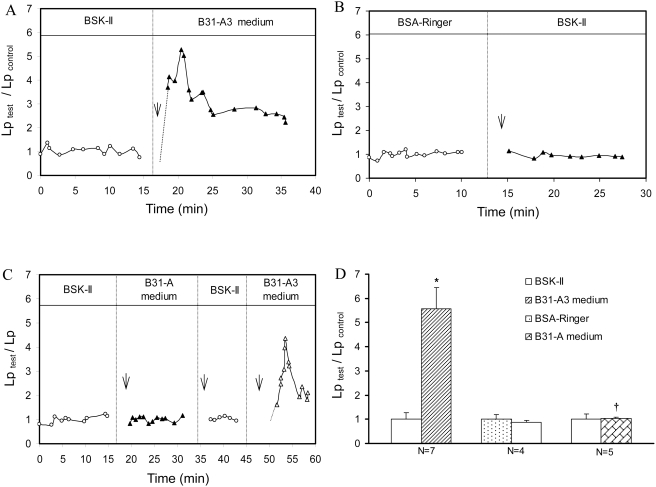
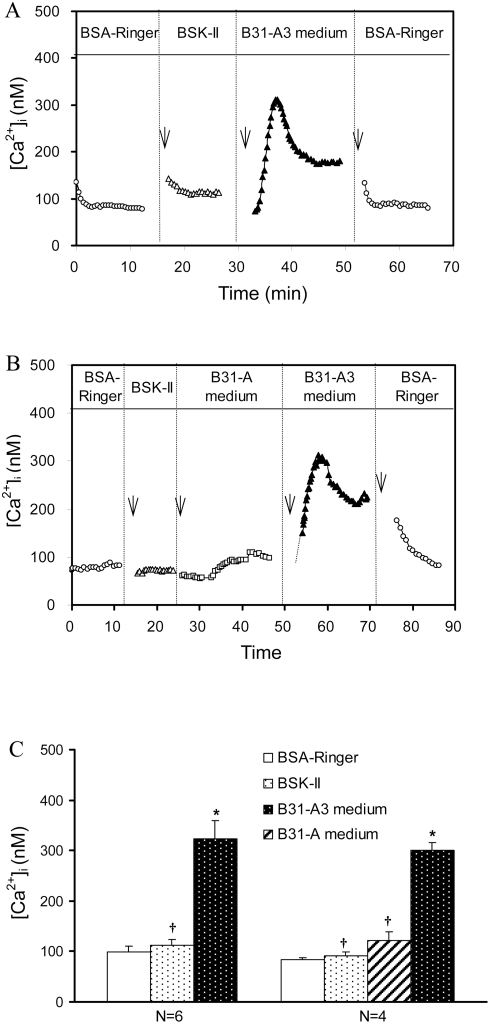
Methodology/principal findings: The effects of cell-free, spent culture medium from virulent (B31-A3) and avirulent (B31-A) B. burgdorferi on microvessel permeability and endothelial calcium concentration, [Ca(2+)](i), were examined in individually perfused rat mesenteric venules. Microvessel permeability was determined by measuring hydraulic conductivity (Lp). Endothelial [Ca(2+)](i), a necessary signal initiating hyperpermeability, was measured in Fura-2 loaded microvessels. B31-A3 spent medium caused a rapid and transient increase in Lp and endothelial [Ca(2+)](i). Within 2-5 min, the mean peak Lp increased to 5.6+/-0.9 times the control, and endothelial [Ca(2+)](i) increased from 113+/-11 nM to a mean peak value of 324+/-35 nM. In contrast, neither endothelial [Ca(2+)](i) nor Lp was altered by B31-A spent medium.
Conclusions/significance: A mediator(s) produced by virulent Bb under culture conditions directly activates endothelial cells, resulting in increases in microvessel permeability. Most importantly, the production of this mediator is associated with Bb virulence and is likely produced by one or more of the 8 plasmid(s) missing from strain B31-A.
Conflict of interest statement
Figures
Similar articles
-
Endothelial [Ca2+]i and caveolin-1 antagonistically regulate eNOS activity and microvessel permeability in rat venules.Cardiovasc Res. 2010 Jul 15;87(2):340-7. doi: 10.1093/cvr/cvq006. Epub 2010 Jan 15. Cardiovasc Res. 2010. PMID: 20080986 Free PMC article.
-
H2O2-induced endothelial NO production contributes to vascular cell apoptosis and increased permeability in rat venules.Am J Physiol Heart Circ Physiol. 2013 Jan 1;304(1):H82-93. doi: 10.1152/ajpheart.00300.2012. Epub 2012 Oct 19. Am J Physiol Heart Circ Physiol. 2013. PMID: 23086988 Free PMC article.
-
Calcium influx-dependent differential actions of superoxide and hydrogen peroxide on microvessel permeability.Am J Physiol Heart Circ Physiol. 2009 Apr;296(4):H1096-107. doi: 10.1152/ajpheart.01037.2008. Epub 2009 Feb 6. Am J Physiol Heart Circ Physiol. 2009. PMID: 19201997 Free PMC article.
-
Vascular permeability modulation at the cell, microvessel, or whole organ level: towards closing gaps in our knowledge.Cardiovasc Res. 2010 Jul 15;87(2):218-29. doi: 10.1093/cvr/cvq115. Epub 2010 Apr 23. Cardiovasc Res. 2010. PMID: 20418473 Free PMC article. Review.
-
Leucocyte/endothelium interactions and microvessel permeability: coupled or uncoupled?Cardiovasc Res. 2010 Jul 15;87(2):281-90. doi: 10.1093/cvr/cvq140. Epub 2010 May 13. Cardiovasc Res. 2010. PMID: 20472564 Free PMC article. Review.
Cited by
-
Analysis of the HD-GYP domain cyclic dimeric GMP phosphodiesterase reveals a role in motility and the enzootic life cycle of Borrelia burgdorferi.Infect Immun. 2011 Aug;79(8):3273-83. doi: 10.1128/IAI.05153-11. Epub 2011 Jun 13. Infect Immun. 2011. PMID: 21670168 Free PMC article.
-
Endothelial cells and fibroblasts amplify the arthritogenic type I IFN response in murine Lyme disease and are major sources of chemokines in Borrelia burgdorferi-infected joint tissue.J Immunol. 2012 Sep 1;189(5):2488-501. doi: 10.4049/jimmunol.1201095. Epub 2012 Jul 30. J Immunol. 2012. PMID: 22851707 Free PMC article.
-
Analysis of the Borrelia burgdorferi cyclic-di-GMP-binding protein PlzA reveals a role in motility and virulence.Infect Immun. 2011 May;79(5):1815-25. doi: 10.1128/IAI.00075-11. Epub 2011 Feb 28. Infect Immun. 2011. PMID: 21357718 Free PMC article.
-
Borrelia burgdorferi Secretes c-di-AMP as an Extracellular Pathogen-Associated Molecular Pattern to Elicit Type I Interferon Responses in Mammalian Hosts.bioRxiv [Preprint]. 2024 Aug 20:2024.08.13.607721. doi: 10.1101/2024.08.13.607721. bioRxiv. 2024. Update in: J Immunol. 2025 Jul 03:vkaf133. doi: 10.1093/jimmun/vkaf133. PMID: 39185169 Free PMC article. Updated. Preprint.
-
Analysis of a Borrelia burgdorferi phosphodiesterase demonstrates a role for cyclic-di-guanosine monophosphate in motility and virulence.Mol Microbiol. 2010 Jul 1;77(1):128-42. doi: 10.1111/j.1365-2958.2010.07191.x. Epub 2010 Apr 27. Mol Microbiol. 2010. PMID: 20444101 Free PMC article.
References
-
- Pinto DS. Cardiac manifestations of Lyme disease. Med Clin North Am. 2002;86:285–296. - PubMed
-
- Wormser GP. Clinical practice. Early Lyme disease. N Engl J Med. 2006;354:2794–2801. - PubMed
-
- Steere AC. Lyme borreliosis in 2005, 30 years after initial observations in Lyme Connecticut. Wien Klin Wochenschr. 2006;118:625–633. - PubMed
-
- Dame TM, Orenzoff BL, Palmer LE, Furie MB. IFN-gamma alters the response of Borrelia burgdorferi-activated endothelium to favor chronic inflammation. J Immunol. 2007;178:1172–1179. - PubMed
-
- Burns MJ, Sellati TJ, Teng EI, Furie MB. Production of interleukin-8 (IL-8) by cultured endothelial cells in response to Borrelia burgdorferi occurs independently of secreted [corrected] IL-1 and tumor necrosis factor alpha and is required for subsequent transendothelial migration of neutrophils. Infect Immun. 1997;65:1217–1222. - PMC - PubMed
Publication types
MeSH terms
Substances
Grants and funding
LinkOut - more resources
Full Text Sources
Miscellaneous
