. 2009 May;219(2):402-14.
doi: 10.1002/jcp.21684.

Dystrophin glycoprotein complex-associated Gbetagamma subunits activate phosphatidylinositol-3-kinase/Akt signaling in skeletal muscle in a laminin-dependent manner

Affiliations

Dystrophin glycoprotein complex-associated Gbetagamma subunits activate phosphatidylinositol-3-kinase/Akt signaling in skeletal muscle in a laminin-dependent manner

Yongmin Xiong et al. J Cell Physiol. 2009 May.

Abstract

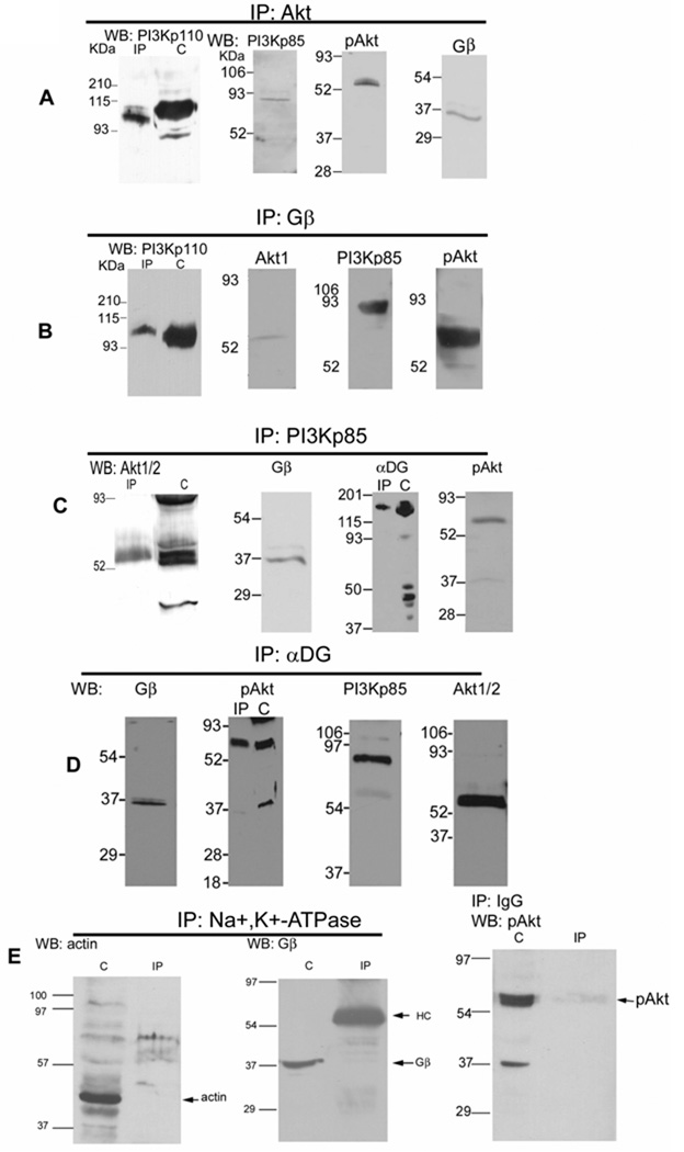
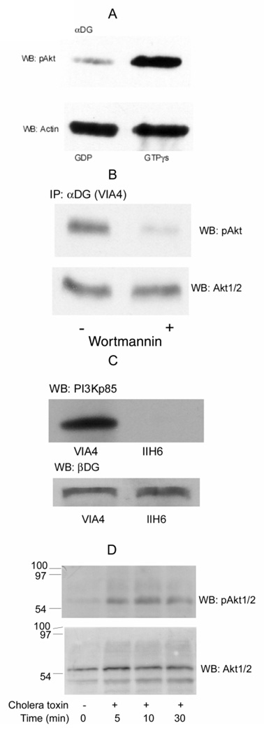
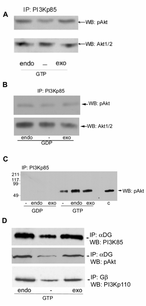
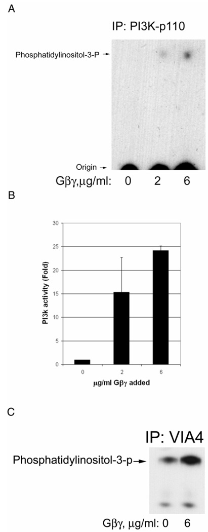
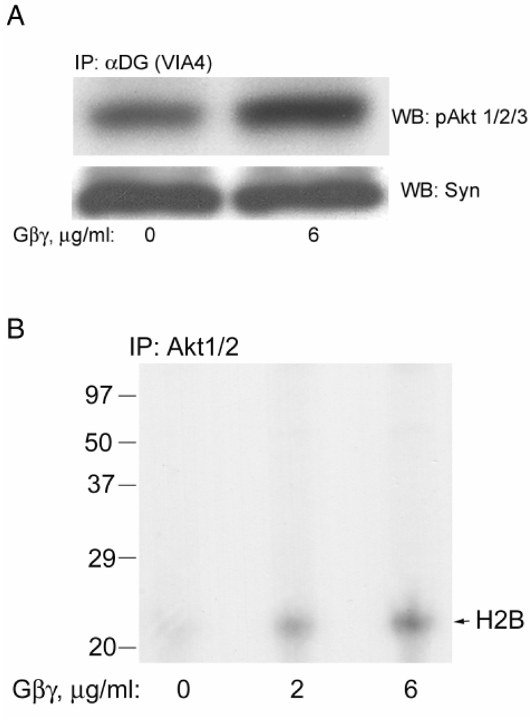
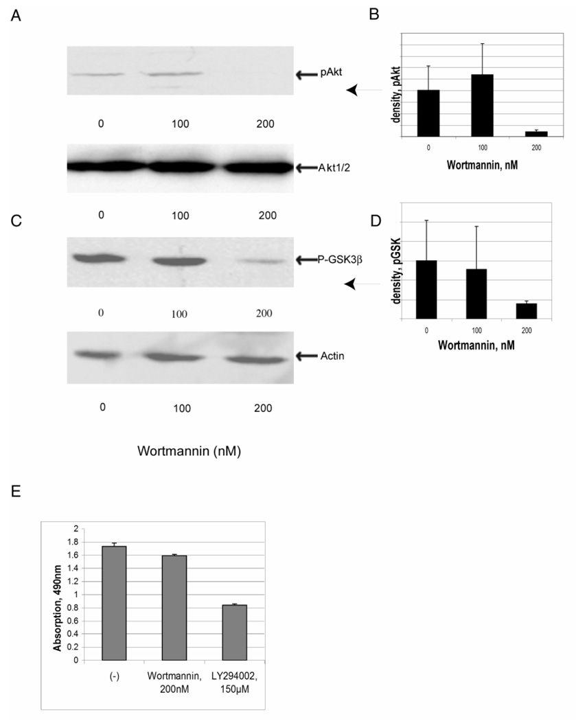
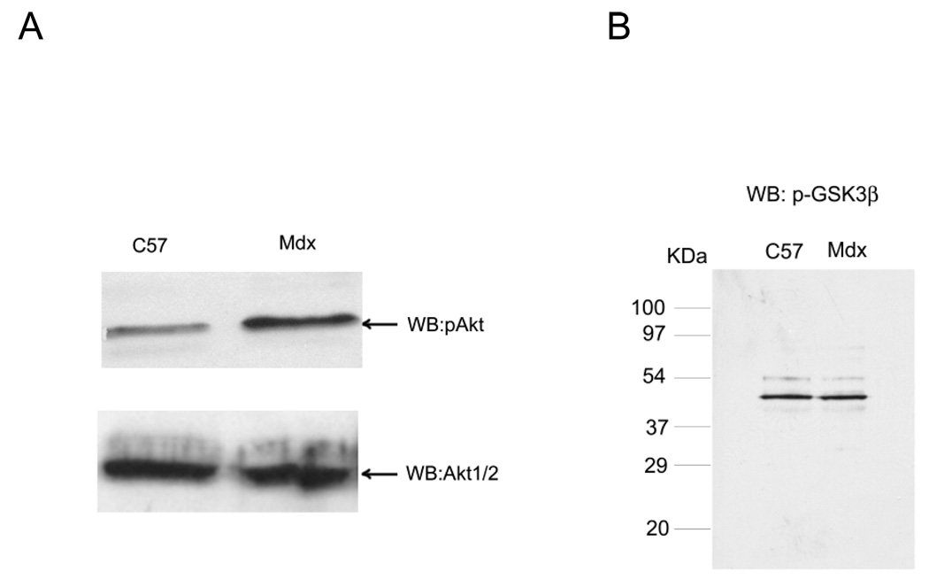
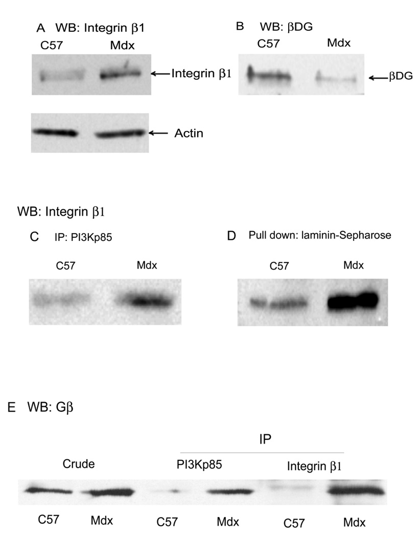
Previously, we showed that laminin-binding to the dystrophin glycoprotein complex (DGC) of skeletal muscle causes a heterotrimeric G-protein (Galphabetagamma) to bind, changing the activation state of the Gsalpha subunit. Others have shown that laminin-binding to the DGC also leads to Akt activation. Gbetagamma, released when Gsalpha is activated, is known to bind phosphatidylinositol-3-kinase (PI3K), which activates Akt in other cells. Here, we investigate whether muscle Akt activation results from Gbetagamma, using immunoprecipitation and immunoblotting, and purified Gbetagamma. In the presence of laminin, PI3K-binding to the DGC increases and Akt becomes phosphorylated and activated (pAkt), and glycogen synthase kinase is phosphorylated. Antibodies, which specifically block laminin-binding to alpha-dystroglycan, prevent PI3K-binding to the DGC. Purified bovine brain Gbetagamma also caused PI3K and Akt activation. These results show that DGC-Gbetagamma is binding PI3K and activating pAkt in a laminin-dependent manner. Mdx mice, which have greatly diminished amounts of DGC proteins, display elevated pAkt signaling and increased expression of integrin beta1 compared to normal muscle. This integrin binds laminin, Gbetagamma, and PI3K. Collectively, these suggest that PI3K is an important target for the Gbetagamma, which normally binds to DGC syntrophin, and activates PI3K/Akt signaling. Disruption of the DGC in mdx mouse is causing dis-regulation of the laminin-DGC-Gbetagamma-PI3K-Akt signaling and is likely to be important to the pathogenesis of muscular dystrophy. Upregulating integrin beta1 expression and activating the PI3K/Akt pathway in muscular dystrophy may partially compensate for the loss of the DGC. The results suggest new therapeutic approaches to muscle disease.


Figures

Fig. 1
Fig. 1. DGC-Gβγ binds PI3K and Akt in rabbit skeletal muscle
A, Rabbit muscle microsomes were isolated and the protein concentration (Bradford, 1976) adjusted so that equal amounts were used for all experiments. Microsomes were incubated with antibodies cross-reactive with both Akt 1 and 2 (Akt 1/2), detergent solubilized upon addition of protein G-Sepharose, electrophoresed, electroblotted, and Western blotted for PI3Kp85, pAkt 1/2/3, PI3Kp110, and Gβ. Where shown “C” denotes crude microsome prior to IP, added in equal concentration. IP: Immunoprecipitation, WB: Western blot. B, Microsomes were immunoprecipited with a Gβ antibody, and Western blotted for PI3Kp110, PI3Kp85, Akt 1 and pAkt 1/2/3. C, Microsomes were immunoprecipitated with a PI3Kp85 antibody, and Western blotted for Akt1/2, α-dystroglycan (αDG, VIA4 monoclonal antibody), Gβ, and pAkt1/2/3. D, Microsomes were immunoprecipited with an α-dystroglycan monoclonal antibody (VIA4), and Western blotted for Gβ, PI3Kp85, pAkt and Akt1/2. These show that DGC-Gβγ binds a complex containing PI3K and Akt, as well as the phosphorylated Akt. E, Microsomes (C) or detergent immunoprecipited with Na+/K+ ATPase or pre-immune IgG antibodies, were probed with the antibodies against actin, Gβ and pAkt.
Fig. 2
Fig. 2. Activation of PI3K/Akt by Gβγ-dimers in skeletal muscles
A, Rabbit skeletal muscle microsomes were isolated and incubated for 1 h at 37° in buffer K containing 1 mM CaCl2 and 1 mM ATP and either 1 mM GTP-γ S o r 1 m M G D P, and then were immunoprecipitated with VIA4-1 α-dystroglycan (α-DG) antibody and Western blotted for pAkt. Membrane were then stripped and re-probed with an actin antibody, as indicated. pAkt was decreased in the presence of GDP compared to GTP-γS, but actin was not significantly altered and provides a loading control. B, C2C12 myotubes were pretreated with 200 nM wortmannin overnight at 37°C (+) or with buffer alone (−). The myotubes were counted and adjusted to equal cell counts, microsomes prepared and immunoprecipited with αDG antibody (VIA4). After SDS-PAGE and electroblotting, the blot was probed with antibody against pAkt. The same samples loaded on adjacent wells of the same gel was probed for total Akt1/2 as a loading control. C, the myotube, grown in either antibody, were used to prepare microsomes, which were then pretreated with the same antibody against αDG (VIA4 or IIH6). After immunoprecipitation with protein G-Sepharose, the proteins were separated on SDS-PAGE and transferred to nitrocellulose membranes. The membrane was probed with antibody against PI3Kp85 (upper panel). The same blot was stripped and re-probed with a βDG antibody (lower panel) to confirm equal loading. D, An equal number of C2C12 cells were cultured with 100 ng/ml cholera toxin for 0, 5, 10, 30, 60 min at 37°. Clarified cell lysates were adjusted to the same protein concentration (Bradford, 1976) and equal amounts separated on 12% SDS-PAGE and transferred to nitrocellulose membranes. The membranes were probed with antibodies against pAkt (upper panel). The stripped blot from was reprobed for total Akt1/2 to confirm equal loading (lower panel).
Fig. 3
Fig. 3. Laminin binding alters PI3K/Akt signaling
A and B, rabbit skeletal muscle microsomes were treated with Sepharose 4B (endo) or with heparin-Sepharose to deplete laminin (−) for 1 hour at 4°. The microsomes were then adjusted to equal protein concentration (Bradford, 1976) and were made 1 mM CaCl2, 10 mM MgCl2, 1 mM ATP, and 1 mM GTP-γS (panel A) or 1 mM GDP (panel B). To the laminin-depleted portions of the microsome 3 µg exogenous laminin-1 (exo) was added for 1 hour at 4°, with gentle mixing. The microsomes were immunoprecipitated for PI3Kp85α and Western blotted with antibody against phosphorylated Akt (pAkt). Membrane were then stripped and re-probed with antibody to total Akt to show total (phosphorylated and unphosphorylated) protein levels in equal loads. The level of Akt phosphorylation was reduced in the laminin-depleted immunoprecipitate, and laminin addition induces the phosphorylation of Akt. C, Microsomes were treated, incubated and immuno-precipitated as in panels A and B, except that the GTP-γS and GDP treated samples and an unequal amount of untreated microsomes (C) were applied to the same gel, and Western blotted for pAkt on the same blot. The data show that much greater amounts of pAkt are immunoprecipitated from microsomes incubated with GTP-γS than GDP and that laminin is also affecting the phosphorylation of pAkt.. D, Rabbit skeletal muscle microsomes were treated as in panel A. Immunoprecipitates with the α-dystroglycan (α-DG) VIA4 or Gβ antibody as indicated were probed with antibodies against PI3Kp85, PI3Kp110 or pAkt. Laminin binding increased αDG-Gβ-PI3K-pAkt interactions. Depletion of laminin resulted in decreased DGC-Gβγ-PI3K-Akt interactions, and exogenous laminin-1 increases PI3K binding and activation of pAkt.
Fig. 4
Fig. 4. Purified Gβγ activates PI3K, which is associated with the DGC
A, Rabbit skeletal muscle microsomes were incubated with GDP and the concentrations of purified Gβγ shown for 15 min. Then digitonin was added for solubilization followed by immunoprecipitation for PI3Kp110 catalytic subunit. The immunoprecipitate was added to a PI3K assay where phosphatidylinositol is added along with γ-32P-ATP as described in Methods. The reaction mixture is extracted for lipids and analyzed by silica thin layer chromatography and autoradiography. The position of product phosphatidylinositol-3-phosphate, obtained from a standard also applied to the plate, is shown to the left. B, The same as panel A except that densitometry was performed and the data normalized so that the absence of Gβγ was one-fold activation. The bars represent the mean of two independent experiments and the standard deviation is shown. C, The same as panel A except that VIA4 antibody was used to immunoprecipitate the DGC. Only the upper portion of the chromatogram is shown and at a longer exposure to emphasize an additional 3-phospholipid also detected.
Fig. 5
Fig. 5. Purified Gβγ activates Akt, which is associated with the DGC
A, Microsomes were treated as described in Fig. 4A and immunoprecipitated with VIA4 antibody and Western blotted. The pAkt antibody shows that pAkt was activated. The same blot was stripped and probed with the syntrophin antibody to provide a loading control. B, Immunoprecipitates were prepared as in panel A except with the Akt1/2 antibody and added to an Akt assay using histone as substrate. The autoradiogram is shown and the expected position of histone H2B is shown to the right.
Fig. 6
Fig. 6. Effects of Wortmannin on PI3K activation in C2C12 myotube cultures
Myotube cultures were maintained in 1% FBS DMEM medium for 6 days before addition of inhibitors. Myotubes were incubated in the absence or presence of different concentrations of Wortmannin as indicated in the figure. After 24 h, cells were lysed and subjected to Western blot assay. A, Western blots for pAkt are shown above with total Akt (on the same blot after stripping) below providing a loading control. B, Densitometry of the pAkt staining. C, Western blots for pGSK3β are shown above with actin shown below providing a loading control. D, Densitometry of the pGSK staining. E, The effect of Wortmannin and LY294002 on C2C12 myotube viability was measured using the MTS assay. The error bars throughout are standard deviation with n=2 for panels B and D, and n=3 for panel E.
Fig. 7
Fig. 7. Activation (phosphorylation) of Akt is increased in dystrophin-deficient skeletal muscles
A, Skeletal muscle microsomes from 4-week-old normal and mdx mice were prepared from the same wet weight of gastocnemius muscle, separated on 12% SDS-PAGE and transferred to nitrocellulose. The blots were probed with antibody against pAkt, and membranes were then stripped and reprobed with the Akt 1/2 antibody to detect total Akt levels. pAkt (Ser 473) was elevated in 4-week-old muscle from mdx mice compared to muscle isolate from age-matched normal mice (C57) while total Akt was not. B, Skeletal muscle (gastrocnemius) microsomes from adult (6 month old) normal and mdx mice were probed for the phosphorylated, p-GSK3β, as indicated. pGSK was not different between normal and mdx mice.
Fig. 8
Fig. 8. Enhanced expression of integrin β1 and Gβ in mdx muscle induces PI3K recruitment
A, Skeletal muscle (gastrocnemius) crude microsomes from adult (6 month old) normal (C57bl) and mdx mice, prepared from the same wet weight of muscle, were Western blotted with anti-integrin β1 or (after stripping the same blot) anti-actin antibodies. Integrin β1 protein expression was found to be higher in mdx mouse muscle compared with control while actin was not. B, Same as panel A except Western blotting with anti-βDG (shown) or anti-actin antibodies (data not shown). βDG was decreased in mdx muscle compared to muscle isolate from age-matched C57bl mice (shown) while actin was not significantly changed (data not shown). C, microsomes from 6 mo. old C57bl and mdx mice were immunoprecipitated with antibodies against PI3Kp85 and analyzed by Western blotting with antibody to integrin β1. Interaction of PI3K-integrin β1 was enhanced in mdx muscle compared with normal muscle. D, Same as panel C except laminin-Sepharose was used instead of immunoprecipitation and again Western blotting with integrin β1 antibody. Laminin-bound integrin β1 increases in skeletal muscle of mdx compared to normal mice. E, Skeletal muscle microsomes (crude) from adult C57 and mdx mice and its immune precipitates (IP) with antibodies against PI3Kp85 and integrin β1 were analyzed by Western blotting with antibody against Gβ. The results show that Gβ was increased in mdx muscle microsome compared with normal, and interactions of PI3K-Gβ and integrin β1-Gβ were enhanced in mdx mice.

Similar articles

Cited by

References

    1. Ananthanarayanan B, Ni Q, Zhang J. Signal propagation from membrane messengers to nuclear effectors revealed by reporters of phosphoinositide dynamics and Akt activity. Proc Natl Acad Sci U S A. 2005;102(42):15081–15086. - PMC - PubMed
    1. Baar K, Esser K. Phosphorylation of p70(S6k) correlates with increased skeletal muscle mass following resistance exercise. Am J Physiol. 1999;276(1 Pt 1):C120–C127. - PubMed
    1. Bradford MM. A rapid and sensitive method for the quantitation of microgram quantities of protein utilizing the principle of protein-dye binding. Anal Biochem. 1976;72:248–254. - PubMed
    1. Brock C, Schaefer M, Reusch HP, Czupalla C, Michalke M, Spicher K, Schultz G, Nurnberg B. Roles of G beta gamma in membrane recruitment and activation of p110 gamma/p101 phosphoinositide 3-kinase gamma. J Cell Biol. 2003;160(1):89–99. - PMC - PubMed
    1. Brown SC, Fassati A, Popplewell L, Page AM, Henry MD, Campbell KP, Dickson G. Dystrophic phenotype induced in vitro by antibody blockade of muscle alpha-dystroglycan-laminin interaction. J Cell Sci. 1999;112(Pt 2):209–216. - PubMed

Publication types

MeSH terms

LinkOut - more resources