. 2008 Dec 30;47(52):13966-73.

Conformational Closure of the Catalytic Site of Human CD38 Induced by Calcium

Affiliations

Conformational Closure of the Catalytic Site of Human CD38 Induced by Calcium

Qun Liu et al. Biochemistry. .

Abstract

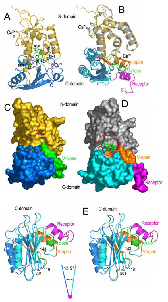
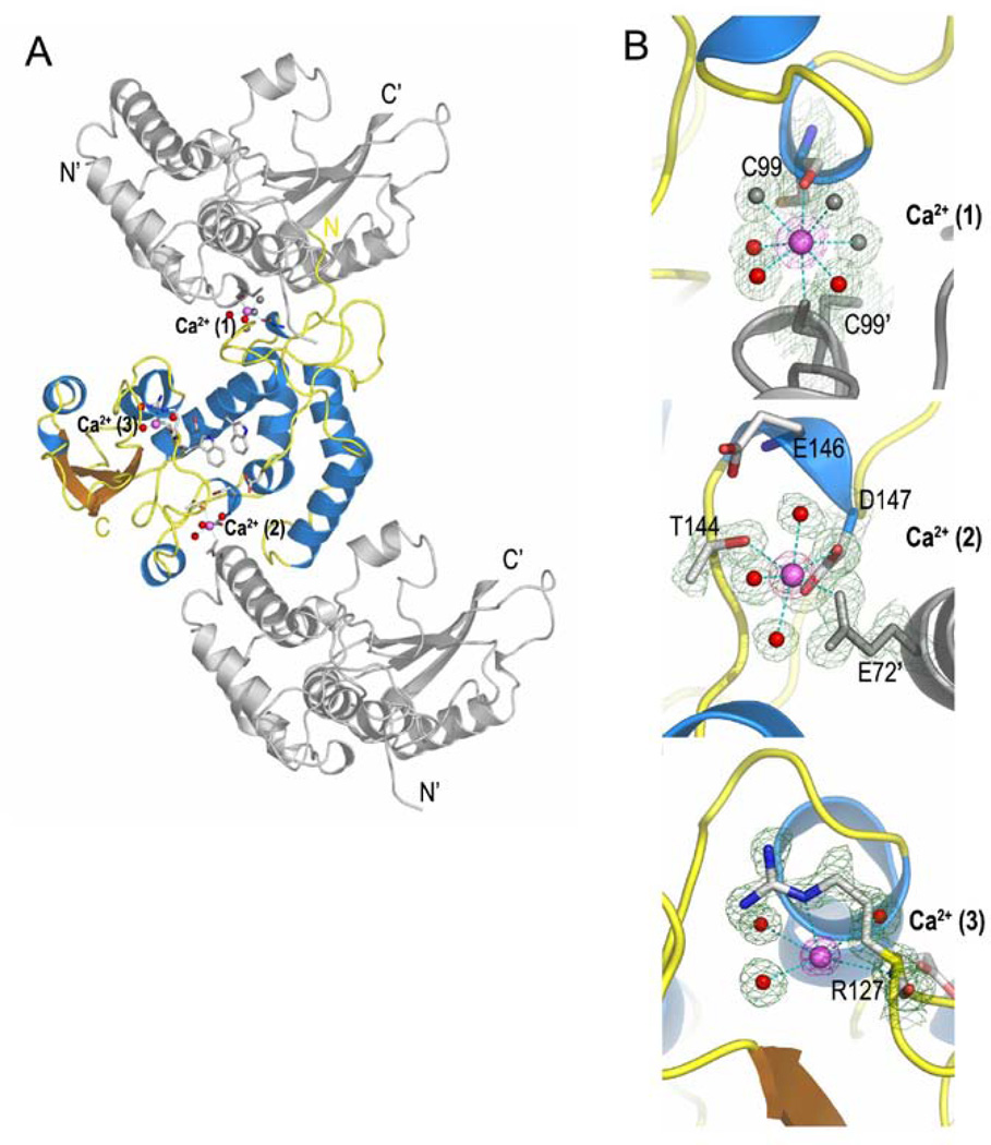
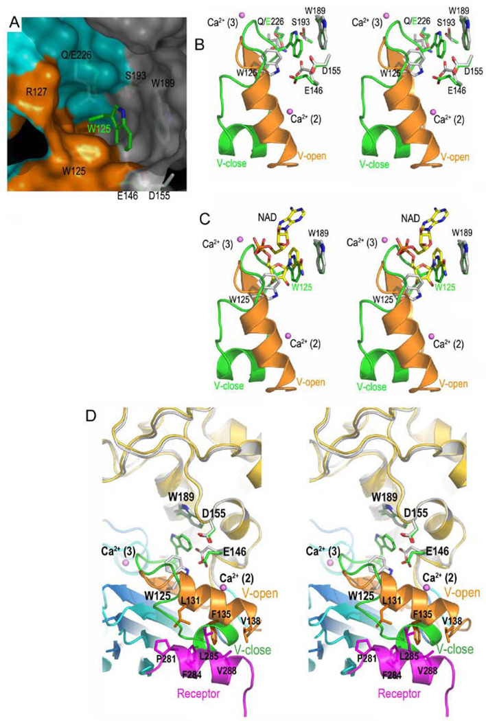
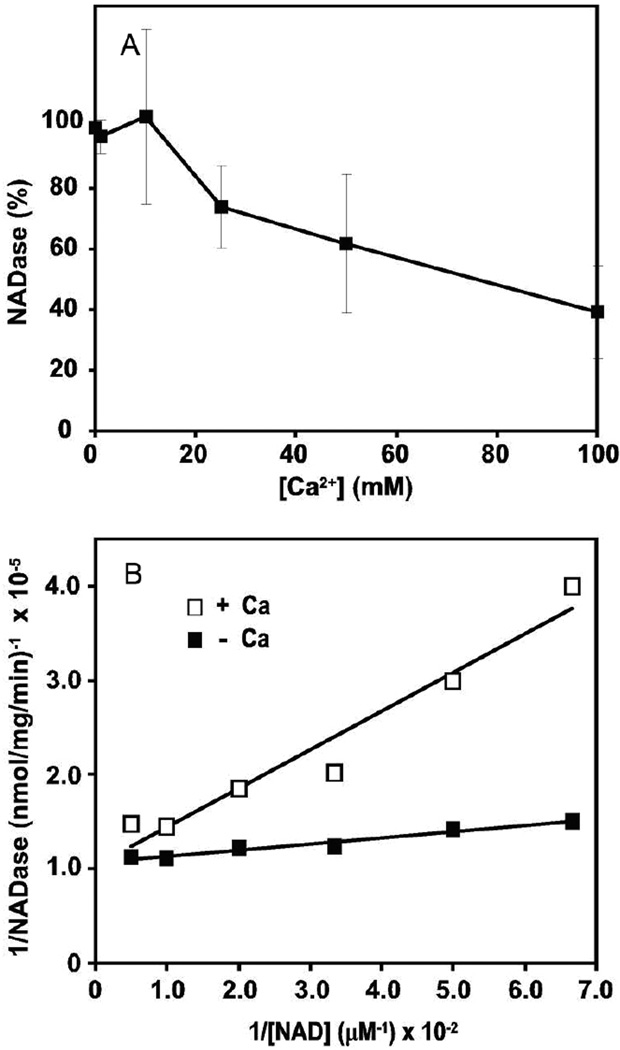
First identified on the surface of lymphoids as a type II transmembrane protein, CD38 has now been established to have dual functions not only as a receptor but also as a multifunctional enzyme,catalyzing the synthesis of and hydrolysis of a general calcium messenger molecule, cyclic ADP-ribose(cADPR). The receptorial functions of CD38 include the induction of cell adhesion, differentiation,apoptosis, and cytokine production upon antibody ligation. Here we determined the crystal structure of calcium-loaded human CD38 at 1.45 A resolution which reveals that CD38 undergoes dramatic structural changes to an inhibited conformation in the presence of calcium. The structural changes are highly localized and occur in only two regions. The first region is part of the active site and consists of residues 121-141.In the presence of calcium, W125 moves 5 A into the active site and forms hydrophobic interactions with W189. The movement closes the active site pocket and reduces entry of substrates, resulting in inhibition of the enzymatic activity. The structural role of calcium in inducing these conformational changes is readily visualized in the crystal structure. The other region that undergoes calcium-induced changes is at the receptor region, where a highly ordered helix is unraveled to a random coil. The results suggest a novel conformational coupling mechanism, whereby protein interaction targeted at the receptor region can effectively regulate the enzymatic activity of CD38.


Figures

FIGURE 1
FIGURE 1
Structure of Calcium loaded CD38. (A) Ribbon view of the overall structure of calcium loaded CD38. The N-domain (yellow) and C-domain (marine) are separately colored. The catalytic site is located between two domains. Residues in the catalytic site critical for the enzymatic activities of CD38 are shown as green sticks. (B) Comparison of the calcium loaded structure (“off”) with calcium free structure (“on”) of CD38. The “on” and “off” structures are aligned against their N-domains. The receptor region in the “on” structure is colored in magenta and consists of a helix; while in the “off” structure, this receptor region is disordered. The region of residues 121–141 is also adopting very different conformations, and thus is labeled as V-open in the “on” structure and V-close in the “off” structures. (C) and (D) are surface representations of the “off” (C) and “on” structures (D). The color scheme of (C) and (D) is the same as (B). Dashed red circles highlight the active sites in “off” (C) and “on” (D) structures. Substrate NAD is shown as green sticks in the active site of the “on” structure. (E) Stereo view showing that the C-domains of the “off” (marine and green) and “on” (cyan, orange, and magenta) structures are related by a rotation of 15.5°.
FIGURE 2
FIGURE 2
Structure of the calcium binding sites. (A) The locations of three bound calcium ions in the calcium-loaded CD38. Two calcium ions, Ca2+(1) and Ca2+(2), are located between the symmetry related molecules. The third one is bound within the molecule and is close to the catalytic site of CD38. Symmetry generated molecules are shown as gray ribbons. (B) Structures of the three calcium binding sites. The calcium ions are shown as magenta spheres. Interacting water molecules are shown as red or gray (symmetry generated) spheres with a smaller size. Protein residues from symmetry generated molecules are shown as gray sticks.
FIGURE 3
FIGURE 3
Structural basis for the calcium-induced enzyme inactivation. (A) The surface view of the catalytic site of the “on” structure. The corresponding residues in the “off” structure are superimposed as green sticks. The color scheme for the surface is: gray for the N-domain; Cyan for the C-domain; and orange for the variable region of the C-domain. W125 (green sticks) of the “off” structure moves into the active site of the “on” structure. (B) Structural alignment of the “on” and “off” structures around their active sites. The large movement of W125,which moves from its white position in the “on” structure to the green position in the “off” structure, is shown The movement of W125 closes the catalytic site, preventing the access of substrates to the catalytic residue E226. The variable region (residues 121–141) of the “off” structure is partially unfolded. (C) Another comparison of the catalytic sites with NAD included. In the “on” structure, the nicotinamide moiety of the bound NAD is parallel to the indole ring of W189. In the “off” structure, loading of calcium induces the movement of W125 to a position overlapping with the nicotinamide moiety of the bound NAD in the “on” structure. (D) Stereo diagram showing structural basis for conformational coupling between the catalytic site and the receptor region. The disordering of the V-open and receptor regions disrupts the hydrophobic interactions between them, resulting in the movement of the variable region (residues 121–141) from its orange position to green position and the disorder of the receptor region. The overall effect is the movement of W125 toward Trp189. Such movement introduces hydrophobic interactions between two tryptophan indole rings, hides the catalytic residue E226, and thus turns the enzyme off.
FIGURE 4
FIGURE 4
Inhibition of the NADase activity of CD38 by calcium. The NADase activity of CD38 was measured as described in the text in the presence of various concentrations of calcium. (A) Calcium inhibits the NADase activity in a concentration dependent manner. (B) The double reciprocal plot showing that the inhibition by calcium is competitive in nature, decreasing the affinity of CD38 for the substrate NAD. From the plot, the Ki value for calcium was determined to be 19 mM.

References

    1. Jackson DG, Bell JI. Isolation of a cDNA encoding the human CD38 (T10) molecule, a cell surface glycoprotein with an unusual discontinuous pattern of expression during lymphocyte differentiation. J Immunol. 1990;144:2811–2815. - PubMed
    1. Howard M, Grimaldi JC, Bazan JF, Lund FE, Santos-Argumedo L, Parkhouse RM, Walseth TF, Lee HC. Formation and hydrolysis of cyclic ADP-ribose catalyzed by lymphocyte antigen CD38. Science. 1993;262:1056–1059. - PubMed
    1. Aarhus R, Graeff RM, Dickey DM, Walseth TF, Lee HC. ADP-ribosyl cyclase and CD38 catalyze the synthesis of a calcium-mobilizing metabolite from NADP. J Biol Chem. 1995;270:30327–30333. - PubMed
    1. Lee HC. Mechanisms of calcium signaling by cyclic ADP-ribose and NAADP. Physiol Rev. 1997;77:1133–1164. - PubMed
    1. Graeff R, Liu Q, Kriksunov IA, Hao Q, Lee HC. Acidic residues at the active sites of CD38 and ADP-ribosyl cyclase determine nicotinic acid adenine dinucleotide phosphate (NAADP) synthesis and hydrolysis activities. J Biol Chem. 2006;281:28951–28957. - PubMed

Publication types

MeSH terms

LinkOut - more resources