Comparative Study
. 2008 Dec 31:5:119.
doi: 10.1186/1742-4690-5-119.

Human prostate supports more efficient replication of HIV-1 R5 than X4 strains ex vivo

Affiliations
Comparative Study

Human prostate supports more efficient replication of HIV-1 R5 than X4 strains ex vivo

Anna Le Tortorec et al. Retrovirology. .

Abstract

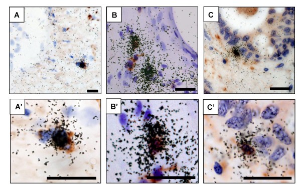
Background: In order to determine whether human prostate can be productively infected by HIV-1 strains with different tropism, and thus represent a potential source of HIV in semen, an organotypic culture of prostate from men undergoing prostatic adenomectomy for benign prostate hypertrophy (BPH) was developed. The presence of potential HIV target cells in prostate tissues was investigated using immunohistochemistry. The infection of prostate explants following exposures with HIV-1 R5, R5X4 and X4 strains was analyzed through the measure of RT activity in culture supernatants, the quantification of HIV DNA in the explants and the detection of HIV RNA+ cells in situ.

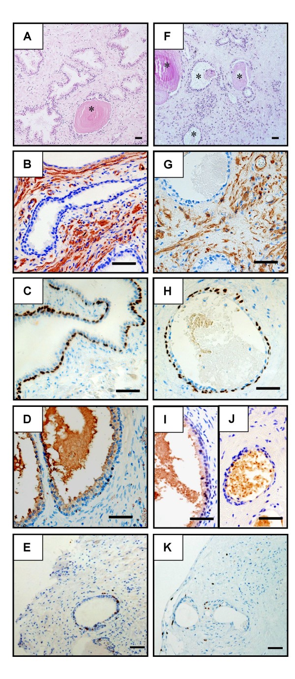
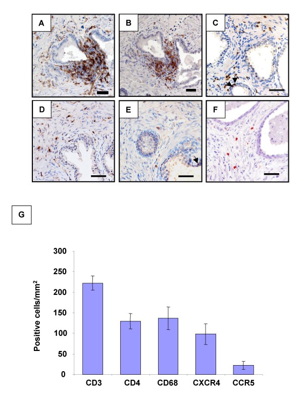
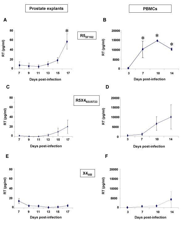
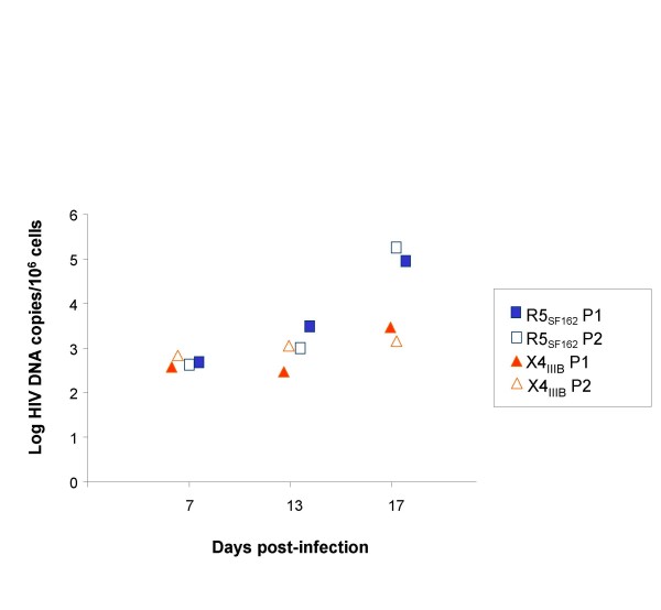
Results: The overall prostate characteristics were retained for 21/2 weeks in culture. Numerous potential HIV-1 target cells were detected in the prostate stroma. Whilst HIV-1 R5SF162 strain consistently productively infected prostatic T lymphocytes and macrophages, the prototypic X4IIIB strain and a primary R5X4 strain showed less efficient replication in this organ.

Conclusion: The BPH prostate is a site of HIV-1 R5 replication that could contribute virus to semen. A limited spreading of HIV-1 X4 and R5X4 in this organ could participate to the preferential sexual transmission of HIV-1 R5 strains.


Figures

Figure 1
Figure 1
Characterization of human prostate in organotypic culture. Paraformaldehyde-fixed paraffin sections were examined morphologically and immunostained for several prostatic cell markers before culture (A-E) and after 4 days (I) or 17 days (F-H, J, K) of culture. (A, F): a mix of normal and hyperplasic glands (identified by stars) were observed in the tissue sections before and after 17 days of culture. The overall morphology of the organ was preserved after 17 days of culture despite a loss of stromal cells in some areas (F). The markers used for the characterization of prostatic cell types were: – α smooth actin for smooth muscle and myofibroblastic stromal cells (B, G); – p63 for basal epithelial cells (C, H); – PSA for secretory luminal epithelial cells (D, I, J); – Ki67 for proliferating cells (E, K). The results are representative of a minimum of three independent cultures performed on prostates collected from different donors. Scale bars = 50 μm.
Figure 2
Figure 2
Presence of potential HIV target cells in the human prostate. Immunohistochemistry on uninfected prostate sections before culture showed the presence of periglandular foci of HLA-DR+ (A) and CD4+ cells (B, serial section with A) as well as scattered stromal cells staining positive for HLA-DR (A), CD4 (B), CD3 (C), CD68 (D), CCR5 (E) and CXCR4 (F). The arrows point out immune cells inserted within the epithelium-Scale bars = 50 μm; (G): the respective proportions of CD3, CD4, CD68, CXCR4 and CCR5+ cells per surface unit were evaluated on whole prostate sections from a minimum of 3 donors whose explants were subsequently exposed to HIV-1 strains.
Figure 3
Figure 3
HIV-1 R5, R5X4 and X4 infection of human prostate in organotypic culture. RT activity was measured in supernatants of human prostate explants exposed to HIV-1 R5SF162 (n = 3 donors) (A), R5X492US723 (n = 3 donors) (C), or X4IIIB (n = 5 donors) (E); and in supernatants of activated PBMCs exposed to day 17 supernatants of prostate explant cultures exposed to R5SF162 (n = 3) (B), R5X492US723(n = 3) (D) or X4IIIB (n = 5) (F). RT activity was never detected in the supernatants of PBMCs infected with an inoculum of the respective viral stocks maintained at 37°C in medium for 17 days. Each dot represents the mean ± SEM of three to five independent cultures (Dunnett test; *, P < 0.05; control: day 7).
Figure 4
Figure 4
Accumulation of HIV-1 DNA in prostate explants following exposure to either R5SF162 or X4IIIB, as assayed for LTR DNA by quantitative real time PCR. Each dot represents the mean value of 2 paired explants (each tested in duplicate PCR) from one individual. For each virus strain, prostate explants from 2 patients were analyzed.
Figure 5
Figure 5
Localization and characterization of HIV-1 RNA positive cells in the human prostate infected ex vivo. Localization of HIV RNA+ cells (black silver grains) in prostate explants exposed for 17 days to R5SF162 (A, C) or R5X492US723 (B) using in situ hybridization for HIV-1 gag. Combined ISH with immunostaining for cell markers was used to assess co-localization of HIV RNA+ cells with either CD68 (A) or CD3 (B, C). A'-C' correspond to higher magnification of A-C showing cells co-labelled for HIV RNA (black silver grains) and cell marker (brown cells). Scale bars = 20 μm.

References

    1. Dejucq-Rainsford N, Jégou B. Viruses in semen and male genital tissues: consequences for the reproductive system and therapeutic perspectives. Current Pharmaceutical Design. 2004;10:557–575. - PubMed
    1. Zhang H, Dornadula G, Beumont M, Livornese LJ, Van Uitert B, Henning K, Pomerantz RJ. Human immunodeficiency virus type 1 in the semen of men receiving highly active antiretroviral therapy. N Engl J Med. 1998;339:1803–1809. - PubMed
    1. Kiessling AA, Fitzgerald LM, Zhang D, Chhay H, Brettler D, Eyre RC, Steinberg J, McGowan K, Byrn RA. Human immunodeficiency virus in semen arises from a genetically distinct virus reservoir. AIDS Res Hum Retroviruses. 1998;14:S33–S41. - PubMed
    1. Mayer KH, Boswell S, Goldstein R, Lo W, Xu C, Tucker L, DePasquale MP, D'Aquila R, Anderson DJ. Persistence of human immunodeficiency virus in semen after adding indinavir to combination antiretroviral therapy. Clin Infect Dis. 1999;28:1252–1259. - PubMed
    1. Lafeuillade A, Solas C, Halfon P, Chadapaud S, Hittinger G, Lacarelle B. Differences in the detection of three HIV-1 protease inhibitors in non-blood compartments: Clinical correlations. HIV Clin Trials. 2002;3:27–35. - PubMed

Publication types