. 2009 Jan 7;9(1):23-34.
doi: 10.1016/j.cmet.2008.11.008.

The glycogen-binding domain on the AMPK beta subunit allows the kinase to act as a glycogen sensor

Affiliations

The glycogen-binding domain on the AMPK beta subunit allows the kinase to act as a glycogen sensor

Andrew McBride et al. Cell Metab. .

Abstract

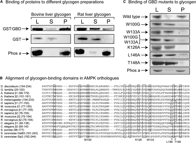
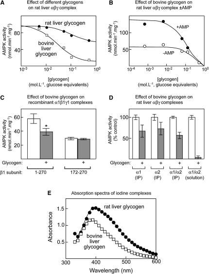
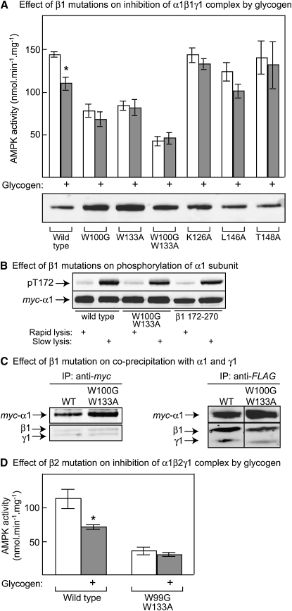
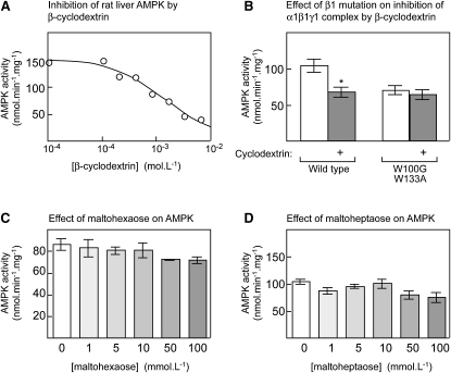
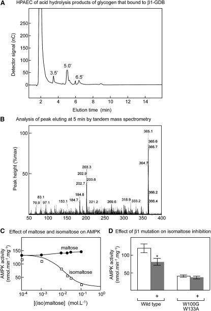
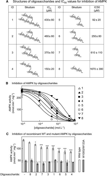
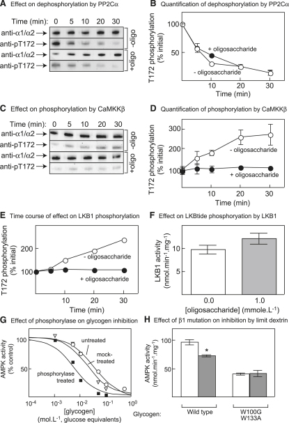
AMPK beta subunits contain a conserved domain that causes association with glycogen. Although glycogen availability is known to affect AMPK regulation in vivo, the molecular mechanism for this has not been clear. We now show that AMPK is inhibited by glycogen, particularly preparations with high branching content. We synthesized a series of branched oligosaccharides and show that those with a single alpha1-->6 branch are allosteric inhibitors that also inhibit phosphorylation by upstream kinases. Removal of the outer chains of glycogen using phosphorylase, thus exposing the outer branches, renders inhibition of AMPK more potent. Inhibition by all carbohydrates tested was dependent on the glycogen-binding domain being abolished by mutation of residues required for carbohydrate binding. Our results suggest the hypothesis that AMPK, as well as monitoring immediate energy availability by sensing AMP/ATP, may also be able to sense the status of cellular energy reserves in the form of glycogen.


Figures

Figure 1
Figure 1
Studies on Binding of Glycogen to Bacterially Expressed β1-GBD (A) Binding of GST:GBD fusion, free GST, and phosphorylase a to glycogen. Samples of each protein were incubated with bovine or rat liver glycogen bound to ConA-Sepharose, the Sepharose beads were recovered by centrifugation, and samples of the load (L), supernatant (S), and pellet (P, resuspended in the original volume) were analyzed by SDS-PAGE. (B) Alignment of GBD sequences from various eukaryotes made using ALIGNX. Residues identical in all species are boxed, as are conserved residues in mammalian species directly involved in carbohydrate binding; the latter are identified at the bottom (rat β1 numbering). (C) Binding to glycogen of GST:GBD fusions (wild-type rat β1 or the point mutations shown). The binding assay was as in (A) using bovine liver glycogen, and binding of phosphorylase a was analyzed as a positive control (bottom panel).
Figure 2
Figure 2
Allosteric Inhibition of AMPK by Different Glycogen Preparations (A) Concentration dependence of inhibition of native rat liver AMPK by preparations of bovine and rat liver glycogen; glycogen concentrations expressed as glucose produced after total hydrolysis. Data were fitted to an IC50 equation (see Supplemental Experimental Procedures), and curves were generated using the estimated best-fit parameters. (B) Concentration dependence of inhibition of native rat liver AMPK by bovine liver glycogen in the presence and absence of 200 μM AMP; curves were generated as in (A). (C) Inhibition by bovine liver glycogen of recombinant AMPK complex (myc-α1:β1:γ1) containing full-length (1–270) or truncated β1 subunit lacking the GBD (172–270). Complexes were expressed in CCL13 cells and purified by immunoprecipitation prior to assay with or without glycogen (200 mM glucose equivalents). Data are means ± SEM (n = 3). Activity different from control without glycogen by t test (p < 0.05). (D) Inhibition by bovine liver glycogen (200 mM glucose equivalents) of purified, native rat liver AMPK (a mixture of α1β1γ1 and α2β1γ1 complexes) that had been recovered by immunoprecipitation (IP) using anti-α1, anti-α2, or a mixture of anti-α1/-α2 antibodies or assayed in solution without immunoprecipitation. Results are expressed as percentages of the activity in controls without glycogen, and data are mean ± SEM (n = 3). (E) Absorption spectra of bovine and rat liver glycogen preparations in complex with iodine.
Figure 3
Figure 3
Effect of β Subunit Mutations on Inhibition of Recombinant AMPK Complexes by Glycogen (A) Myc-α1, γ1, and β1 with the wild-type sequence or the indicated mutations were expressed in CCL13 cells, immunoprecipitated, and assayed ± bovine liver glycogen (200 mM glucose equivalents); results are mean ± SEM (n = 3). The lower panel shows expression of the α1 subunit assessed by probing blots using anti-myc antibody; the activities in the upper panel were corrected for small variations in expression. (B) Myc-α1, γ1, and either wild-type β1, a W100G/W133A mutant, or an N-terminally truncated mutant (β1 172–270) were expressed in CCL13 cells, extracted using the rapid or slow lysis procedures, and subject to western blotting using anti-myc or anti-pT172 antibodies. (C) Myc-α1, γ1, and β1 with the wild-type sequence or with W100G/W133A mutations were expressed in CCL13 cells, immunoprecipitated using anti-myc (left) or anti-FLAG (right) antibodies, and analyzed by western blotting using anti-myc antibodies (top panels) or a mixture of anti-β1 and -γ1 antibodies (bottom panels). (D) Myc-α1, γ1, and human β2 with the wild-type sequence or with W99G/W133A mutations were coexpressed, immunoprecipitated, and assayed ± bovine liver glycogen (200 mM glucose equivalents); results are mean ± SEM (n = 3). Activities were corrected for small variations in the observed level of expression as in (A). Activity different from control without glycogen by t test (p < 0.05).
Figure 4
Figure 4
Effect of Unbranched Oligosaccharides on AMPK Activity (A) Inhibition of native rat liver AMPK by β-cyclodextrin. The curve was generated as in Figure 2A. (B) Effect of GBD double mutation on inhibition of recombinant AMPK by 2 mM β-cyclodextrin. Myc-α1, γ1, and β1 with the wild-type sequence or with W100G/W133A mutations were coexpressed, immunoprecipitated, and assayed ± β-cyclodextrin (2 mM). The activities were corrected for small variations in the observed level of expression as in Figure 2A. Activity different from control without β-cyclodextrin (p < 0.05). (C and D) Effect of maltohexaose (C) and maltoheptaose (D) on AMPK activity. Native rat liver AMPK was assayed in the presence and absence of the indicated concentrations of oligosaccharide. Results in (B) through (D) are mean ± SEM (n = 3).
Figure 5
Figure 5
Isolation of Isomaltose as an Inhibitor of AMPK (A) Bovine liver glycogen was subjected to partial acid hydrolysis and the hydrolysate passed through a glutathione Sepharose column to which the GST:GBD fusion had been prebound. The column was eluted with propionic acid and bound oligosaccharides analyzed by HPAEC. The peak eluting at 5 min was the only one found to contain carbohydrate. (B) Analysis of the peak eluting at 5 min by tandem ES-MS using collision-induced dissociation. Oligosaccharide fragments are observed as adducts with Na+ ions, increasing their mass by 23. (C) Effect of maltose and isomaltose on AMPK activity. The curve for isomaltose was generated using the best-fit parameters as in Figure 2A. (D) Effect of GBD double mutation on inhibition of recombinant AMPK by 20 mM isomaltose; results are mean ± SEM (n = 3). Activity different from control without isomaltose by t test (p < 0.05).
Figure 6
Figure 6
Inhibition of Rat Liver AMPK by Synthetic Branched Oligosaccharides (A) Table showing the identification (ID) number, structure, and estimated IC50 ± SEM. (B) Inhibition of purified rat liver AMPK by oligosaccharides. These data were used to generate the IC50 values shown in (A), and the curves were drawn using the best-fit parameters. A key to the ID of each oligosaccharide is shown on the right. (C) Inhibition of recombinant wild-type and W100G/W133A mutant AMPK (α1β1γ1 complex) by synthetic oligosaccharides, numbered as in (A) and (B). The data are presented with the oligosaccharides in order of increasing potency from left to right; results are mean ± SEM (n = 3). Significant differences ± oligosaccharide for the wild-type by two-way ANOVA. p < 0.05; ∗∗∗p < 0.001.
Figure 7
Figure 7
Effect of Oligosaccharide Number 5, See Figure 6, on Dephosphorylation of AMPK by PP2Cα and on Phosphorylation by CaMKKβ and LKB1 (A) Effect of oligosaccharide on dephosphorylation by PP2Cα. Rat liver AMPK was incubated with PP2Cα in the presence and absence of 1 mM oligosaccharide; at various times, levels of total α subunit and phosphorylation on Thr-172 were assessed by western blotting. (B) Quantification of results as in (A); means ± SEM of two experiments. (C) Effect of oligosaccharide on phosphorylation by CaMKKβ. Rat liver AMPK was dephosphorylated using PP1, okadaic acid was added to inhibit the phosphatase, and the protein was incubated with MgATP and CaMKKβ in the presence and absence of 1 mM oligosaccharide. At various times, levels of total α subunit and of phosphorylation on Thr-172 were assessed by western blotting. (D) Quantification of results as in (C); means ± SEM (n = 2). (E) Time course of effect of oligosaccharide on phosphorylation by LKB1, performed as in (C) but with LKB1 in place of CaMKKβ. (F) Effect of oligosaccharide on phosphorylation of a synthetic peptide substrate of LKB1 (mean ± SD; n = 2). (G) Inhibition of AMPK by bovine liver glycogen, a phosphorylase limit dextrin, and a mock-treated sample. IC50 values were determined as in Figure 2A and curves generated from the best-fit parameters. (H) Effect of limit dextrin on recombinant α1β1γ1 complex with wild-type and W100G/W133A mutant β1. Results are mean ± SEM (n = 3). Activity different from control without limit dextrin (p < 0.05).

Comment in

References

    1. Aschenbach W.G., Suzuki Y., Breeden K., Prats C., Hirshman M.F., Dufresne S.D., Sakamoto K., Vilardo P.G., Steele M., Kim J.H. The muscle-specific protein phosphatase PP1G/R(GL)(G(M)) is essential for activation of glycogen synthase by exercise. J. Biol. Chem. 2001;276:39959–39967. - PubMed
    1. Carling D., Hardie D.G. The substrate and sequence specificity of the AMP-activated protein kinase. Phosphorylation of glycogen synthase and phosphorylase kinase. Biochim. Biophys. Acta. 1989;1012:81–86. - PubMed
    1. Danforth W.H. Glycogen synthetase activity in skeletal muscle. Interconversion of two forms and control of glycogen synthesis. J. Biol. Chem. 1965;240:588–593. - PubMed
    1. Davies S.P., Helps N.R., Cohen P.T.W., Hardie D.G. 5′-AMP inhibits dephosphorylation, as well as promoting phosphorylation, of the AMP-activated protein kinase. Studies using bacterially expressed human protein phosphatase-2Cα and native bovine protein phosphatase-2AC. FEBS Lett. 1995;377:421–425. - PubMed
    1. Derave W., Ai H., Ihlemann J., Witters L.A., Kristiansen S., Richter E.A., Ploug T. Dissociation of AMP-activated protein kinase activation and glucose transport in contracting slow-twitch muscle. Diabetes. 2000;49:1281–1287. - PubMed

Publication types

MeSH terms

Substances

LinkOut - more resources