. 2009 Mar 13;284(11):7007-16.
doi: 10.1074/jbc.M805736200. Epub 2008 Dec 31.

The HLA-DRalpha chain is modified by polyubiquitination

Affiliations

The HLA-DRalpha chain is modified by polyubiquitination

Nicolas Lapaque et al. J Biol Chem. .

Abstract

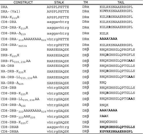
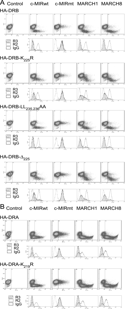
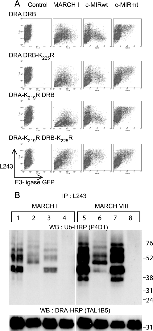
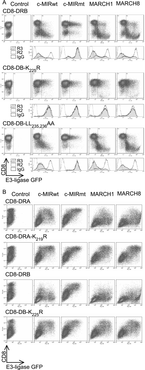
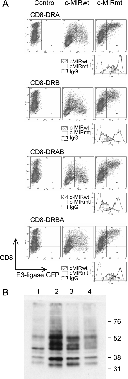
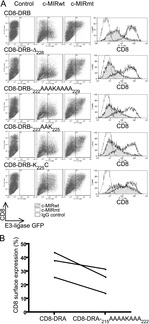
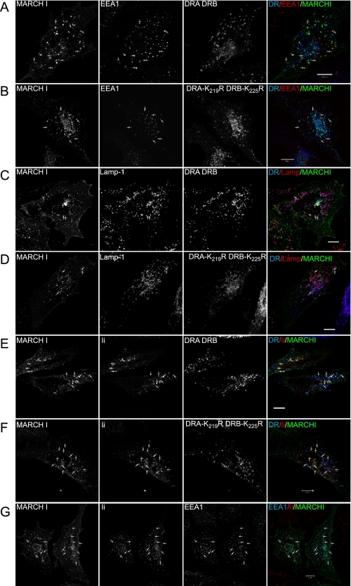
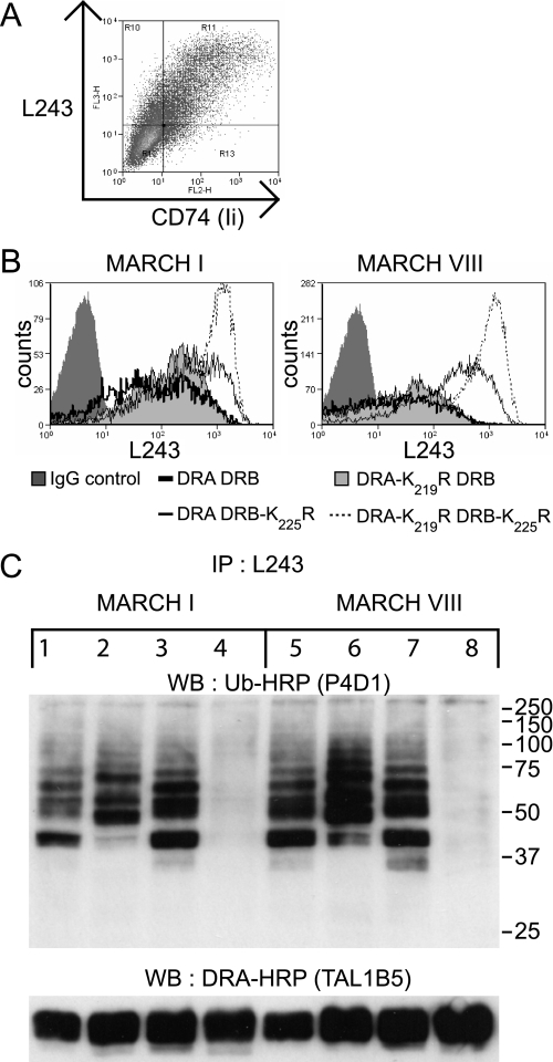
Ubiquitination plays a major role in regulating cell surface and intracellular localization of major histocompatibility complex class II molecules. Two E3 ligases, MARCH I and MARCH VIII, have been shown to polyubiquitinate lysine residue 225 in the cytoplasmic tail of I-Abeta and HLA-DRbeta. We show that lysine residue 219 in the cytoplasmic tail of DRalpha is also subject to polyubiquitination. Each chain of the HLA-DR heterodimer is independently recognized and ubiquitinated, but DRbeta is more extensively modified. In the cytoplasmic tail of DRbeta lysine, residue 225 is the only residue that is absolutely required for ubiquitination; all other residues can be deleted or substituted without loss of function. In contrast, although lysine 219 is absolutely required for modification of DRalpha, other features of the DRalpha tail act to limit the extent of ubiquitination.


Figures

FIGURE 1.
FIGURE 1.
Summary of the amino acid composition of constructs used in this study. The numbering of residues is taken from the mature protein after signal sequence removal. Single-letter amino codes are used. Sequences derived from CD8 are in italic type, and substituted residues are in boldface type.
FIGURE 2.
FIGURE 2.
MARCH-induced down-regulation of HA-DRB-K225R. Mel JuSo cells, stably expressing various HA-tagged DRα and DRβ constructs, were transiently transfected with MARCH I, MARCH VIII, c-MIRwt, and c-MIRmt, and surface HA expression was assessed by FACS. Dot plots show GFP (FL1) on the x axis and PE-HA (FL2) on the y axis. The R3 gate, which represents cells expressing the E3 ligase, as assessed by GFP expression, was set against IgG control at 0.1%. The IgG control was set using cells expressing GFP (R3), to enable direct comparison between with E3 ligase-expressing transfectants. Data are representative of at least three independent experiments. A, expression of MARCH I, MARCH VIII, and c-MIRwt induced down-regulation of surface HA-DRB, HA-DRB-K225R, HA-DRB-L235A,L236A, and HA-DRB-Δ225. No reduction in surface expression was observed in cells transfected with c-MIRmt. When compared with IgG control antibody staining, MARCH-induced down-regulation of HA-DRB-K225R and HA-DRB-Δ225 was substantial, but less than observed for HA-DRB and HA-DRB-L235A,L236A. B, MARCH I, MARCH VIII, and c-MIRwt induced down-regulation of surface HA-DRA and DRA-K219R. No reduction in surface expression was observed in cells transfected with c-MIRmt. Data are representative of at least three experiments.
FIGURE 3.
FIGURE 3.
Lysine residues Lys219 and Lys225 on HLA-DRα and HLA-DRβ, respectively, are subject to polyubiquitination. A, 293T cells were transiently co-transfected with wild-type or mutated forms of HLA-DRα and HLA-DRβ, together with either MARCH I, c-MIRwt, or c-MIRmt. The levels of surface HLA-DR were assessed by FACS using PE-L243 in the FL2 channel. E3 ligase expression was monitored indirectly by measuring GFP expression in the FL1 channel. All combinations of DRα and DRβ resulted in MARCH I- and c-MIRwt (MARCH VIII)-induced DR down-regulation, except for DRA-K219R/DRB-K225R. No change in surface HLA-DR expression was seen in the presence of c-MIRmt. Data are representative of at least three independent experiments. B, HLA-DR was immunoprecipitated (IP) from lysates of the transfected cells depicted above using L243 monoclonal antibody directly conjugated to Sepharose. After standard SDS-PAGE separation and Western transfer (WB), DRα was detected with HRP-TAL1B5, and ubiquitinated HLA-DR was detected with the anti-ubiquitin antibody HRP-P4D1. Lanes 1–4 were transfected with MARCH I; lanes 5–8 were transfected with MARCH VIII. Lanes 1 and 5 expressed DRA and DRB; lanes 2 and 6 expressed DRA and DRB-K225R; lanes 3 and 7 expressed DRA-K219R and DRB; and lanes 4 and 8 expressed DRA-K219R and DRB-K225R. The upper panels show ubiquitinated HLA-DR, as detected with HRP-P4D1, the lower panels show DRα as detected by HRP-TAL1B5. No additional bands suggestive of ubiquitinated DRα were evident in the lower panels, even after long exposure. The upper panel shows that both DRα and DRβ are subject to polyubiquitination. The signal for DRβ is stronger than for DRα. Data are representative of at least three independent experiments.
FIGURE 4.
FIGURE 4.
Transmembrane and cytoplasmic domains from both DRα and DRβ are independently targeted by MARCH I and MARCH VIII. CD8 reporter constructs comprising the CD8 extracellular domain and DRα or DRβ transmembrane and cytoplasmic tails were stably expressed in Mel JuSo cells (A) or transiently expressed in 293T cells (B). Dot plots show GFP (FL1) on the x axis and anti-CD8 (PE-OKT8) binding (FL2) on the y axis. The R3 gate, which represents cells expressing the E3 ligase, as assessed by GFP expression, was set against IgG control at 0.1%. The IgG control was set using cells expressing GFP (R3), to enable direct comparison between E3 ligase-expressing transfectants. Data are representative of at least three independent experiments. A, a CD8 chimera containing the transmembrane and cytoplasmic tail of DRβ is subject to MARCH I- and MARCH VIII-induced down-regulation. This was abolished by substitution of lysine residue Lys225 for arginine (CD8-DRB-K225R). The dileucine motif at positions 235 and 236 was not required for MARCH-induced down-regulation, since the behavior of CD8-DRB-L235A,L236A was indistinguishable from that of CD8-DRB. B, CD8-DRA was also targeted by MARCH I and MARCH VIII, and this was dependent upon a lysine residue, Lys219, in its cytoplasmic tail. The extent of surface down-regulation of CD8-DRA was less than for CD8-DRB. Data are representative of at least three independent experiments.
FIGURE 5.
FIGURE 5.
Efficient ubiquitination correlates with the presence of the DRβ cytoplasmic tail. To determine which regions of DRα and DRβ were important for ubiquitination, 293T cells were transfected with CD8-DRA, CD8-DRB, CD8-DRAB, and CD8-DRBA, together with c-MIRwt or c-MIRmt. CD8-DRAB was generated by replacing the cytoplasmic tail of CD8-DRA with that from DRβ. CD8-DRBA is the reciprocal exchange, involving CD8-DRB and DRA. Data are representative of at least three independent experiments. A, FACS analysis of the 293T cell transfectants, described above, stained with the anti-CD8 antibody, PE-OKT8. Similar levels of surface CD8 expression and control IgG staining were seen in all four transfectants. Greater c-MIR-induced down-regulation was seen in CD8-DRB and CD8-DRAB transfectants compared with CD8-DRA and CD8-DRBA. No down-regulation was seen in the presence of c-MIRmt; in fact, surface CD8 appears to increase. B, CD8 chimeras were immunoprecipitated from lysates of the transfected cells depicted above using OKT8 and protein A-Sepharose. After standard SDS-PAGE separation and Western transfer, the presence of the ubiquitinated CD8-chimeras was detected with the anti-ubiquitin antibody HRP-P4D1. Lane 1, CD8-DRA; lane 2, CD8-DRB; lane 3, CD8-DRAB; lane 4, CD8-DRBA. All molecules were subject to polyubiquitination, and the strength of signal was highest for CD8-DRB and CD8-DRAB and lowest for CD8-DRA and CD8-DRBA.
FIGURE 6.
FIGURE 6.
A minimal CD8-DRB cytoplasmic tail, RAAK, is an efficient target for ubiquitination. 293T cells were transfected with CD8-DRB or CD8-DRA reporter molecules containing deletions and substitutions in the DRβ cytoplasmic tail, coupled with either c-MIRwt or c-MIRmt. After 24 h, cells were subjected to FACS analysis with PE-OKT8. Dot plots show c-MIR expression on the x axis (FL1), as monitored by GFP expression and CD8 expression on the y axis (FL2). Histograms showing CD8 expression are presented. Data are representative of at least three independent experiments. A, all CD8-DRB constructs were efficiently down-regulated by c-MIR, including a “minimal” cytoplasmic tail CD8-DRB-222RAAK225. In addition, substitution of lysine 225 for cysteine resulted in significant, if reduced, down-regulation. B, CD8-DRA-215AAAAKAAA222 showed enhanced down-regulation in the presence of c-MIRwt compared with CD8-DRA, suggesting that elements in the tail of DRα have an antagonistic effect upon ubiquitination of DRα. 293T cells were transfected with CD8-DRA-215AAAAKAAA222 or CD8-DRA, together with either c-MIRwt or c-MIRmt and cell surface CD8 determined by FACS. The plot shows mean fluorescence values for cell surface CD8-DRA-215AAAAKAAA222 and CD8-DRA in the presence of c-MIRwt, expressed as a percentage of the expression observed in the presence of c-MIRmt. In each of three independent experiments, there is less CD8-DRA-215AAAAKAAA222 at the plasma membrane compared with CD8-DRA. CD8-DRA-215AAAAKAAA222 is therefore more efficiently removed from the cell surface compared with CD8-DRA.
FIGURE 7.
FIGURE 7.
Intracellular distribution of MHC class II and MARCH I. HeLa cells were transiently transfected with wild-type and mutated DRα and DRβ constructs, together with MARCH I, in the presence or absence of Ii. Intracellular distribution was analyzed by confocal microscopy. A and B show colocalization of MARCH I and EEA1 (arrows), in cells transfected with either DRA/DRB (A) or DRA-K219R/DRB-K225R (B). The merged image shows DR in blue, EEA1 in red, and MARCH I in green; co-localized MARCH and EEA1 is yellow. C and D show colocalization of MARCH I and Lamp-1 (arrows), in cells transfected with either DRA/DRB (C) or DRA-K219R/DRB-K225R (D). The merged images show DR in blue, Lamp-1 in red, and MARCH Iin green. Note that the majority of class II is present in Lamp-1-positive compartments (purple), some of which co-localize with MARCH I. E–G show localization of class II in the presence of Ii. The merged image shows DR or EEA1 in blue, Ii in red, and MARCH I in green. E and F show colocalization of Ii and MARCH I (arrows) in cells transfected with DRA/DRB (E) or DRA-K219R/DRB-K225R (F). In both cases, Ii shows good colocalization with MARCH I, whereas DR is mainly in MARCH I-negative vesicles. G shows a high degree of colocalization between Ii and EEA1. Together, this shows that Ii is in MARCH I-positive, EEA1-positive early endosomes. Bar, 10 μm.
FIGURE 8.
FIGURE 8.
Ubiquitination of MHC class II in the presence and absence of Ii. To determine if ubiquitination was influenced by the presence of Ii, 293T cells were transiently co-transfected with Ii and wild-type or mutated forms of HLA-DRα and HLA-DRβ, together with either MARCH I or MARCH VIII. A, FACS analysis of 293T cells transfected with DRA/DRB and Ii demonstrates that the majority of transfected cells express both class II and Ii. B, 293T cells were transiently co-transfected with Ii and wild-type or mutated forms of DRα and DRβ, together with either MARCH I or MARCH VIII. FACS profiles of surface L243-reactive class II expression demonstrate that all combinations of DRα and DRβ are down-regulated by MARCH I and MARCH VIII, except for DRA-K219R/DRB-K225R, as previously seen in the absence of Ii (Fig. 3). C, HLA-DR was immunoprecipitated (IP) from lysates of the transfected cells depicted above, using L243 monoclonal antibody directly conjugated to Sepharose. After standard SDS-PAGE separation and Western transfer (WB), DRα was detected with HRP-TAL1B5 (bottom), and ubiquitinated HLA-DR was detected with the anti-ubiquitin antibody HRP-P4D1 (top). Lanes 1–4 were transfected with MARCH I, and lanes 5–8 were transfected with MARCH VIII. Lanes 1 and 5 expressed DRA and DRB; lanes 2 and 6 expressed DRA and DRB-K225R; lanes 3 and 7 expressed DRA-K219R and DRB; and lanes 4 and 8 expressed DRA-K219R and DRB-K225R. Comparison with Fig. 3C shows that the pattern of ubiquitination of DRα and DRβ is similar in the presence or absence of Ii. Data are representative of two independent experiments.

References

    1. Peters, P. J., Neefjes, J. J., Oorschot, V., Ploegh, H. L., and Geuze, H. J. (1991) Nature 349 669-676 - PubMed
    1. Cresswell, P. (1992) Curr. Opin. Immunol. 4 87-92 - PubMed
    1. Denzin, L. K., and Cresswell, P. (1995) Cell 82 155-165 - PubMed
    1. Watts, C. (2004) Nat. Immunol. 5 685-692 - PubMed
    1. Hiltbold, E. M., and Roche, P. A. (2002) Curr. Opin. Immunol. 14 30-35 - PubMed

Publication types