. 2009 Jan 1;69(1):37-44.
doi: 10.1158/0008-5472.CAN-08-1648.

TGFBI deficiency predisposes mice to spontaneous tumor development

Affiliations

TGFBI deficiency predisposes mice to spontaneous tumor development

Ye Zhang et al. Cancer Res. .

Abstract

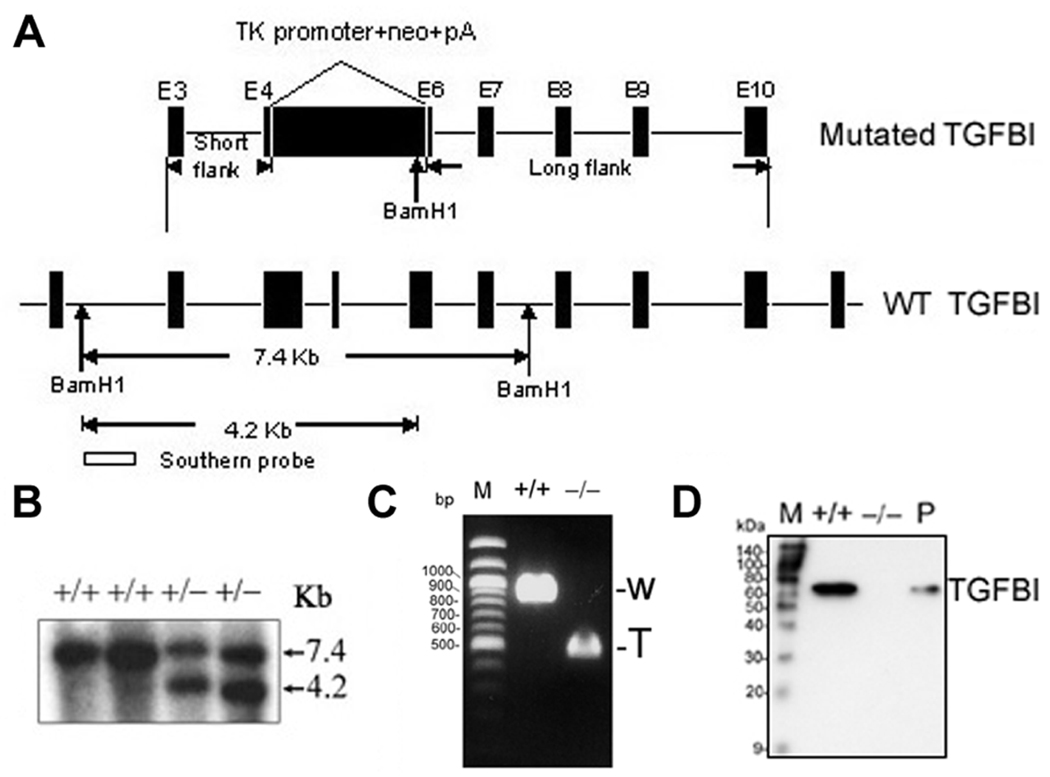
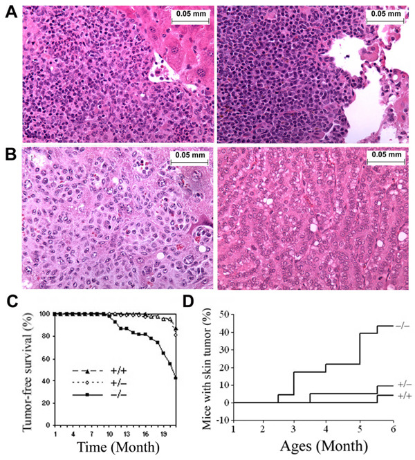
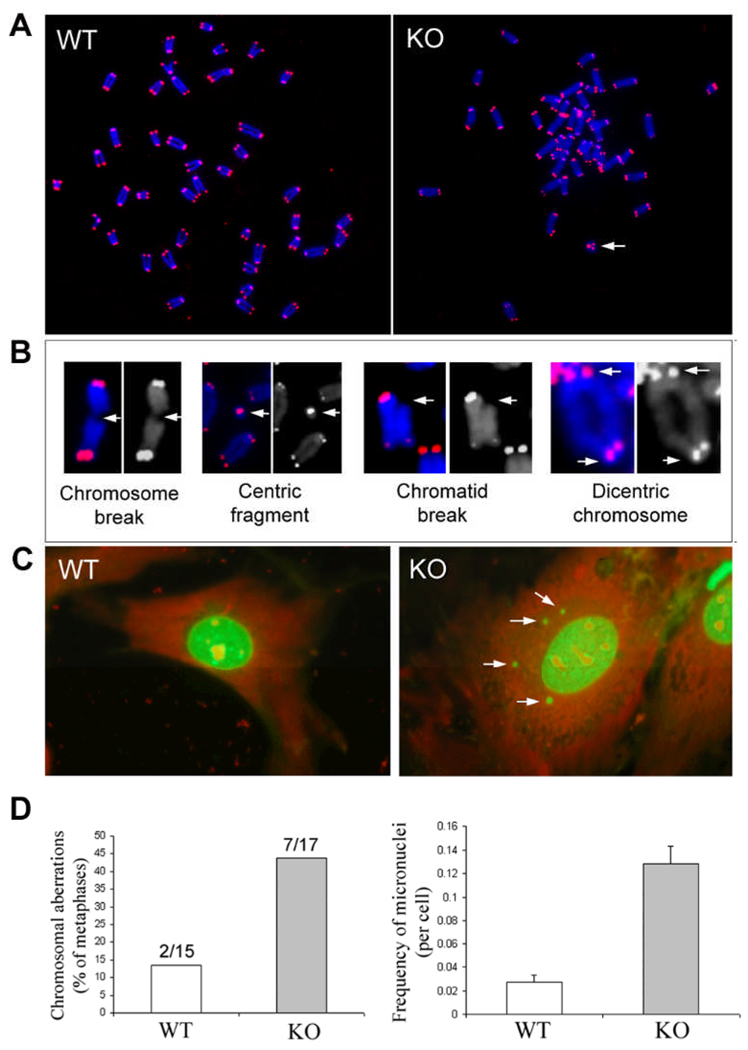
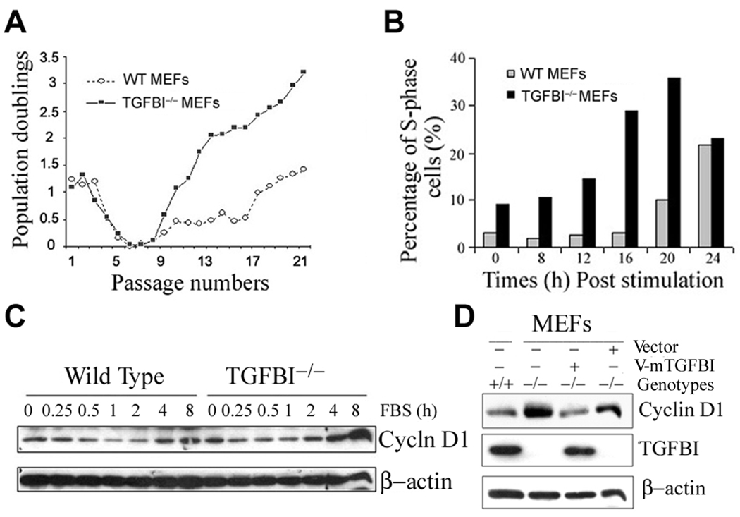
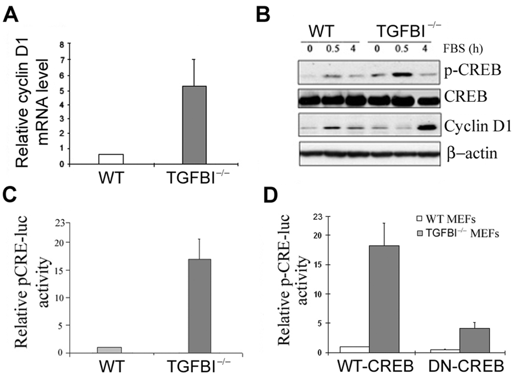
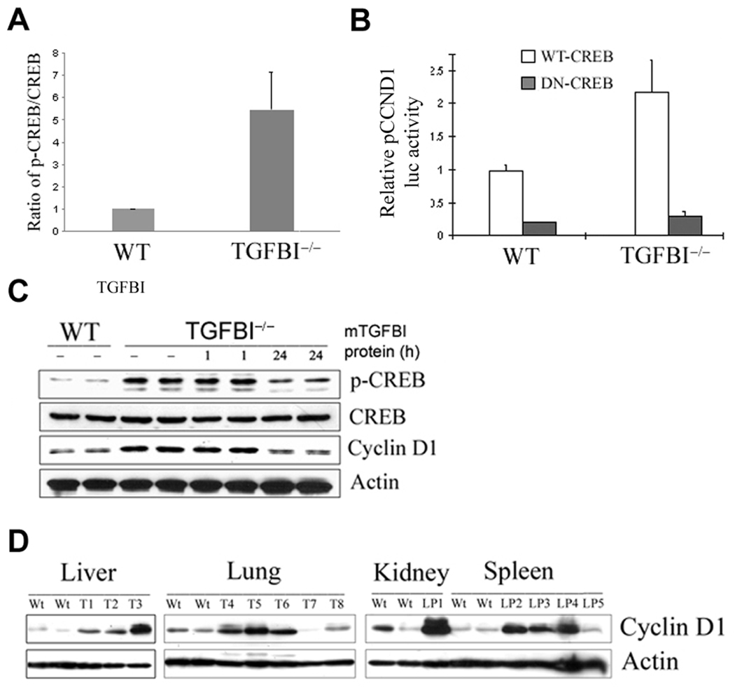
Loss of TGFBI, a secreted protein induced by transforming growth factor-beta, has been implicated in cell proliferation, tumor progression, and angiogenesis by in vitro studies. However, in vivo antitumor functions of TGFBI as well as the underlying molecular mechanism are not well understood. To these aims, we have generated a mouse model with disruption of TGFBI genomic locus. Mice lacking TGFBI show a retarded growth and are prone to spontaneous tumors and 7,12-dimethylbenz(a)anthracene-induced skin tumors. In relation to wild-type (WT) mouse embryonic fibroblasts (MEF), TGFBI(-/-) MEFs display increased frequencies of chromosomal aberration and micronuclei formation and exhibit an enhanced proliferation and early S-phase entry. Cyclin D1 is up-regulated in TGFBI(-/-) MEFs, which correlates with aberrant activation of transcription factor cyclic AMP-responsive element binding protein (CREB) identified by chromatin immunoprecipitation and luciferase reporter assays. TGFBI reconstitution in TGFBI(-/-) cells by either retroviral infection with WT TGFBI gene or supplement with recombinant mouse TGFBI protein in the culture medium leads to the suppression of CREB activation and cyclin D1 expression, and further inhibition of cell proliferation. Cyclin D1 up-regulation was also identified in most of the tumors arising from TGFBI(-/-) mice. Our studies provide the first evidence that TGFBI functions as a tumor suppressor in vivo.


Figures

Figure 1
Figure 1. Targeted disruption of TGFBI in mice
(A) Strategy for generating the targeted TGFBI allele. Exons 4–6 were replaced by neomycin-resistance cassette (neo) with introduction of one BamH1 restriction site at 3’ terminal. Targeting construct and wild type allele are shown. Successful targeting will yield a 4.2 kb BamH1-restricted fragment in the neo allele. (B) Germinal transmission of the targeted TGFBI allele was identified by Southern blot. (C) Identification of deletion of exons 4–6 in KO MEFs by RT-PCR using a pair of primers specific for the upstream and downstream regions of exons 4–6. W: wild type; T: truncated. (D) Western blot of conditioned medium prepared from MEFs with indicated genotypic backgrounds. Mouse TGFBI recombinant protein was used as positive control (P).
Figure 2
Figure 2. TGFBI−/− mice showed an increased tumor incidence
(A) Images of Low grade lymphoma in Live (left panel) and Lung (right panel). (B) Image of metastasized tumor in liver (left panel) and lung adenocarcinoma (right panel). Magnification of images (A–B): ×400. (C) Tumor-free survival of TGFBI−/− mice compared with wild type and heterozygots. (D) Incidence of DMBA-induced skin tumors in wild type and TGFBI−/− mice.
Figure 3
Figure 3. Increased frequency of chromosomal aberrations and micronuclei in early passage (P2) of TGFBI−/− MEFs
(A) Digital images of Cy-3 (identify telomeres) and DAPI (identify chromosomes)-stained chromosomal metaphases in wild type and TGFBI KO MEFs. Arrow: centric ring. (B) Various types of chromosomal aberrations (Arrows) found in KO MEFs. (C) Multiple micronuclei (Arrows) identified in KO MEFs. (D) Frequency of chromosomal aberrations and micronuclei in wild type and TGFBI−/− MEFs.
Figure 4
Figure 4. Characterization of growth property and cyclin D1 expression in TGFBI-null MEFs
(A) Cell proliferation on a 3T3 protocol. MEFs were isolated from 13.5-d embryos, and grown at 5% CO2 in DMEM (Invitrogen) supplemented with 10% FCS. For 3T3 protocol, 9 ×10⁵ cells were plated into 10cm-dish and cell numbers were counted at 3 days interval. At least three independent lines per genotype with two independent cultures per line were examined. (B) Kinetics of S-phase entry upon serum stimulation of quiescent TGFBI−/− and wild type MEFs. (C) Cyclin D1 induction in serum-stimulated quiescent TGFBI−/− and wild type MEFs determined by Western blots. (D) Western blots result of cyclin D1 level in exponentially-grown MEFs with TGFBI−/− and wild type backgrounds, and in TGFBI−/− MEFs after reconstitution of TGFBI by infection with retroviral V-mTGFBI vectors (pMSCV-mTGFBI).
Figure 5
Figure 5. Aberrant activation of CREB in TGFBI−/− MEFs
(A) mRNA level of cyclin D1 in TGFBI−/− and wild type MEFs determined by real time RT-PCR. (B) Levels of p-CREB and cyclin D1 in serum-stimulated wild type and TGFBI−/− MEFs examined by Western blots. (C) Relative pCRE-luc activity in wild type and TGFBI−/− MEFs. (D) Relative pCRE-luc activity in wild type and TGFBI−/− MEFs after co-transfection of pCRE-luc with WT-CREB or DN-CREB vectors.
Figure 6
Figure 6. Correlation between CREB activation and cyclin D1 upregulation in TGFBI−/− MEFs
(A) Ratio of pCREB/CREB binding to cyclin D1 promoter region quantified by a quantitative PCR-based ChIP assay. (B) Significant suppression of relative pCCND1 promoter luciferase activity in TGFBI−/− MEFs after cotransfection of pCCND1 promoter with wild type CREB or dominant negative CREB vectors. (C) Suppression of CREB phosphorylation and cyclin D1 expression in TGFBI−/− cells after incubation with recombinant mouse TGFBI protein at 0.5 µg/ml for 24 h. (D) Western blot results of cyclin D1 protein level in tumor tissues arising from TGFBI−/− mice compared to the tissues from wild type littermates. Wt: wild type; T: tumors; LP: lymphoma.

References

    1. Tlsty TD. Cell-adhesion-dependent influences on genomic instability and carcinogenesis. Curr Opin Cell Biol. 1998;10:647–653. - PubMed
    1. Giancotti FG, Ruoslahti E. Integrin signaling. Science. 1999;285:1028–1032. - PubMed
    1. Danen EH. Integrins: regulators of tissue function and cancer progression. Curr Pharm Des. 2005;11:881–891. - PubMed
    1. Ramsay AG, Marshall JF, Hart IR. Integrin trafficking and its role in cancer metastasis. Cancer Metastasis Rev. 2007;26:567–578. - PubMed
    1. Skonier J, Neubauer M, Madisen L, Bennett K, Plowman GD, Purchio AF. cDNA cloning and sequence analysis of beta ig-h3, a novel gene induced in a human adenocarcinoma cell line after treatment with transforming growth factor-beta. DNA Cell Biol. 1992;11:511–522. - PubMed

Publication types

MeSH terms

Substances