. 2009 Jan 1;69(1):369-78.
doi: 10.1158/0008-5472.CAN-08-2724.

Paracrine signaling by platelet-derived growth factor-CC promotes tumor growth by recruitment of cancer-associated fibroblasts

Affiliations

Paracrine signaling by platelet-derived growth factor-CC promotes tumor growth by recruitment of cancer-associated fibroblasts

Charlotte Anderberg et al. Cancer Res. .

Abstract

Cancer results from the concerted performance of malignant cells and stromal cells. Cell types populating the microenvironment are enlisted by the tumor to secrete a host of growth-promoting cues, thus upholding tumor initiation and progression. Platelet-derived growth factors (PDGF) support the formation of a prominent tumor stromal compartment by as of yet unidentified molecular effectors. Whereas PDGF-CC induces fibroblast reactivity and fibrosis in a range of tissues, little is known about the function of PDGF-CC in shaping the tumor-stroma interplay. Herein, we present evidence for a paracrine signaling network involving PDGF-CC and PDGF receptor-alpha in malignant melanoma. Expression of PDGFC in a mouse model accelerated tumor growth through recruitment and activation of different subsets of cancer-associated fibroblasts. In seeking the molecular identity of the supporting factors provided by cancer-associated fibroblasts, we made use of antibody arrays and an in vivo coinjection model to identify osteopontin as the effector of the augmented tumor growth induced by PDGF-CC. In conclusion, we establish paracrine signaling by PDGF-CC as a potential drug target to reduce stromal support in malignant melanoma.


Figures
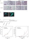
Figure 1
Figure 1. PDGF-CC and PDGFR-α are expressed in human malignant melanoma
(a) Representative pictures of immunostaining for PDGF-CC and PDGFR-α in normal human skin and in malignant melanoma (400x magnification). (b) Immunostaining for PDGF-CC in B16 cells transfected with either human PDGF-C (B16/PDGF-C) or empty vector (B16/mock) (630x magnification). (c) Immunoprecipitation (IP) of PDGFR-α followed by western blot (WB) for phosphorylated proteins from a receptor stimulation assay using PDGF-CC to stimulate B16/PDGF-C and B16/mock cells. Porcine aortic endothelial cells stably transfected with PDGFR-α (α-PAE) were used as a positive control. (d) Left panel, assessment of in vitro growth rate of B16/mock and B16/PDGF-C cells in medium supplemented with serum. Similar results were obtained using serum-free conditions (data not shown). Right panel, in vivo growth rate of B16/mock and B16/PDGF-C cells injected subcutaneously into syngeneic C57Bl/6J mice (n=6). *p<0.05, Student’s t-test.
Figure 2
Figure 2. B16/PDGF-C tumors display an increased proliferation, decreased apoptosis and enhanced angiogenic response
(a) Proportion of BrdU positive proliferating cells (upper panel) and representative pictures of BrdU immunostaining of B16/mock and B16/PDGF-C tumor sections (lower panel; 400x magnification). (b) Proportion of TUNEL positive apoptotic cells (upper panel) and representative pictures of apoptotic labeling of B16/mock and B16/PDGF-C tumor sections (lower panel; 400x magnification). Arrows denote apoptotic cells. (c) Number of CD31+ blood vessels per field (upper panel) and representative pictures of CD31 immunostaining of B16/mock and B16/PDGF-C tumor sections (lower panel; 400x magnification). (d) Pericyte coverage assessed as NG2+ vessels in relation to number of CD31+ vessels (upper panel). Representative pictures of NG2 immunostaining of B16/mock and B16/PDGF-C tumor sections (lower panel; 400x magnification). All quantifications were made on at least 4 tumors of each type using at least 10 randomly selected fields of vision per tumor. *p <0.05; *** p<0.001, Student’s t-test.
Figure 3
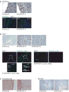
Figure 3. PDGF-C expression promotes recruitment of CAFs
(a) Top panel, immunostaining for PDGFR-α in B16/mock and B16/PDGF-C tumor sections (400x magnification). Bars represent the thickness of the fibrous capsule surrounding the tumor. Bottom panel, visualization of infiltrating PDGFR-α+ cells in PDGFR-α/GFP mice. Representative pictures from B16/mock and B16/PDGF-C tumors (100x magnification). Dotted line denotes boundary between skin and tumor. (b) Top panel, immunostaining for FSP-1 in B16/mock, B16/PDGF-C and B16/PDGF-B tumor sections (400x magnification). Bottom panel, immunostaining for FSP-1 (blue) of tumor sections from B16/PDGF-C tumors grown in PDGFR-α/GFP mice (PDGFR-α, green) at the tumor edge and tumor center (400x magnification). Arrows denote CAFs of the PDGFR-αhigh subclass, arrowheads denote the PDGFR-αlow/FSP-1 subclass of CAFs, and stars denote the FSP-1 subclass of CAFs. (c) Picrosirius red staining for collagen in B16/mock and B16/PDGF-C tumor sections (200x magnification). (d) Immunostaining for the macrophage marker F4/80 of B16/mock and B16/PDGF-C tumor sections (200x magnification).
Figure 4
Figure 4. B16/PDGF-C tumors show an increased abundance of FGF-2 and osteopontin
(a) Pooled protein lysates from 3 separate B16/mock and B16/PDGF-C tumors were applied to antibody arrays. Densitometry was used to quantify the intensity of each spot following normalization to internal standards. (b) Validation of increased osteopontin (OPN) and FGF-2 levels in B16/PDGF-C tumors using immunoprecipitation (IP) and western blot (WB) on separate protein lysates from 3 tumors of each type. WB for PDGF-C verifying increased levels. Calnexin was used as an independent loading control. (c) Relative expression of transcripts for osteopontin and FGF-2, respectively, as measured by quantitative RT-PCR of RNA derived from B16/mock and B16/PDGF-C cell lines.
Figure 5
Figure 5. Osteopontin is expressed by CAFs, and is functionally important for tumor growth
(a) Immunostaining for osteopontin (OPN) in B16/PDGF-C tumor sections (400x magnification). (b) Immunostaining for FSP-1 (blue), osteopontin (red) in tumor sections from B16/PDGF-C tumors grown in PDGFR-α/GFP mice (PDGFRα, green) (400x magnification). Arrowheads denote the PDGFR-αlow/FSP-1 subclass of CAFs; stars denote the FSP-1 subclass of CAFs. (c) Relative expression of osteopontin transcript, as measured by quantitative RT-PCR, following stimulation of NIH/3T3 fibroblasts with PDGF-CC. (d) Left panel, latency of B16 tumors injected alone (n=10), or in combination with a 1:1 ratio of wt (n=10) or osteopontin deficient (OPN KO) (n=9) mouse embryonic fibroblasts (MEFs). Right panel, growth of tumors established from B16 cells co-injected with a 1:1 ratio of wt or osteopontin deficient MEFs, compared to injection of each cell type separately (MEFs alone, n=3)). *p<0.05; **p<0.01; ***p<0.001, Student’s t-test.
Figure 6
Figure 6. Schematic illustration of the effect of PDGF-C expression by tumor cells
Malignant cells (dark grey) express PDGF-C that signals in a paracrine manner to recruit fibroblasts (light grey), thereby promoting formation of a thicker fibrous capsule surrounding the tumor and migration of fibroblasts into the tumor mass. Cancer-associated fibroblasts produce osteopontin (OPN) and fibroblast growth factor-2 (FGF-2), which in turn exert tumor growth-stimulatory and pro-angiogenic effects (endothelial cells, white).

References

    1. Joyce JA. Therapeutic targeting of the tumor microenvironment. Cancer Cell. 2005;7:513–20. - PubMed
    1. Walker RA. The complexities of breast cancer desmoplasia. Breast Cancer Res. 2001;3:143–5. - PMC - PubMed
    1. Mahadevan D, Von Hoff DD. Tumor-stroma interactions in pancreatic ductal adenocarcinoma. Mol Cancer Ther. 2007;6:1186–97. - PubMed
    1. Bhowmick NA, Neilson EG, Moses HL. Stromal fibroblasts in cancer initiation and progression. Nature. 2004;432:332–7. - PMC - PubMed
    1. Tuxhorn JA, Ayala GE, Rowley DR. Reactive stroma in prostate cancer progression. J Urol. 2001;166:2472–83. - PubMed

Publication types

MeSH terms

Substances