. 2009 Jan 1;15(1):48-59.
doi: 10.1158/1078-0432.CCR-08-1805.

Intracellular clusterin inhibits mitochondrial apoptosis by suppressing p53-activating stress signals and stabilizing the cytosolic Ku70-Bax protein complex

Affiliations

Intracellular clusterin inhibits mitochondrial apoptosis by suppressing p53-activating stress signals and stabilizing the cytosolic Ku70-Bax protein complex

Ioannis P Trougakos et al. Clin Cancer Res. .

Abstract

Purpose: Secretory clusterin (sCLU)/apolipoprotein J is an extracellular chaperone that has been functionally implicated in DNA repair, cell cycle regulation, apoptotic cell death, and tumorigenesis. It exerts a prosurvival function against most therapeutic treatments for cancer and is currently an antisense target in clinical trials for tumor therapy. However, the molecular mechanisms underlying its function remained largely unknown.

Experimental design: The molecular effects of small interfering RNA-mediated sCLU depletion in nonstressed human cancer cells were examined by focusing entirely on the endogenously expressed sCLU protein molecules and combining molecular, biochemical, and microscopic approaches.

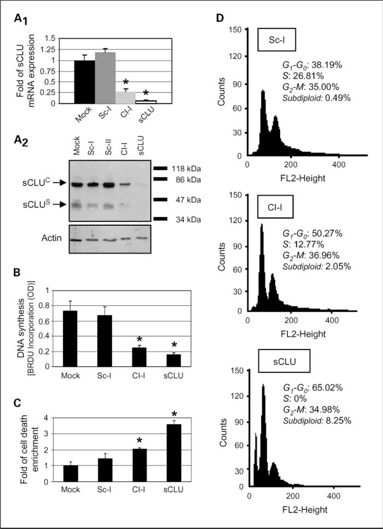
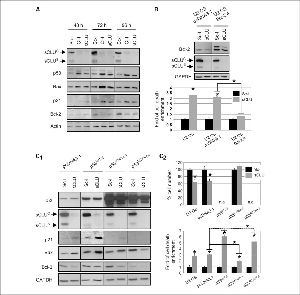
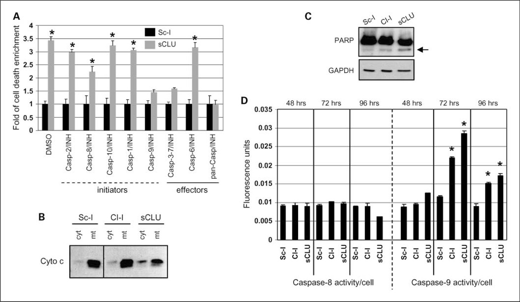
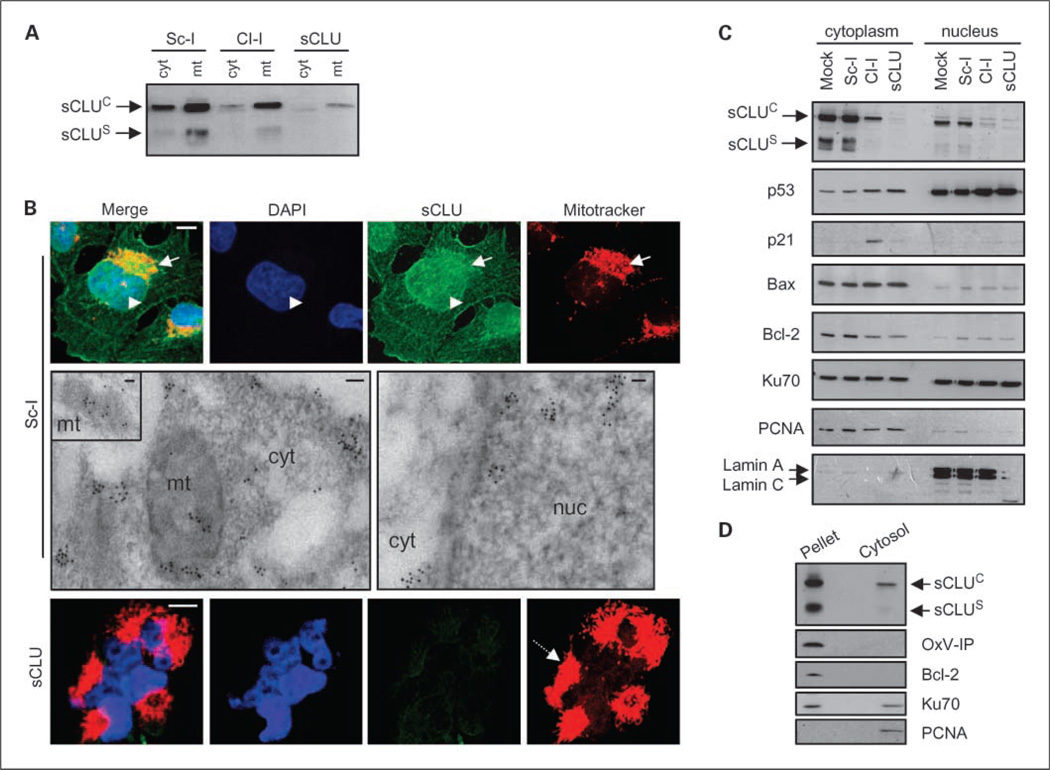
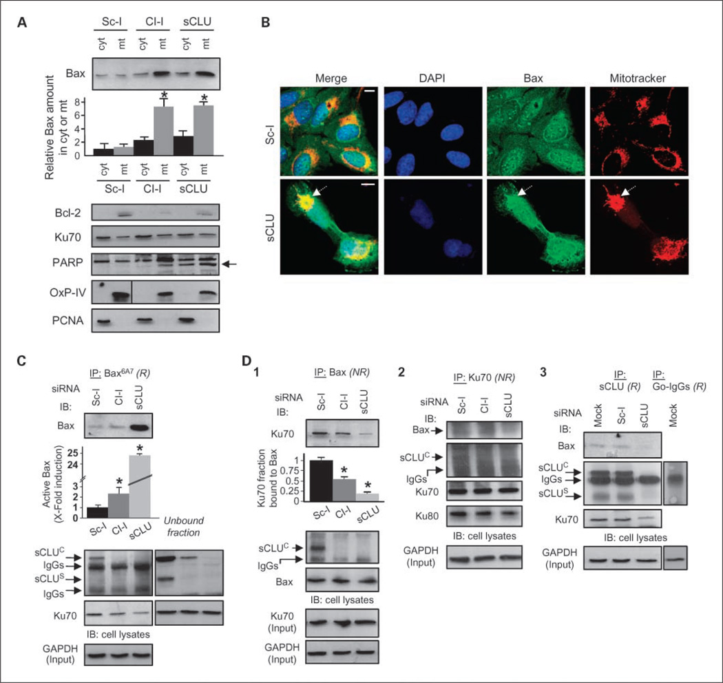
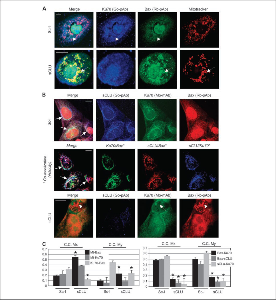
Results: We report here that sCLU depletion in nonstressed human cancer cells signals stress that induces p53-dependent growth retardation and high rates of endogenous apoptosis. We discovered that increased apoptosis in sCLU-depleted cells correlates to altered ratios of proapoptotic to antiapoptotic Bcl-2 protein family members, is amplified by p53, and is executed by mitochondrial dysfunction. sCLU depletion-related stress signals originate from several sites, because sCLU is an integral component of not only the secretory pathway but also the nucleocytosolic continuum and mitochondria. In the cytoplasm, sCLU depletion disrupts the Ku70-Bax complex and triggers Bax activation and relocation to mitochondria. We show that sCLU binds and thereby stabilizes the Ku70-Bax protein complex serving as a cytosol retention factor for Bax.

Conclusions: We suggest that elevated sCLU levels may enhance tumorigenesis by interfering with Bax proapoptotic activities and contribute to one of the major characteristics of cancer cells, that is, resistance to apoptosis.


Conflict of interest statement

Disclosure of Potential Conflicts of Interest

No potential conflicts of interest were disclosed.

Figures

Fig. 1
Fig. 1
sCLU depletion in U-2 OS cells induces growth arrest and increased cell death responses. A1 quantitative PCR analyses (n = 3) of sCLU mRNA levels after cell transfection with indicated siRNA oligonucleotides. A2, immunoblot analyses of sCLU protein levels in whole-cell lysates following transfection with shown siRNAs. Actin probing was used as reference for equal protein loading; molecular weight markers are shown to the right of the blot. B, DNA synthesis measurement (n = 2) after bromodeoxyuridine incorporation. C, cumulative cell death quantitation (n = 3) of DNA fragments following cell transfection with the indicated siRNAs. D, comparative fluorescence-activated cell sorting analysis (representative of two independent experiments) of control (Sc-I), Cl-I, or sCLU siRNA-treated cells. Assays were done 72 h after siRNA cell transfection. Bars, SD. *, P < 0.05.
Fig. 2
Fig. 2
Down-regulation of Bcl-2 family prosurvival proteins and p53 activation are essential for cell death and growth retardation, respectively, in sCLU-depleted U-2 OS cells. A, immunoblot analyses of whole-cell lysates 48 to 96 h post-transfection with the shown siRNAs. B, whole-cell lysate immunoblot analyses of empty vector (pcDNA3.1) or Bcl-2 stably transfected cells (top) and quantitation of cumulative cell death (bottom; n = 3) in transgenic cells after treatment with either Sc-I or sCLU siRNAs. C, immunoblot analyses of cells stably transfected with vector (pcDNA3.1), WT-p53 (p53WT.5), V143A-p53 (p53v143A.1), and R273H-p53 (p53R273H.5) plasmids (C1) as well as measurement (n = 3) of cellular growth (C2, top) and cumulative cell death (C2, bottom) in the corresponding cell lines after transfection with the indicated siRNAs. All p53WT stable clones showed moderate transgene expression (p53WT.5; C1, top) due to prodeath effect of high p53WT levels. Growth was not assayed (n.a.) in the sCLU-depleted U-2 OS-p53WT.5 and U-2 OS-p53R273H.5 cells due to high death rates. Assays were done 72 h after siRNA transfection unless otherwise indicated. Actin or GAPDH probing was used as protein loading controls. Bars, SD. *, P < 0.05, compared with respective controls.
Fig. 3
Fig. 3
Induction of the mitochondrial pathway of apoptosis in sCLU-depleted U-2 OS cells. A, cumulative cell death measurement (n = 3) after cell transfection with the Sc-I or sCLU siRNAs in the presence of the indicated caspase inhibitors. B, immunoblotting of cytochrome c in cytosolic and mitochondrial cellular fractions following cell transfection with shown siRNAs (for fraction purity, see Fig. 5A). C, immunoblot analysis of control or sCLU-depleted whole-cell lysates probed with antibodies against PARP. Arrow, cleaved 85-kDa apoptotic fragment of PARP. GAPDH probing was used as a protein loading control. D, caspase-8 and caspase-9 activity measurement (n = 2) after incubating whole-cell lysates of sCLU-depleted cells with the caspase-8, IETD-AFC, or the caspase-9, LEHD-AMC, substrates, respectively. Assays were done 72 h after siRNA transfection unless otherwise indicated. Bars, SD. *, P < 0.05.
Fig. 4
Fig. 4
Endogenous sCLU localizes in the mitochondria and the nucleocytosolic continuum of U-2 OS cells. Immunoblot analyses of isolated cytosolic (cyt; A), mitochondrial (mt; A), and cytoplasmic and nuclear fractions (C) probed with an anti-sCLU antibody. Blots in C were also probed with antibodies against p53, p21, Bax, Bcl-2, and Ku70. Fraction purity in A was verified by anti-OxPhos complex IV (OxP-IV) probing (mitochondrial marker; shown in Fig. 5A), and in C by blot probing with anti-proliferating cell nuclear antigen (exclusively cytosolic in osteosarcoma cells) and anti-lamin A/C (nuclear marker). B, CLSM immunofluorescence (top and bottom) or TEM immunogold (middle) localization of sCLU protein in control and sCLU knocked-down cells. For CLSM immunolocalization, cells were costained with an anti-sCLU (green), MitoTracker (mitochondria specific stain; red), and 4’,6-diamidino-2-phenylindole dihydrochloride (blue). Captured images were merged to reveal codistribution sites (yellow for green-red and cyan for green-blue). Top, arrows, mitochondria; arrowheads, nuclei. Bottom, dashed arrow, aggregated mitochondria in sCLU-depleted cells. Middle, sCLU-related gold particles after immunogold TEM localization were found in mitochondria (mt; see also inset), cytosol (cyt), and nucleus (nuc). Bar, 10 µm (CLSM) and 100 nm (TEM). D, immunoblotting of nonextractable pelleted material and the soluble cytosol after hypotonic lysis of control cells. Blots were probed with antibodies against sCLU, OxPhos complex IV, Bcl-2 (membrane bound), Ku70 (nuclear and cytosolic distribution), and proliferating cell nuclear antigen. All assays were done 72 h post-siRNA transfection.
Fig. 5
Fig. 5
sCLU knockdown in U-2 OS cells promotes Bax activation and translocation to the mitochondria due to destabilization of the cytoplasmic Ku70-Bax complex. A, immunoblot analyses of Bax, Bcl-2, Ku70, and PARP in isolated cytosolic and mitochondrial fractions of control or sCLU knocked-down cells; arrow in PARP immunoblot denotes the apoptotic 85-kDa PARP fragment. Second from top, quantitation (n = 2) of Bax copurification with cytosol or mitochondria. Fraction purity was verified by probing with anti-OxPhos Complex IV and anti-proliferating cell nuclear antigen. B, CLSM immunolocalization of Bax after staining control or sCLU-depleted cells with anti-Bax, MitoTracker, and 4’,6-diamidino-2-phenylindole dihydrochloride. The captured images were merged to reveal antigen colocalization with mitochondria (dashed arrows, bottom). Bar, 10 µm. C and D, quantitative immunoprecipitation analyses of the sCLU, Ku70, and Bax proteins interaction in control and sCLU knocked-down cells. Cells were lysed in CHAPS and lysates were immunoprecipitated (IP; R, reducing conditions; NR, nonreducing conditions) with antibodies against activated Bax (antibody Bax6A7; C), total Bax (D1), Ku70 (D2), and sCLU (D3); the immunoprecipitation with goat IgG (D3) was used to exclude sCLU binding to IgG. Immunoprecipitates were probed (IB) with anti-Bax, anti-sCLU, anti-Ku70, and anti-Ku80. The unbound fraction shown in C depicts the sCLU protein molecules that were not coimmunoprecipitated with the anti-Bax6A7-bead complexes. Quantitative analysis of either Bax activation or Ku70-Bax dissociation (n = 2) after sCLU depletion is shown in C and D1, respectively (second from top). GAPDH or Ku70 probing was used to show equal protein input in the immunoprecipitations. Assays were done 72 h post-transfection with the indicated siRNAs. Bars, SD. *,P <0.05.
Fig. 6
Fig. 6
sCLU depletion-mediated mitochondrial relocation of Bax in U-2 OS cells relates to the disruption of a cytoplasmic sCLU-Ku70-Bax nexus. A, CLSM staining of control and sCLU-depleted cells with anti-Ku70 (goat polyclonal antibody; blue), anti-Bax (rabbit polyclonal antibody; green), and MitoTracker. Captured images were merged to reveal codistribution sites (yellow for green-red, cyan for green-blue, magenta for red-blue, and white for blue-green-red). Top, arrowheads, cytoplasmic colocalization of Ku70 and Bax in Sc-l-treated cells; bottom, dashed arrows, Bax colocalization with mitochondria after sCLU depletion. B, triple staining of Sc-I or sCLU siRNA-transfected cells with anti-sCLU (goat polyclonal antibody; blue), anti-Ku70 (mouse monoclonal antibody; green), and anti-Bax (rabbit polyclonal antibody; red) antibodies. Middle, digitations of each pair of antigen colocalization in control cells and subsequent superimposition of the three images; the analysis was done by using the Volocity software. Top and middle, arrows, antigen colocalization in the perinuclear cytoplasmic region of control cells; bottom, dashed arrows, elimination of Ku70 and Bax colocalization in sCLU-depleted cells. Assays were done 72 h post-siRNA transfection. Bars, 10 µm (A and B). C, CLSM-derived quantitative analyses of fluorochromes colocalization in preparations of Ku70-Bax-mitochondria (left) and sCLU-Ku70-Bax (right) triple staining. Data were deduced by the Volocity software and values in the graphs indicate the overlapping degree of the two fluorochomes; maximum overlap was set to 1. C.C. Mx and C.C. My are the colocalization coefficients Mx and My (23) and refer to the first and second fluorochromes analyzed, respectively. Thus, for example, the C.C. Mx value for the Ku70-Bax pair in the left denotes the percentage of stained Ku70 that colocalizes with Bax, whereas C.C. My value indicates the percentage of stained Bax that colocalizes with Ku70. Average values of 10 micrographs containing >50 cells are presented in the graphs. Bars, SD. *, P < 0.05.

References

    1. de Silva HV, Harmony JA, Stuart WD, Gil CM, Robbins J. Apolipoprotein J: structure and tissue distribution. Biochemistry. 1990;29:5380–5389. - PubMed
    1. Wilson MR, Easterbrook-Smith SB. Clusterin is a secreted mammalian chaperone. Trends Biochem Sci. 2000;25:95–98. - PubMed
    1. Trougakos IP, Gonos ES. Clusterin/apolipoprotein J in human aging and cancer. Int J Biochem Cell Biol. 2002;34:1430–1448. - PubMed
    1. Leskov KS, Klokov DY, Li J, Kinsella TJ, Boothman DA. Synthesis and functional analysis of nuclear clusterin: a cell death protein. J Biol Chem. 2003;278:11590–11600. - PubMed
    1. Caccamo AE, Scaltriti M, Caporali A, et al. Ca2+ depletion induces nuclear clusterin, a novel effector of apoptosis in immortalized human prostate cells. Cell Death Differ. 2005;12:101–104. - PubMed

Publication types