Review
. 2009 Jan;20(1):23-35.
doi: 10.1681/ASN.2008050456. Epub 2008 Dec 31.

Nephronophthisis: disease mechanisms of a ciliopathy

Affiliations
Review

Nephronophthisis: disease mechanisms of a ciliopathy

Friedhelm Hildebrandt et al. J Am Soc Nephrol. 2009 Jan.

Abstract

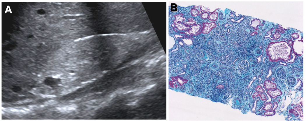
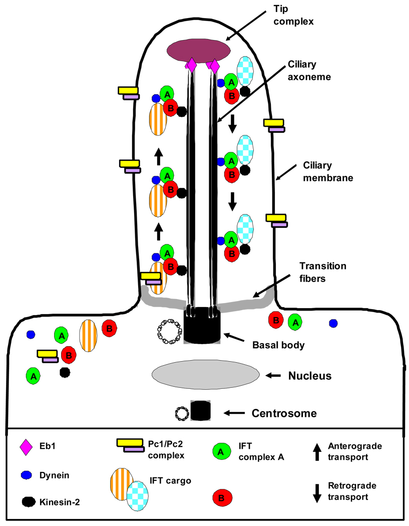
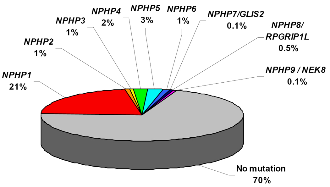
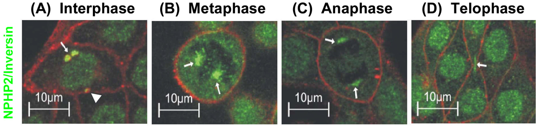
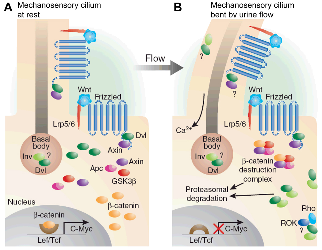
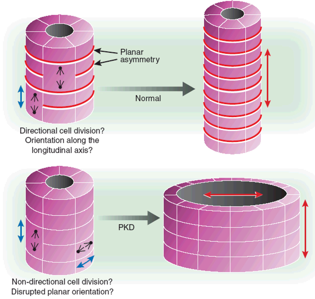
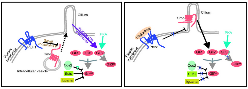
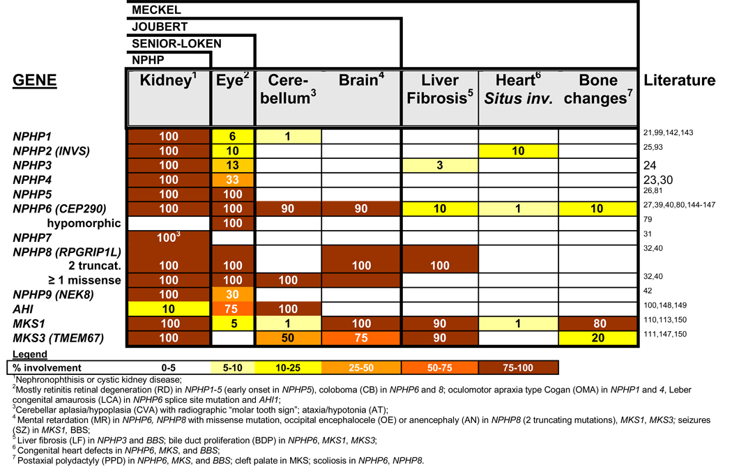
Nephronophthisis (NPHP), a recessive cystic kidney disease, is the most frequent genetic cause of end-stage kidney disease in children and young adults. Positional cloning of nine genes (NPHP1 through 9) and functional characterization of their encoded proteins (nephrocystins) have contributed to a unifying theory that defines cystic kidney diseases as "ciliopathies." The theory is based on the finding that all proteins mutated in cystic kidney diseases of humans or animal models are expressed in primary cilia or centrosomes of renal epithelial cells. Primary cilia are sensory organelles that connect mechanosensory, visual, and other stimuli to mechanisms of epithelial cell polarity and cell-cycle control. Mutations in NPHP genes cause defects in signaling mechanisms that involve the noncanonical Wnt signaling pathway and the sonic hedgehog signaling pathway, resulting in defects of planar cell polarity and tissue maintenance. The ciliary theory explains the multiple organ involvement in NPHP, which includes retinal degeneration, cerebellar hypoplasia, liver fibrosis, situs inversus, and mental retardation. Positional cloning of dozens of unknown genes that cause NPHP will elucidate further signaling mechanisms involved. Nephrocystins are highly conserved in evolution, thereby allowing the use of animal models to develop future therapeutic approaches.


Figures

Figure 1
Figure 1. Morphology of nephronophthisis
(A) Renal ultrasound demonstrates increased echogenicity, loss of corticomedullary differentiation, and the presence of corticomedullary cysts. In contrast to polycystic kidney disease kidneys are not enlarged. (B) Renal histology in NPHP shows the characteristic triad of renal tubular cysts, tubular membrane disruption, and tubulointerstitial cell infiltrates with interstitial fibrosis and periglomerular fibrosis (B is courtesy of D. Bockenhauer, London).
Figure 2
Figure 2. Cilia structure and intraflagellar transport
The cilium is a hair-like structure that extends from the cell surface into the extracellular space. Virtually all vertebrate cell types can produce cilia. Cilia consist of a microtubule-based axoneme covered by a specialized plasma membrane. The axoneme has nine peripheral microtubule doublets. There may be two central microtubules (9+2 versus 9+0 axoneme). 9+2 cilia usually have dynein arms that link the microtubule doublets and are motile, while most 9+0 cilia lack dynein arms and are non-motile (“primary cilia”) with a few exceptions. The ciliary axoneme is anchored in the basal body, a microtubule-organizing center derived from the mother centriole. The transition zone at the junction of the basal body acts as a filter for the molecules that can pass into or out of the cilium. Nephrocystin-1 is localized at the transition zone of epithelial cells. During ciliogenesis, cilia elongate from the basal body by the addition of new axonemal subunits to the distal tip, the plus end of the microtubules. Axonemal and membrane components are transported in raft macromolecular particles (complex A and B) by so-called intraflagellar transport (IFT) along the axonemal doublet microtubules. Anterograde transport towards the tip is driven by heterotrimeric kinesin 2, which contains motor subunits Kif3a and Kif3b and a non-motor subunit. Mutations of Kif3a cause renal cysts and cerebellar vermis aplasia in mice. Retrograde transport back to the cell body occurs via the motor protein cytoplasmic dynein 1B (modified from Bisgrove and Yost, 2006).
Figure 3
Figure 3. Distribution of causative mutations in the NPHP1NPHP9 genes in a worldwide cohort of 1,079 families with NPHP
Note that, whereas mutations in NPHP1 account for >21% of NPHP, all other genes are in the range of 3%.
Figure 4
Figure 4. Subcellular localization of nephrocystins to primary cilia, basal bodies, the mitotic spindle, focal adhesions and adherens junctions, and functional interaction with other proteins mutated in renal “ciliopathies”
"Cystoproteins” are proteins of genes mutated in cystic kidney diseases of humans, mice, or zebrafish. Depending on cell cycle stage, cystoproteins are localized at different subcellular organelles (shown in grey), including primary cilia, basal bodies, endoplasmic reticulum, the mitotic spindle, centrosomes, adherens junctions or focal adhesions. Arrows in the primary cilium indicate the direction of anterograde transport along the microtubule system mediated by kinesin-2 and retrograde transport by cytoplasmic dynein 1b. A) Most nephrocystins (blue) are located at cilia, the basal body, and centrosome in a cell cycle dependent manner. NPHP1 is also at the transition zone, focal adhesions and adherens junctions. B) Sensory cilia perceive and process cell external signals, and “cystoproteins” are involved in signaling mechanisms downstream of cilial signal recognition. Downstream of cilia (pink), Wnt signaling (Figure 6) and hedgehog signaling (Figure 8) play a role in planar cell polarity, which is mediated (C) partially through orientation of centrosomes and the mitotic spindle poles (Figure 7). D) Cilia-dependent mechanisms of planar cell polarity seem to be the central to the pathogenesis of the ciliopathies, the most prominent of which are listed on the right. Wht, the Wnti signaling pathway; Shh, the sonic hedgehog signaling pathway.
Figure 5
Figure 5. Inversin/NPHP2 localizes to cilia, centrosomes, and the mitotic spindle in a cell cycle dependent manner
Inversin/NPHP2 is found in interphase (A) in the cilial axoneme (not shown), close to the centrioles of the basal body complex (arrow) and at the centrosome (arrow head). In metaphase (B) and anaphase (C) it is at the mitotic spindle (arrows), and in telophase (D) at the midbody (arrow) of the separating cells and in the nucleus. This reflects a centriole-associated function of inversin/NPHP2 throughout the cell cycle (from Morgan et al. Hum Mol Genet 11:3345, 2002).
Figure 6
Figure 6. Inversin/NPHP2 mediates a switch from the canonical the non-canonical Wnt signaling pathway, which plays a role in planar cell polarity maintenance
(A) This cartoon of arenal tubular epithelial cell shows how Wnt signaling occurs primarily through β-catenin–dependent pathways in the absence of urine flow. Ligand binding by the frizzled receptor results in inactivation of the β-catenin destruction complex through the presence of disheveled (Dvl), increased β-catenin levels, and upregulation of effector gene expression of the canonical Wnt signaling pathway. (B) Stimulation of the primary cilium, e.g. by urine flow, results in increased expression of inversin (Inv), which then reduces levels of cytoplasmic Dvl by increasing its proteasomal degradation. This allows reassembly and activation of the β-catenin destruction complex, thereby switching from the canonical to the non-canonical Wnt signaling pathway. The model is consistent with the finding that overexpression of β-catenin (equivalent to canonical Wnt signaling) leads to renal cysts in a mouse model (from: Germino Nature Genet 37:455, 2005).
Figure 7
Figure 7. Defects of cystoproteins lead to disruption of planar cell polarity, and thereby to renal cysts through to malorientation of the centrosome or mitotic spindle complex
Correct orientation of the mitotic spindle and centrosomes with respect to the longitudinal axis of the tubule is critical for proper planar cell polarity (i.e., the orientation of an epithelial cell layer in 3-dimensional space). Non-canonical Wnt signaling (see Figure 6) is involved in regulation of planar cell polarity during renal tubular morphogenesis, when in rodents 2 weeks post partum the tubules still elongate. The structure that would result from disruption of this longitudinal orientation is a dilated tubule or cyst (from Germino Nature Genet 37:455, 2005).
Figure 8
Figure 8. The hedgehog signaling pathway may be involved in renal cystogenesis
In the hedgehog pathway of vertebrates, upon binding of the Shh ligand to the patched (Ptch1) receptor, repression of the smoothened (Smo) receptor to be inserted into the cilium membrane is relieved, and posttranslational modification of the Gli transcription factors within the cilium induces both their activator (GliAct) and repressor (GliR) functions. Several studies in mice have demonstrated that ciliary proteins are needed for hedgehog signaling. Recently, the related transcription factor Gli-similar 2 (GLIS2) was found to be mutated in NPHP type 7 (from: Huangfu & Anderson Development, 2005).
Figure 9
Figure 9. Ciliopathies feature a broad spectrum of organ involvement, shown here for the nephronophthisis-related ciliopathies
There is overlap between different syndromes: Exlusive kidney involvement is called nephronophthisis. Associated retinal degeneration in known as Senior-Loken syndrome. Involvement of the cerebellum represents Joubert syndrome. In the most severe form, Meckel syndrome, there are brain malformations, liver fibrosis, heart defects, polydactyly and perinatal mortality associated. It has recently become evident that the spectrum can vary by at least 2 mechanisms: First, multiple allelism, in which a hypomorphic mutation may cause a milder phenotype. For example, a splice site mutation of NPHP6 may cause Leber congenital optical atrophy (LCA) only. In another example, the presence of one non-truncating mutation in NPHP8 can rescue the phenotype from Meckel syndrome to Joubert syndrome. Secondly, NPHP genes can modify each other. For instance, NPHP6 and AHI1 modify recessive NPHP1 mutations to express a more severe phenotype.

References

    1. Hildebrandt F, Otto EA. Primary cilia: a unifying pathogenic concept for cystic kidney disease? Nature Reviews Genetics. 2005 - PubMed
    1. Smith C, Graham J. Congenital medullary cysts of the kidneys with severe refractory anemia. Am J Dis Child. 1945;69:369–377.
    1. Fanconi G, Hanhart E, Albertini A. Die familiäre juvenile Nephronophthise. Hel Pediatr Acta. 1951;6:1–49. - PubMed
    1. Hildebrandt F. Juvenile nephronophthisis. In: Barratt TM, Avner ED, Harmon WE, editors. Pediatric nephrology. Baltimore: Williams & Wilkins; 1999.
    1. Ala-Mello S, Kivivuori SM, Ronnholm KA, Koskimies O, Siimes MA. Mechanism underlying early anaemia in children with familial juvenile nephronophthisis. Pediatr Nephrol. 1996;10:578–581. - PubMed

Publication types

MeSH terms