. 2009 Mar;296(3):C422-32.
doi: 10.1152/ajpcell.00381.2008. Epub 2008 Dec 31.

Nox4 NADPH oxidase mediates oxidative stress and apoptosis caused by TNF-alpha in cerebral vascular endothelial cells

Affiliations

Nox4 NADPH oxidase mediates oxidative stress and apoptosis caused by TNF-alpha in cerebral vascular endothelial cells

Shyamali Basuroy et al. Am J Physiol Cell Physiol. 2009 Mar.

Abstract

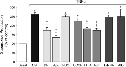
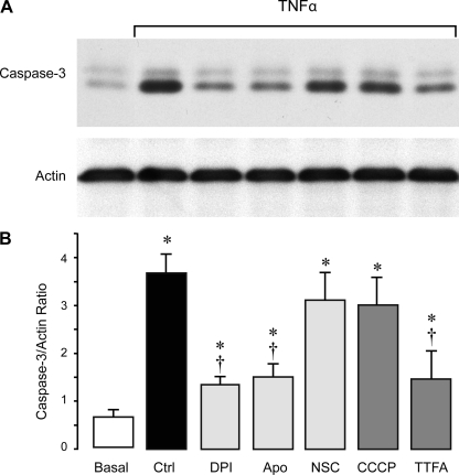
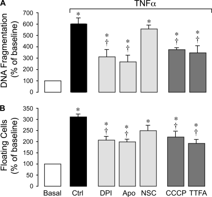
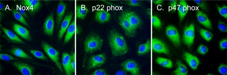
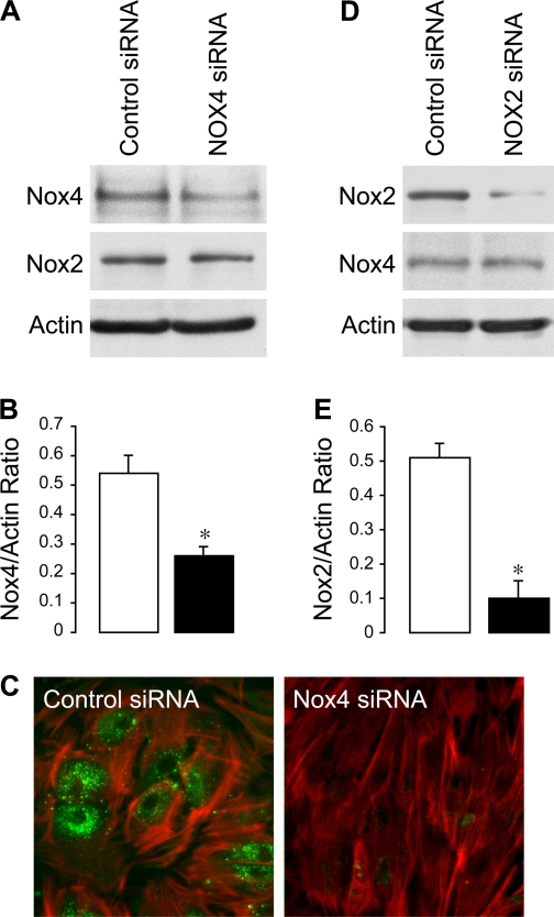
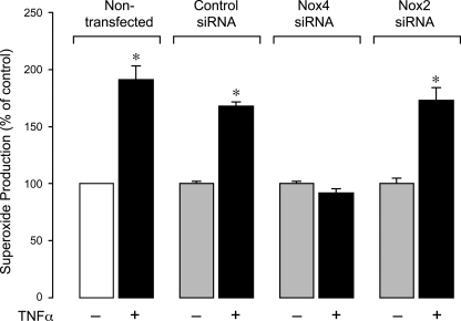
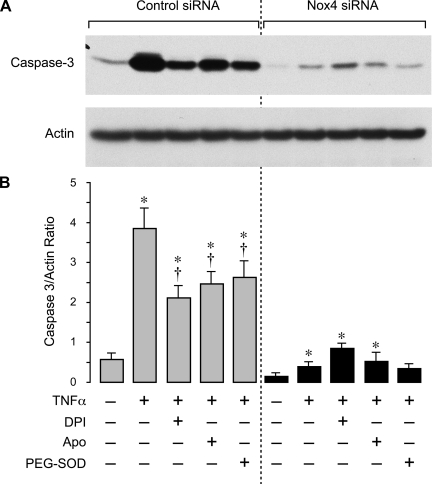
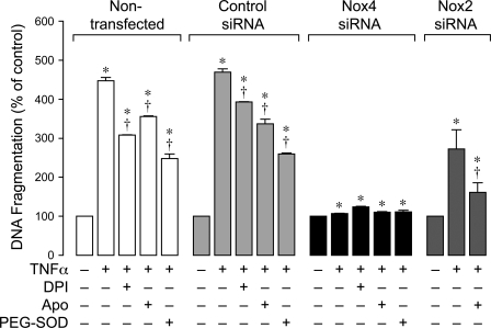
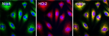
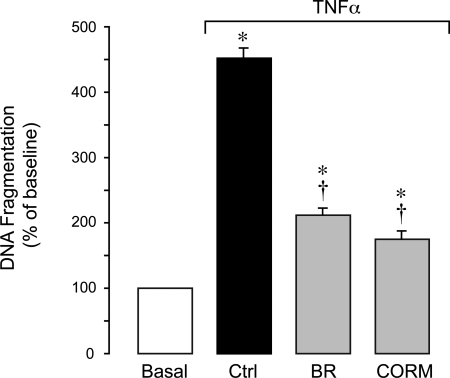
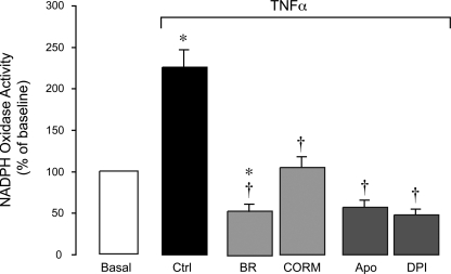
Inflammatory brain disease may damage cerebral vascular endothelium leading to cerebral blood flow dysregulation. The proinflammatory cytokine TNF-alpha causes oxidative stress and apoptosis in cerebral microvascular endothelial cells (CMVEC) from newborn pigs. We investigated contribution of major cellular sources of reactive oxygen species to endothelial inflammatory response. Nitric oxide synthase and xanthine oxidase inhibitors (N(omega)-nitro-l-arginine and allopurinol) had no effect, while mitochondrial electron transport inhibitors (CCCP, 2-thenoyltrifluoroacetone, and rotenone) attenuated TNF-alpha-induced superoxide (O(2)(*-)) and apoptosis. NADPH oxidase inhibitors (diphenylene iodonium and apocynin) greatly reduced TNF-alpha-evoked O(2)(*-) generation and apoptosis. TNF-alpha rapidly increased NADPH oxidase activity in CMVEC. Nox4, the cell-specific catalytic subunit of NADPH oxidase, is highly expressed in CMVEC, contributes to basal O(2)(*-) production, and accounts for a burst of oxidative stress in response to TNF-alpha. Nox4 small interfering RNA, but not Nox2, knockdown prevented oxidative stress and apoptosis caused by TNF-alpha in CMVEC. Nox4 is colocalized with HO-2, the constitutive isoform of heme oxygenase (HO), which is critical for endothelial protection against TNF-alpha toxicity. The products of HO activity, bilirubin and carbon monoxide (CO, as a CO-releasing molecule, CORM-A1), inhibited Nox4-generated O(2)(*-) and apoptosis caused by TNF-alpha stimulation. We conclude that Nox4 is the primary source of inflammation- and TNF-alpha-induced oxidative stress leading to apoptosis in brain endothelial cells. The ability of CO and bilirubin to combat TNF-alpha-induced oxidative stress by inhibiting Nox4 activity and/or by O(2)(*-) scavenging, taken together with close intracellular compartmentalization of HO-2 and Nox4 in cerebral vascular endothelium, may contribute to HO-2 cytoprotection against inflammatory cerebrovascular disease.


Figures

Fig. 1.
Fig. 1.
Effects of reactive oxygen species (ROS) inhibitors on TNF-α-induced O2•− production in cerebral microvascular endothelial cells (CMVEC). CMVEC from newborn piglets were treated with TNF-α (15 ng/ml, 1 h) in the absence or presence of diphenyliodonium (DPI; 5 μM), apocynin (Apo, 500 μM), NSC-23766 (NSC, 50 μM), CCCP (5 μM), 2-thenoyltrifluoroacetone (TTFA; 5 μM), rotenone (Rot, 5 μM), Nω-nitro-l-arginine (l-NNA; 0.5 mM), or allopurinol (Allo, 50 μM). O2•− production was measured by ethidium fluorescence generated from dihydroethidium (DHE) and is expressed as percentage of the baseline control. Data represent average of 11 independent experiments. Values are means ± SE. *P < 0.05 compared with baseline control values. †P < 0.05 compared with TNF-α alone.
Fig. 2.
Fig. 2.
Effects of ROS inhibitors on TNF-α-induced caspase-3 activation in CMVEC. CMVEC from newborn piglets were treated with TNF-α (15 ng/ml, 3 h) in the absence or presence of NADPH oxidase inhibitors DPI (5 μM), Apo (500 μM), and NSC (50 μM) and mitochondrial inhibitors CCCP (5 μM) and TTFA (5 μM). Caspase-3 activity was immunodetected in cell lysates by formation of active proteolitic fragment of caspase-3 (17 kDa). Blots were reprobed for actin as the loading control. A: representative blot. B: densitometry (n = 3 independent experiments). Values are means ± SE. *P < 0.05 compared with control values. †P < 0.05 compared with TNF-α alone.
Fig. 3.
Fig. 3.
Effects of ROS inhibitors on TNF-α-induced apoptosis in CMVEC. CMVEC from newborn piglets were treated with TNF-α (15 ng/ml, 3 h) in the absence or presence of DPI (5 μM), Apo (500 μM), Rac1 inhibitor NSC (50 μM), CCCP (5 μM), and TTFA (5 μM). A: DNA fragmentation. B: cell detachment. Data represent average of 4 independent experiments. Values are means ± SE. *P < 0.05 compared with baseline control values. †P < 0.05 compared with TNF-α alone.
Fig. 4.
Fig. 4.
Localization of NADPH oxidase subunits in CMVEC. Nox4 (A), p22phox (B), and p47phox (C) were visualized by immunofluorescence in confluent quiescent CMVEC.
Fig. 5.
Fig. 5.
Small interfering RNA (siRNA) knockdown of Nox4 and Nox2 in CMVEC. CMVEC were transfected with control siRNA, Nox4 siRNA (AC), or Nox2 siRNA (D and E). A, B, D, and E: Nox4 and Nox2 detection by immunoblotting. A and D: representative blots. B and E: densitometry (n = 3 independent transfections; *P < 0.05 compared with control). C: Nox4 immunofluorescence in CMVEC transfected with control siRNA or Nox4 siRNA: Nox4 was visualized with FITC-conjugated secondary antibodies (green); F-actin was visualized by rhodamine-phalloidin (red).
Fig. 6.
Fig. 6.
Effects of TNF-α on O2•− production in Nox4 siRNA- and Nox2 siRNA-knockdown CMVEC. CMVEC (nontransfected and transfected with control siRNA, Nox4 siRNA, or Nox2 siRNA) were treated with TNF-α (15 ng/ml, 1 h). O2•− production was measured by ethidium fluorescence generated from DHE and is expressed as a percentage of the baseline control. Values are means ± SE. *P < 0.05 compared with baseline control values.
Fig. 7.
Fig. 7.
Effects of TNF-α on caspase-3 activity in Nox4 knockdown CMVEC. CMVEC from newborn piglets transfected with control siRNA or Nox4 siRNA were treated with TNF-α (15 ng/ml, 3 h) in the absence or presence of DPI (5 μM), Apo (500 μM), or polyethylene glycol (PEG)-SOD (1,000 units). Active fragment of caspase-3 (17 kDa) was detected by immunoblotting and normalized to actin. A: representative blot. B: densitometry (n = 2 independent experiments). Values are means ± SE. *P < 0.05 compared with control values. †P < 0.05 compared with TNF-α alone.
Fig. 8.
Fig. 8.
Effects of TNF-α on DNA fragmentation in Nox4 siRNA- and Nox2 siRNA-knockdown CMVEC. CMVEC (nontransfected or transfected with control siRNA, Nox4 siRNA, or Nox2 siRNA) were treated with TNF-α (15 ng/ml, 3 h) in the absence or presence of DPI (5 μM), Apo (500 μM), or PEG-SOD (1,000 units). DNA fragmentation was detected by ELISA. Values are means ± SE (n = 2 independent experiments in triplicates). *P < 0.05 compared with baseline control values. †P < 0.05 compared with TNF-α alone.
Fig. 9.
Fig. 9.
Colocalization of Nox4 and heme oxygenase isoform 2 (HO-2) in CMVEC. Quiescent CMVEC were immunostained for Nox4 (green fluorescence) and HO-2 (red fluorescence); nuclei were stained with 4′,6-diamidino-2-phenylindole (DAPI; blue).
Fig. 10.
Fig. 10.
Protective effects of bilirubin (BR) and a CO-releasing molecule, CORM-A1 (CORM), on TNF-α-induced apoptosis in CMVEC. Confluent quiescent CMVEC from newborn piglets were treated with TNF-α (15 ng/ml, 3 h) in the absence or presence of BR (1 μM) or CORM (50 μM). Apoptosis was determined by DNA fragmentation. Values are means ± SE. *P < 0.05 compared with baseline control values. †P < 0.05 compared with TNF-α alone.
Fig. 11.
Fig. 11.
Effects of BR and a CO-releasing molecule (CORM-A1) on TNF-α-induced NADPH oxidase-derived O2•− production in CMVEC. Fractioned confluent quiescent CMVEC were treated with TNF-α (15 ng/ml, 1 h) in the absence or presence of BR (1 μM), CORM (50 μM), Apo (500 μM), and DPI (5 μM). NADPH oxidase activity was determined as O2•− production from NADPH (100 μM) measured by enhanced lucigenin luminescence, was normalized to the protein amount, and is expressed as percentage of the baseline control. Values are means ± SE. *P < 0.05 compared with control values. †P < 0.05 compared with TNF-α alone.

Similar articles

Cited by

References

    1. Abraham NG, Kappas A. Pharmacological and clinical aspects of heme oxygenase. Pharmacol Rev 60: 79–127, 2008. - PubMed
    1. Abramov AY, Jacobson J, Wientjes F, Hothersall J, Canevari L, Duchen MR. Expression and modulation of an NADPH oxidase in mammalian astrocytes. J Neurosci 40: 9176–9184, 2005. - PMC - PubMed
    1. Ago T, Kitazono T, Ooboshi H, Iyama T, Han YH, Takada J, Wakisaka M, Ibayashi S, Utsumi H, Iida M. Nox4 as the major catalytic component of an endothelial NAD(P)H oxidase. Circulation 109: 227–233, 2004. - PubMed
    1. Barone FC, Arvin B, White RF, Miller A, Webb CL, Willette RN, Lysko PG, Feuerstein GZ. Tumor necrosis factor-alpha. A mediator of focal ischemic brain injury. Stroke 28: 1233–1244, 1997. - PubMed
    1. Basuroy S, Bhattacharya S, Tcheranova D, Qu Y, Regan RF, Leffler CW, Parfenova H. HO-2 provides endogenous protection against oxidative stress and apoptosis caused by TNF-α in cerebral vascular endothelial cells. Am J Physiol Cell Physiol 291: C897–C908, 2006. - PubMed

Publication types

MeSH terms

LinkOut - more resources