. 2008 Dec 31;28(53):14537-45.
doi: 10.1523/JNEUROSCI.2692-08.2008.

Picomolar amyloid-beta positively modulates synaptic plasticity and memory in hippocampus

Affiliations

Picomolar amyloid-beta positively modulates synaptic plasticity and memory in hippocampus

Daniela Puzzo et al. J Neurosci. .

Abstract

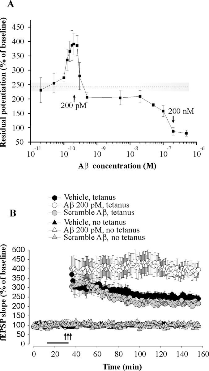
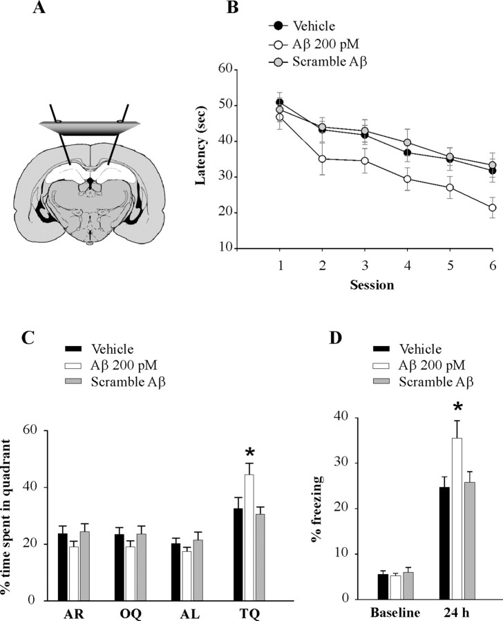
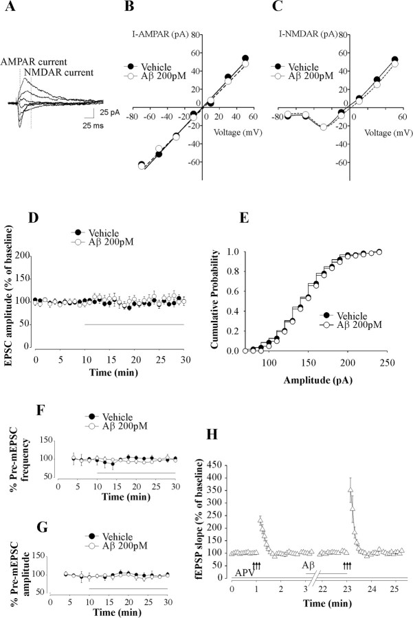
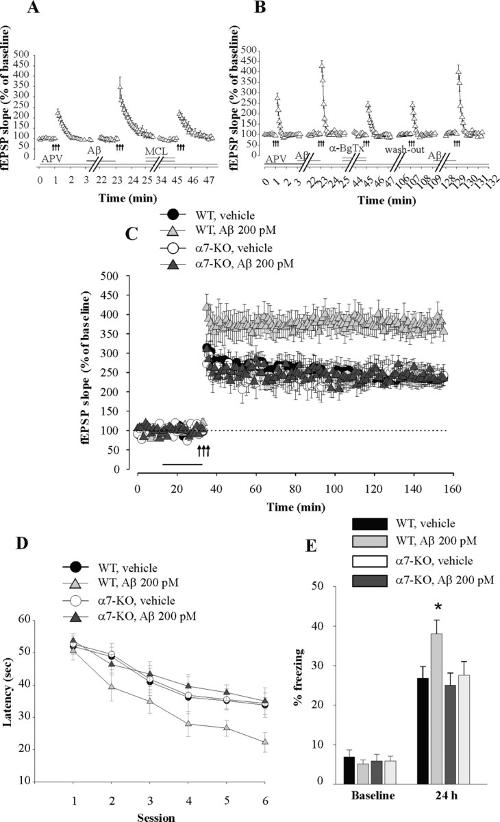
Amyloid-beta (Abeta) peptides are produced in high amounts during Alzheimer's disease, causing synaptic and memory dysfunction. However, they are also released in lower amounts in normal brains throughout life during synaptic activity. Here we show that low picomolar concentrations of a preparation containing both Abeta(42) monomers and oligomers cause a marked increase of hippocampal long-term potentiation, whereas high nanomolar concentrations lead to the well established reduction of potentiation. Picomolar levels of Abeta(42) also produce a pronounced enhancement of both reference and contextual fear memory. The mechanism of action of picomolar Abeta(42) on both synaptic plasticity and memory involves alpha7-containing nicotinic acetylcholine receptors. These findings strongly support a model for Abeta effects in which low concentrations play a novel positive, modulatory role on neurotransmission and memory, whereas high concentrations play the well known detrimental effect culminating in dementia.


Figures

Figure 1.
Figure 1.
42 has two opposite effects on LTP in hippocampus. A, Concentration–response curve for the effect of Aβ42 on CA1-LTP indicating that the peptide has an enhancing effect with a peak around 200 pm, whereas it has an opposite detrimental effect above 20 nm. The dotted line and the shaded area around it correspond to the amount of potentiation and the SE range in vehicle-treated slices. The residual potentiation was calculated by averaging the last 5 min of LTP. B, Perfusion of hippocampal slices with a preparation containing human Aβ42 (200 pm), but not scramble Aβ42 (200 pm), for 20 min before a theta-burst stimulation increases LTP without affecting baseline transmission. The horizontal bar indicates the period during which Aβ was added to the bath solution. Each bar denotes the mean ± SEM in this and the following figures.
Figure 2.
Figure 2.
Picomolar concentrations of Aβ42 enhance hippocampal-dependent memory. A, Schematic representation showing cannulas implanted bilaterally into the dorsal hippocampi. B, Bilateral injections of human Aβ42 (200 pm), but not scramble Aβ42 (200 pm), into dorsal hippocampi, 20 min before the session improve the performance with the Morris water maze task both as the mice search for the hidden platform (C) and for the probe test. D, Bilateral injections of human Aβ42 (200 pm), but not scramble Aβ42 (200 pm), into dorsal hippocampi, 20 min before training, enhance contextual conditioning performance as the mice are exposed to the context after 24 h. The asterisks indicate statistical significance in this and the following figures.
Figure 3.
Figure 3.
Picomolar concentrations of Aβ42 enhance PTP without affecting NMDA and AMPA receptor currents. A, Example of EPSCs evoked at holding potentials ranging from −70 mV to +50 mV with a step of 20 mV. Dotted lines represent time points at which AMPA receptor (AMPAR) and NMDA receptor (NMDAR) currents were measured. B, C, Perfusion of hippocampal slices with human Aβ42 (200 pm) does not affect current–voltage relationship for NMDAR current (I-NMDAR) (B) and AMPAR current (I-AMPAR) (C). D, Perfusion of hippocampal slices with human Aβ42 (200 pm) for 20 min does not affect EPSC amplitude. Each point of the trace is the average of six consecutive recordings. The horizontal bar indicates the period of perfusion with Aβ. E, Perfusion of hippocampal slices with human Aβ42 (200 pm) does not affect EPSC amplitude distributions. Data from one cell before and after perfusion with Aβ42 are shown. F, G, Perfusion of hippocampal slices with human Aβ42 (200 pm) for 20 min does not affect mEPSC frequency (F) and amplitude (G). Each point represents the average of a 2 min period. Frequency and amplitude were normalized to the average value during the 6 min before Aβ application. The horizontal bar indicates the period of perfusion with Aβ. H, Perfusion of hippocampal slices with human Aβ42 (200 pm) in d-APV (50 μm) enhances PTP. The horizontal bars indicate the period during which Aβ and/or APV were added to the bath solution.
Figure 4.
Figure 4.
The enhancement of synaptic plasticity and memory by picomolar concentrations of Aβ42 involves α7-nAChRs. A, Hippocampal slices perfused with MCL (3 μm) concurrent with human Aβ42 (200 pm) and d-APV (50 μm) no longer display the Aβ-induced PTP enhancement. The horizontal bars indicate the period of perfusion with MCL, Aβ, and APV. B, Hippocampal slices perfused with α-BgTx (0.1 μm) concurrent with human Aβ42 (200 pm) and D-APV (50 μm) no longer display the Aβ-induced PTP enhancement. The enhancement is still present after washout of α-BgTx if slices are perfused again with Aβ42 (200 pm). The horizontal bars indicate the period of perfusion with α-BgTx, Aβ, and APV. C, Perfusion of hippocampal slices with human Aβ42 (200 pm) for 20 min before tetanus does not increase LTP in slices from α7-KO mice, whereas it still enhances potentiation in slices from WT littermates. BST was normal in the α7-KO mice (data not shown). The horizontal bar indicates the period of perfusion with Aβ. D, E, Bilateral injections of human Aβ42 (200 pm) into dorsal hippocampi, 15 min before training, do not enhance reference or contextual memory in α7-KO mice, whereas they still enhance memory in WT littermates.

References

    1. ArakiW, Kitaguchi N, Tokushima Y, Ishii K, Aratake H, Shimohama S, Nakamura S, Kimura J (1991) Trophic effect of beta-amyloid precursor protein on cerebral cortical neurons in culture. Biochem Biophys Res Commun 181:265–271. - PubMed
    1. ArancioO, Chao MV (2007) Neurotrophins, synaptic plasticity and dementia. Curr Opin Neurobiol 17:325–330. - PubMed
    1. CaoX, Südhof TC (2001) A transcriptionally [correction of transcriptively] active complex of APP with Fe65 and histone acetyltransferase Tip60. Science 293:115–120. - PubMed
    1. CirritoJR, May PC, O'Dell MA, Taylor JW, Parsadanian M, Cramer JW, Audia JE, Nissen JS, Bales KR, Paul SM, DeMattos RB, Holtzman DM (2003) In vivo assessment of brain interstitial fluid with microdialysis reveals plaque-associated changes in amyloid-β metabolism and half-life. J Neurosci 23:8844–8853. - PMC - PubMed
    1. CirritoJR, Yamada KA, Finn MB, Sloviter RS, Bales KR, May PC, Schoepp DD, Paul SM, Mennerick S, Holtzman DM (2005) Synaptic activity regulates interstitial fluid amyloid-beta levels in vivo. Neuron 48:913–922. - PubMed

Publication types

MeSH terms

LinkOut - more resources