. 2009 Jan 6;106(1):44-9.
doi: 10.1073/pnas.0803746106. Epub 2008 Dec 31.

Combined targeting of lentiviral vectors and positioning of transduced cells by magnetic nanoparticles

Affiliations

Combined targeting of lentiviral vectors and positioning of transduced cells by magnetic nanoparticles

Andreas Hofmann et al. Proc Natl Acad Sci U S A. .

Abstract

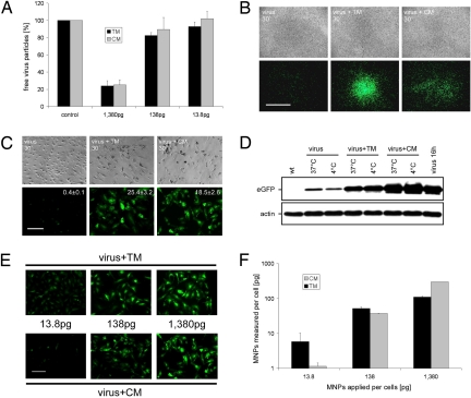
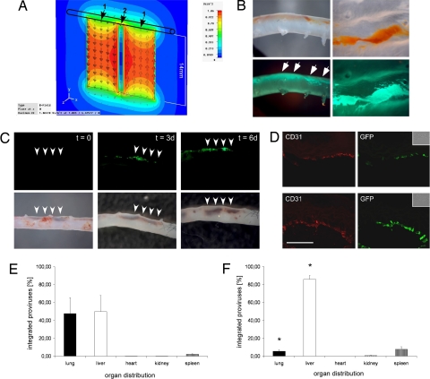
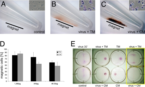
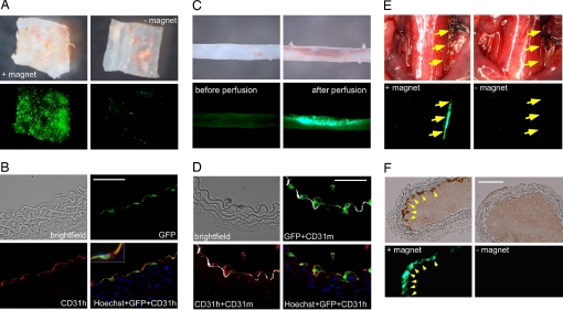
Targeting of viral vectors is a major challenge for in vivo gene delivery, especially after intravascular application. In addition, targeting of the endothelium itself would be of importance for gene-based therapies of vascular disease. Here, we used magnetic nanoparticles (MNPs) to combine cell transduction and positioning in the vascular system under clinically relevant, nonpermissive conditions, including hydrodynamic forces and hypothermia. The use of MNPs enhanced transduction efficiency of endothelial cells and enabled direct endothelial targeting of lentiviral vectors (LVs) by magnetic force, even in perfused vessels. In addition, application of external magnetic fields to mice significantly changed LV/MNP biodistribution in vivo. LV/MNP-transduced cells exhibited superparamagnetic behavior as measured by magnetorelaxometry, and they were efficiently retained by magnetic fields. The magnetic interactions were strong enough to position MNP-containing endothelial cells at the intima of vessels under physiological flow conditions. Importantly, magnetic positioning of MNP-labeled cells was also achieved in vivo in an injury model of the mouse carotid artery. Intravascular gene targeting can be combined with positioning of the transduced cells via nanomagnetic particles, thereby combining gene- and cell-based therapies.


Conflict of interest statement

The authors declare no conflict of interest.

Figures

Fig. 1.
Fig. 1.
Analysis of transduction efficiency and cellular MNP uptake after MNP-assisted lentiviral infection. (A) Analysis of virus-binding capacity of TM and CM nanoparticles. Three different concentrations [1,380 pg of MNPs per 50 virus particles (VPs), 138 pg/50 VPs, and 13.8 pg/50 VPs] were analyzed. Shown are the amounts (±SEM) of unbound virus particles in the supernatants of LV/MNP incubations (n = 3). (B and C) Analysis of transduction efficiency under nonpermissive conditions. (B) Transduction of HUVECs under hydrodynamic flow stress (cells were shaken in the presence of a magnetic gradient field). Note that the area of EGFP expression is close to the site of the magnetic gradient field after LV/TM (virus + TM 30′, Center) and LV/CM (virus + CM 30′, Right) transduction, but not after transduction without MNPs (virus 30′, Left). (C) Hypothermic transduction (4 °C) of HUVECs with LV/MNP complexes (virus + TM 30′, Center and virus + CM 30′, Right). Note that under these conditions lentiviral transduction without MNPs (virus 30′, Left) achieved only low levels of EGFP expression. The copy number (mean ± SEM) of integrated proviruses per cell (n = 3) is indicated in the fluorescence pictures. (D) Western blot analysis of EGFP expression after transduction of HUVECs with LVs (virus) or LV/MNP complexes (virus + TM, virus + CM) for 30 min at 4 °C and 37 °C. Overnight-infected cells (virus 16h) were used as control. (E and F) Analysis of transgene expression and cellular MNP uptake. HUVECs were transduced with equal amounts of lentiviral particles (50 VPs per cell), but different MNP doses (picograms of MNPs applied per cell are indicated). (E) Fluorescence images showing transgene expression 3 days after LV/TM (virus + TM) and LV/CM (virus + CM) transduction. (F) Quantification of nanoparticle uptake by magnetorelaxometry. Error bars are the uncertainty of the magnetic relaxation signal. (Scale bars: B, 5 mm; C and E, 100 μm.)
Fig. 2.
Fig. 2.
Ex vivo and in vivo targeting of lentiviral vectors by magnetic nanoparticles. (A) During ex vivo perfusion, 2 magnets were placed at one side of the aorta. The magnetic flux density of the magnets is shown as a multicolor contour plot in the x–y plane at z = 0 (center plane through the 2 magnets). 1 indicates poles of the magnets; 2, gap between the magnets. (B) Ex vivo perfusion of aortas with a mixture of magnetic nanoparticles and fluorescent LV particles containing gag-EGFP fusion proteins. Note that directly after perfusion (Left), nanoparticles (Upper), and EGFP fluorescence originating from the virus particles (Lower) were clearly visible at the site of the magnetic field (indicated by the arrows, 3.2× magnification). View of the intima layer after opening of the aorta (Right, 8× magnification). (C) Aortas were perfused ex vivo for 30 min with LV/CM complexes while magnets were placed at one side of the aorta (indicated by the arrowheads, 3.2× magnification). At the end of the perfusion (t = 0), accumulation of LV/CM complexes is clearly visible in the brightfield pictures (Lower). Note the EGFP expression after 3 (t = 3d) and 6 (t = 6d) days of culture (Upper). (D) Immunhistochemical analysis of transgene expression in aortic sections after perfusion with LV/TM (Upper) and LV/CM (Lower) complexes. EGFP expression is clearly visible in the CD31-positive endothelial layer (red) of the aortas. (Inset) Brightfield pictures. (Scale bar: 50 μm.) (E and F) In vivo distribution of integrated lentivectors after injection into the carotid artery of mice. Shown is the percental distribution (±SEM) in different organs (n = 3) of mice that were exposed to a strong magnetic field at the right abdominal wall (F, n = 5) and of control mice (E, n = 3). *, P < 0.05 compared with the control group.
Fig. 3.
Fig. 3.
Nanomagnetic cell positioning. (A–C) At 24 h after transduction with LV/MNP complexes, HUVECs were transferred into reaction tubes with a magnet placed at one side (indicated by the black bars). Accumulation of cells at the tube walls was seen only for LV/TM-transduced (B) and LV/CM-transduced (C) cells, but not for control cells (A). Prussian blue stainings of the cells (A–C Insets) demonstrate MNP incorporation (Inset, width ≈35 μm). (D) The experiments shown in A–C were performed with 3 different MNP doses (1,380 pg/50 VPs, 345 pg/50 VPs, and 86.25 pg/50 VPs). Shown are the numbers (mean ± SEM) of “magnetic” cells that adhered to the tube walls (n = 4). (E) Targeted attachment of HUVECs after transduction with LV/MNP complexes (virus + TM and virus + CM) or with MNPs alone (TM and CM). Shown are the plates after staining with sulforhodamine B dye. Note that directed cell positioning in close proximity to the magnets was observed only for cells that were transduced with nanoparticles (4 wells in the middle). Control, untreated cells; virus 30′, cells transduced without MNPs; no magnet, cells cultured without magnets below the plates.
Fig. 4.
Fig. 4.
Ex vivo and in vivo positioning of HUVECs to the vessel walls in the presence of hydrodynamic forces or blood flow. (A) LV/TM-transduced HUVECs were transferred to aortic strips and cultured for 24 h with (+magnet) and without (-magnet) magnets below the strips while being shaken. Shown are brightfield (Upper) and fluorescence pictures (Lower) taken by a stereomicroscope (4-fold magnification). (B) Immunhistochemical analysis of aortas with LV/TM-transduced HUVECs. Sections were stained with antibodies against human PECAM-1 (CD31h, red) and Hoechst dye (blue). Note the colocalization of the HUVEC-derived CD31h signals with the EGFP fluorescence. The inset shows a triple-stained cell (Hoechst plus eGFP plus CD31h). (C) Aortas were perfused ex vivo with medium containing LV/TM-transduced HUVECs while 2 magnets were positioned at one side. Brightfield (Upper) and fluorescence images (Lower) were taken before and directly after perfusion (3.2-fold magnification). Note the accumulation of EGFP-expressing “magnetic” cells. (D) Immunhistochemical analysis of the aorta after ex vivo perfusion. Sections were stained with antibodies against murine (CD31m, white) and human CD31 (CD31h, red), and Hoechst dye (blue). Note that EGFP and CD31h signals, which are both derived from the transduced HUVECs, were colocalized and located in close proximity to the murine aortic endothelial layer (CD31m). (E) In vivo positioning of LV/TM-transduced HUVECs to the intima of injured common carotid arteries by magnetic forces (+magnet). Control experiments were performed under the same conditions, but without placing a magnet (-magnet). Images were taken 10 min after application of the cells and restoration of the blood flow. Yellow arrows indicate retention of MNP-labeled HUVECs in the area of the magnetic field (Left), but not in the control animal (Right). Shown are bright-field (Upper) and fluorescence images (Lower) taken by a stereomicroscope (1.8-fold magnification). (F) Histological analysis of the carotid arteries. After application of a magnetic field to the injured common carotid artery, accumulation of MNP-loaded cells was found at the vessel wall (Left). No cells were found in the control vessel (Right). Shown are bright-field (Upper) and fluorescence (Lower) pictures. (Scale bars: 50 μm.)

References

    1. Verma IM, Weitzman MD. Gene therapy: Twenty-first century medicine. Annu Rev Biochem. 2005;74:711–738. - PubMed
    1. Wiznerowicz M, Trono D. Harnessing HIV for therapy, basic research and biotechnology. Trends Biotechnol. 2005;23:42–47. - PubMed
    1. Dimitrov DS. Virus entry: Molecular mechanisms and biomedical applications. Nat Rev Microbiol. 2004;2:109–122. - PMC - PubMed
    1. Marsh M, Helenius A. Virus entry: Open sesame. Cell. 2006;124:729–740. - PMC - PubMed
    1. Smith AE, Helenius A. How viruses enter animal cells. Science. 2004;304:237–242. - PubMed

Publication types

LinkOut - more resources