Review
. 2009 Jan 15;122(Pt 2):179-86.
doi: 10.1242/jcs.042127.

The mechanical integrin cycle

Affiliations
Review

The mechanical integrin cycle

Eileen Puklin-Faucher et al. J Cell Sci. .

Erratum in

  • J Cell Sci. 2009 Feb 15;122(Pt 4):575

Abstract

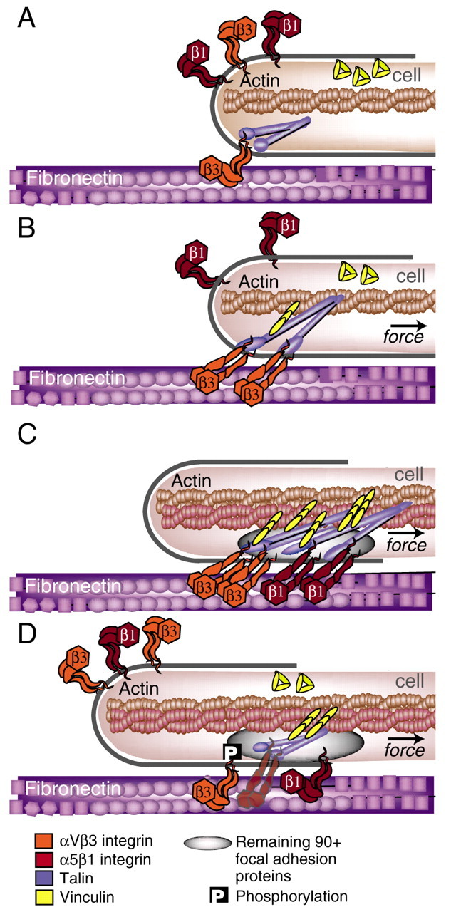
Cells govern tissue shape by exerting highly regulated forces at sites of matrix adhesion. As the major force-bearing adhesion-receptor protein, integrins have a central role in how cells sense and respond to the mechanics of their surroundings. Recent studies have shown that a key aspect of mechanotransduction is the cycle by which integrins bind to the matrix at the leading cell edge, attach to the cytoskeleton, transduce mechanical force, aggregate in the plasma membrane as part of increasingly strengthened adhesion complexes, unbind and, ultimately, are recycled. This mechanical cycle enables the transition from early complexes to larger, more stable adhesions that can then rapidly release. Within this mechanical cycle, integrins themselves exhibit intramolecular conformational change that regulates their binding affinity and may also be dependent upon force. How the cell integrates these dynamic elements into a rigidity response is not clear. Here, we focus on the steps in the integrin mechanical cycle that are sensitive to force and closely linked to integrin function, such as the lateral alignment of integrin aggregates and related adhesion components.


Figures

Fig. 1.
Fig. 1.
The mechanical integrin cycle. (A) Cell-ECM adhesion occurs when actin-dependent protrusions bring integrins at the leading edge (orange) in contact with the matrix (purple) where they can bind. (B) Next, the integrins link to the actin cytoskeleton through adaptor proteins, such as talin (blue), Shp2, filamin or α-actinin. Integrins bind to these adaptor proteins through their β-tails. Rearward actin flow, generated by actin polymerization and actomyosin contractions (see Box 1) induces a pulling force on the integrin-ECM linkage. On sufficiently rigid substrates, this may serve to accelerate an integrin-activating conformational change, as well as a talin stretch, which may expose buried vinculin-binding sites (yellow). Although the bent conformation of the ligand-bound αVβ3-integrin crystal structure produced much controversy in the integrin field (Liddington and Ginsberg, 2002; Mould et al., 2003), it has subsequently been shown in electron microscopy experiments to stably bind fibronectin (Adair et al., 2005). Force might accelerate the switch to high-binding affinity by freeing the ligand-bound integrin head from the constraints of neighboring domains, which would essentially accelerate the allosteric pathway to the activated state (Puklin-Faucher et al., 2006). (C) The cell begins to pull itself over the site of adhesion. Intramolecular conformational changes in α5β1 integrins facilitate their inward translocation, whereas αVβ3 integrins remain anchored at the edge. This segregation of integrins may further facilitate the talin stretch. At this stage of adhesion, a wide variety of intracellular focal-adhesion proteins are accumulated in the adhesive plaque (grey oval). (D) Ultimately, highly clustered integrins switch from high- to low-binding affinity, possibly catalyzed by the phosphorylation of β3-integrin tails. Membrane exocytosis places recycled, low-affinity integrins at the end of microtubules, often 2-4 μm away from the leading edge. The integrin turnover in focal adhesions (from C to D) is ∼1-3 minutes (Hu et al., 2007). For the description of a single integrin see supplementary material Fig. S1.
Fig. 2.
Fig. 2.
Segregation of αVβ3- and α5β1-integrins in focal adhesions. The segregation of αVβ3- and α5β1-integrins in focal adhesions (depicted schematically in Fig. 1D) is shown here by antibody staining (Felsenfeld et al., 1999). (A) Src-deficient cells that expressing a truncated form of Src tagged with GFP (Src-251GFP) were fixed and stained with antibodies recognizing β1 integrins, αVβ3 integrins and vinculin. (B) In higher-magnification views, the αVβ3 subunit can be seen to colocalize at the periphery of focal adhesions, with vinculin and Src-251GFP. Vinculin staining was co-distributed with that of Src-251GFP in all cases. (C). By contrast, β1 integrins distributed in longer peripheral stripes (consistent with the distribution of stress fibers) that did not overlap with the distribution of αVβ3–Src-251GFP. Arrowhead indicates the boundary of staining. (D) Overlap of GFP and vinculin staining without β1 integrins is indicated by yellow pixels. Scale bars: 10 μm (A) and 5 μm (D). Images reproduced with permission (Felsenfeld et al., 1999).

References

    1. Adair, B., Xiong, J.-P., Maddock, C., Goodman, S., Arnaout, M. A. and Yeager, M. (2005). Three-dimensional EM structure of the ectodomain of integrin alphaVbeta3 in a complex with fibronectin. J. Cell Biol. 168, 1109-1118. - PMC - PubMed
    1. Alexandrova, A. Y., Arnold, K., Schaub, S., Vasiliev, J. M., Meister, J. J., Bershadsky, A. D. and Verkhovsky, A. B. (2008). Comparative dynamics of retrograde actin flow and focal adhesions: formation of nascent adhesions triggers transition from fast to slow flow. PLoS ONE 3, e3234. - PMC - PubMed
    1. Alon, R. and Dustin, M. L. (2007). Force as a facilitator of integrin conformational changes during leukocyte arrest on blood vessels and antigen-presenting cells. Immunity 26, 17-27. - PubMed
    1. Arias-Salgado, E. G., Lizano, S., Sarkar, S., Brugge, J., Ginsberg, M. H. and Shattil, S. J. (2003). Src kinase activation by direct interaction with the integrin beta cytoplasmic domain. Proc. Natl. Acad. Sci. USA 100, 13298-13302. - PMC - PubMed
    1. Balaban, N. Q., Schwarz, U. S., Riveline, D., Goichberg, P., Tzur, G., Sabanay, I., Mahalu, D., Safran, S., Bershadsky, A., Addadi, L. et al. (2001). Force and focal adhesion assembly: a close relationship studied using elastic micropatterned substrates. Nat. Cell Biol. 3, 466-472. - PubMed

Publication types