

Mice that lack activity of alphavbeta6- and alphavbeta8-integrins reproduce the abnormalities of Tgfb1- and Tgfb3-null mice
- PMID: 19118215
- PMCID: PMC2714418
- DOI: 10.1242/jcs.035246
Mice that lack activity of alphavbeta6- and alphavbeta8-integrins reproduce the abnormalities of Tgfb1- and Tgfb3-null mice
Abstract
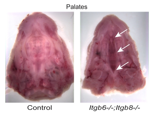
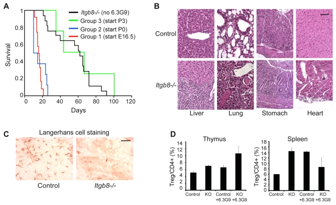
The arginine-glycine-aspartate (RGD)-binding integrins alphavbeta6 and alphavbeta8 activate latent TGFbeta1 and TGFbeta3 in vivo, but it is uncertain whether other RGD-binding integrins such as integrins alphavbeta5 and alphavbeta3 activate these TGFbeta isoforms. To define the combined role of alphavbeta6- and alphavbeta8-integrin in TGFbeta activation, we analyzed mice lacking function of both integrins by means of gene deletion and/or pharmacologic inhibition. Most Itgb6-/-;Itgb8-/- embryos die at mid-gestation; those that survive develop cleft palate-as observed in Tgfb3-/- mice. Itgb8-/- mice treated with an anti-alphavbeta6-integrin antibody develop severe autoimmunity and lack Langerhans cells-similar to Tgfb1-null mice. These results support a model in which TGFbeta3-mediated palate fusion and TGFbeta1-mediated suppression of autoimmunity and generation of Langerhans cells require integrins alphavbeta6 and alphavbeta8 but not other RGD-binding integrins as TGFbeta activators.
Figures

References
-
- Annes, J. P., Rifkin, D. B. and Munger, J. S. (2002). The integrin αVβ6 binds and activates latent TGFβ3. FEBS Lett. 511, 65-68. - PubMed
-
- Annes, J. P., Munger, J. S. and Rifkin, D. B. (2003). Making sense of latent TGFβ activation. J. Cell Sci. 116, 217-224. - PubMed
-
- Asano, Y., Ihn, H., Yamane, K., Jinnin, M., Mimura, Y. and Tamaki, K. (2005a). Increased expression of integrin αvβ3 contributes to the establishment of autocrine TGF-β signaling in scleroderma fibroblasts. J. Immunol. 175, 7708-7718. - PubMed
Publication types
MeSH terms
Substances
Grants and funding
LinkOut - more resources
Full Text Sources
Other Literature Sources
Medical
Molecular Biology Databases
Miscellaneous
