Review
. 2009 Jan;155(Pt 1):9-15.
doi: 10.1099/mic.0.023564-0.

Early molecular-recognition events in the synthesis and export of group 2 capsular polysaccharides

Affiliations
Review

Early molecular-recognition events in the synthesis and export of group 2 capsular polysaccharides

Eric R Vimr et al. Microbiology (Reading). 2009 Jan.

Abstract

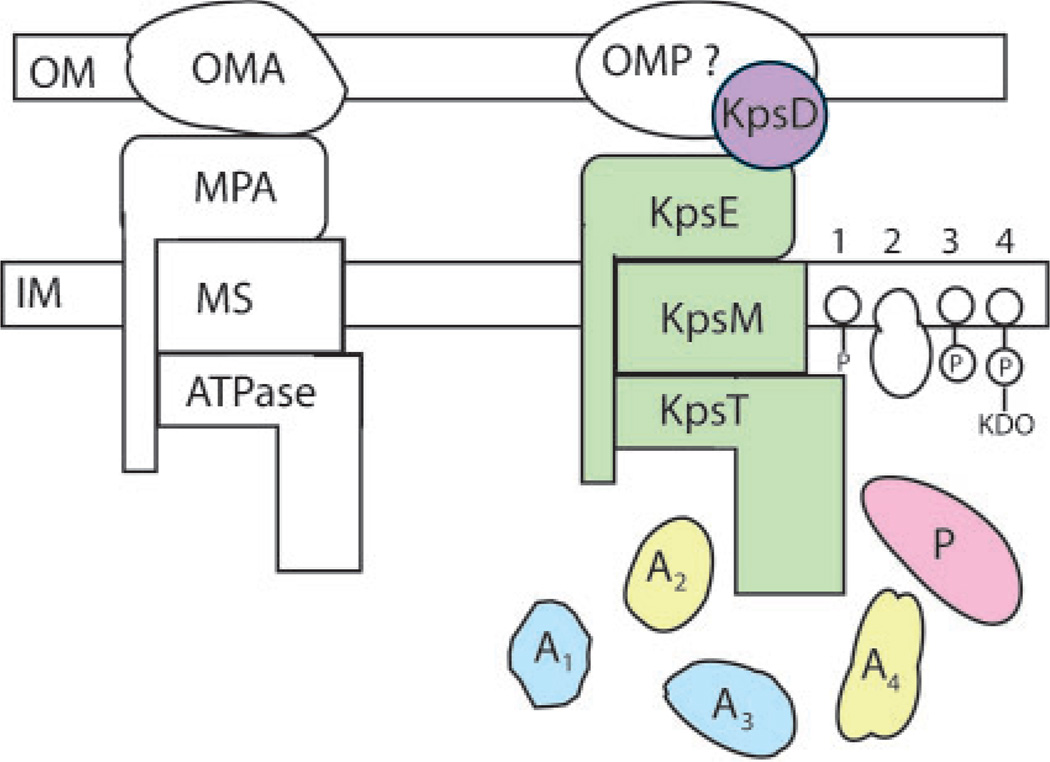
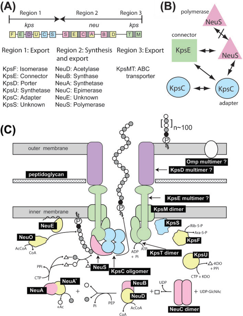
The outer membrane (OM) of almost all Gram-negative bacteria is composed of phospholipids, lipopolysaccharide, proteins and capsular or loosely adherent polysaccharides that together mediate cellular interactions with diverse environments. Most OM components are synthesized intracellularly or at the inner membrane (IM) and thus require an export mechanism. This mini-review focuses on recent progress in understanding how synthesis of one kind of capsular polysaccharide (group 2) is coupled to the export apparatus located in the IM and spanning the periplasmic space, thus providing a transport channel to the cell surface. Although the model system for these investigations is the medically important extraintestinal pathogen Escherichia coli K1 and its polysialic acid capsule, the conclusions are general for other group 2 and group 2-like polysaccharides synthesized by many different bacterial species.


Figures

Fig. 1
Fig. 1
Type I protein and drug efflux exporters (left) compared to group 2 and group 2-like capsular polysaccharide exporters (right). A1–4 indicate accessory proteins and the numbers 1–4 at the far right refer to undecaprenyl phosphate, protein, phospholipid and phospholipid-KDO endogenous acceptors (initiators) or terminators, respectively. Other abbreviations not listed in the text are: OMA, OM auxiliary protein; MPA, membrane–periplasm auxiliary protein; MS and ATPase are the IM-spanning and ATPase modules of the ABC transporter, respectively; OMP?, hypothetical OM protein working in concert with KpsD; P, polymerase. The figure is adapted from Silver et al. (2001). Refer to Fig. 3 legend for colour-coding.
Fig. 2
Fig. 2
Summary of the steps involved in polysaccharide biosynthesis. See text for details.
Fig. 3
Fig. 3
Group 2 capsular polysialic acid biosynthesis in E. coli K1. (A) Genetic and functional organization of the kps–neu gene cluster for polysialic acid biosynthesis adapted from Steenbergen & Vimr (2008); see text for details. (B) Protein–protein interactions between export and synthetic gene products detected by two-hybrid analysis. (C) Model of group 2 capsular polysaccharide biosynthesis. Gene products indicated by black boxes are defined in panel (A) and the text. Open and shaded small circles indicate unacetylated and acetylated sialic acids respectively. The small black circles indicate KDO or unknown moiety while the squiggle-circled P is phosphatidic acid. Triangles indicate CMP; the square is N-acetylmannosamine (ManNAc); Ac, acetyl groups; AcCoA and CoA, acetyl coenzyme A and coenzyme A respectively; Rib-5-P, ribulose 5-phosphate; Ara-5-P, d-arabinose 5-phosphate. Other small molecules have their usual abbreviations. Colours indicate essential or non-essential functions as follows: pink, essential synthetic functions; yellow, non-essential synthetic functions in the K1 system; green, essential exporter functions; blue, essential accessory proteins; purple, atypical OM protein found in some but not all group 2 or group 2-like systems. Specific interactions detected by two-hybrid analysis between NeuS, KpsE, and KpsC are indicated by the overlapping shapes. The specific steps in polysialic acid biosynthesis include the following. Step 1, the first committed step, involves the releasing epimerase, NeuC, producing ManNAc. Step 2: ManNAc interacting with the NeuB–NeuD complex is condensed with phosphoenolpyruvate (PEP) by NeuB while NeuD transfers an acetyl group to most of the nascent sialic acids. Step 3: the N-terminal domain of the synthetase NeuA activates sialic acid for polymerization by coupling it to CMP donated by CTP. Concomitantly with activation the C-terminal synthetase domain (NeuA*) acts as an O-acetyl esterase to remove the acetyl group from most sialic acid substrates. The functional significance of the cyclical acetylation and deacetylation reactions is not presently understood. Step 4: sialic acid residues are polymerized by NeuS while NeuO in most K1 strains reacetylates the growing chain. Step 5: NeuS, NeuC and NeuE interact such that during or shortly after polymerization the completed polysialic acid is exported through the transmembrane channel to the outer membrane where the phospholipid anchors it to the outer leaflet of the OM.
Fig. 4
Fig. 4
Phenotypes of export-deficient acapsular mutants. Thin sections of representative bacteria with the indicated defects in export gene were examined by transmission electron microscopy as previously described by Cieslewicz & Vimr (1996, 1997). Phenotypes and their interpretations are described in the text. (A) Wild-type, where the capsule is not visible due to the fixation procedure, (B) kpsF mutant, (C) kpsT mutant, (D) kpsS mutant, (E) kpsC mutant, and (F) kpsE mutant.

References

    1. Andreischeva EN, Vann WF. Gene products required for de novo synthesis of polysialic acid in Escherichia coli K1. J Bacteriol. 2006;188:1786–1797. - PMC - PubMed
    1. Annunziato PW, Wright LF, Vann WF, Silver RP. Nucleotide sequence and genetic analysis of the neuD and neuB genes in region 2 of the polysialic acid gene cluster of Escherichia coli K1. J Bacteriol. 1995;177:312–319. - PMC - PubMed
    1. Azurmendi HF, Vionnet J, Wrightson L, Trinh LB, Shiloach J, Freedberg DI. Extracellular structure of polysialic acid explored by on cell solution NMR. Proc Natl Acad Sci U S A. 2007;104:11557–11561. - PMC - PubMed
    1. Bos MP, Robert V, Tommassen J. Biogenesis of the gram-negative bacterial outer membrane. Annu Rev Microbiol. 2007;61:191–214. - PubMed
    1. Bronner D, Sieberth V, Pazzanik C, Roberts IS, Boulnois G, Jann B, Jann K. Expression of the capsular K5 polysaccharide of Escherichia coli: biochemical and electron microscopic analyses of mutants with defects in region 1 of the K5 gene cluster. J Bacteriol. 1993;175:5984–5992. - PMC - PubMed

Publication types

MeSH terms

Substances

LinkOut - more resources