. 2009 Jan 2:9:1.
doi: 10.1186/1475-2867-9-1.

Combined xanthorrhizol-curcumin exhibits synergistic growth inhibitory activity via apoptosis induction in human breast cancer cells MDA-MB-231

Affiliations

Combined xanthorrhizol-curcumin exhibits synergistic growth inhibitory activity via apoptosis induction in human breast cancer cells MDA-MB-231

Yew Hoong Cheah et al. Cancer Cell Int. .

Abstract

Background: It has been suggested that combined effect of natural products may improve the treatment effectiveness in combating proliferation of cancer cells. The present study was undertaken to evaluate the possibility that the combination of xanthorrhizol and curcumin might show synergistic growth inhibitory effect towards MDA-MB-231 human breast cancer cells via apoptosis induction. The effective dose that produced 50% growth inhibition (GI50) was calculated from the log dose-response curve of fixed-combinations of xanthorrhizol and curcumin generated from the sulforhodamine B (SRB) assay. The experimental GI50 value was used to determine the synergistic activity of the combination treatment by isobolographic analysis and combination-index method. Further investigation of mode of cell death induced by the combination treatment was conducted in the present study.

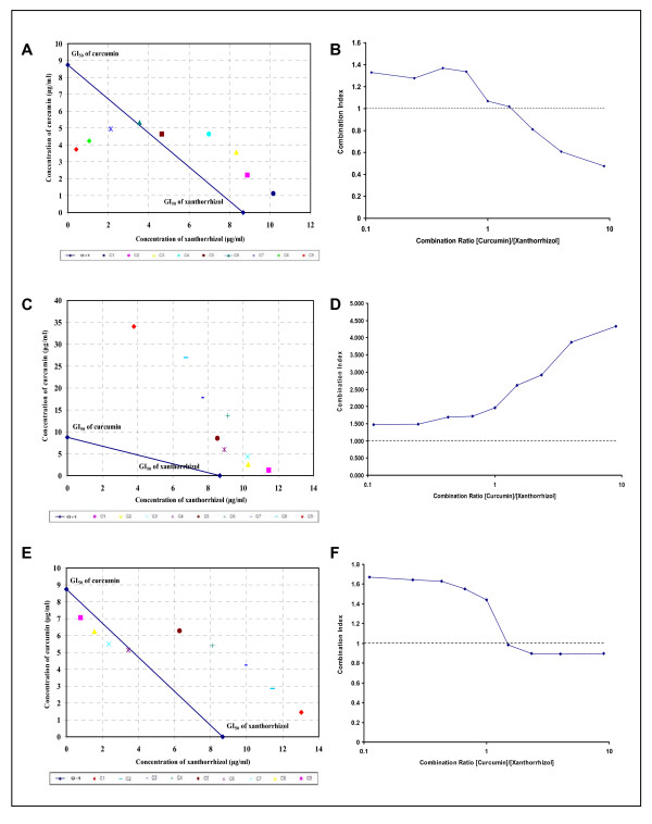
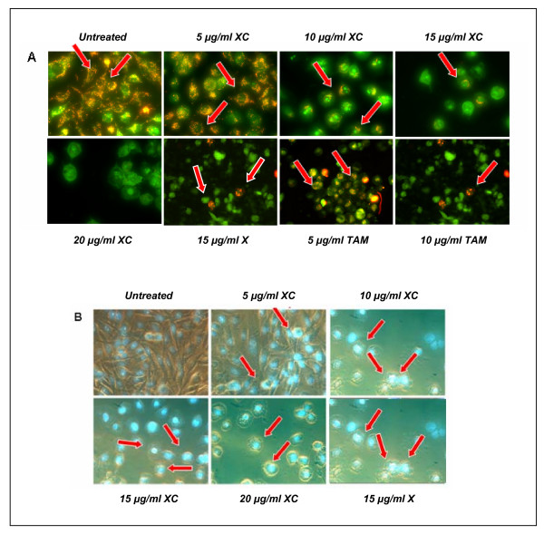
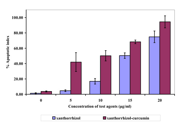
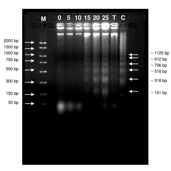
Results: Isobole analysis revealed that substances interaction was synergistic when xanthorrhizol and curcumin were added concurrently to the cultures but merely additive when they were added sequentially. The synergistic combination treatment was then applied to the cultures to investigate the mode of cell death induced by the treatment. Immunofluorescence staining using antibody MitoCapturetrade mark revealed the possibility of altered mitochondrial transmembrane potential, which is one of the hallmark of apoptosis. Hoechst 33258 nuclear staining assay showed the rate of apoptosis of MDA-MB-231 cells to increase in response to the treatment. Apoptotic cell death was further confirmed by DNA fragmentation assay, where internucleosomal excision of DNA was induced upon treatment with xanthorrhizol-curcumin.

Conclusion: This is the first time the combined cytotoxic effect of xanthorrhizol and curcumin on MDA-MB-231 cells has been documented and our findings provide experimental support to the hypothesis that combined xanthorrhizol-curcumin showed synergistic growth inhibitory activity on MDA-MB-231 cells via apoptosis induction.


Figures

Figure 1
Figure 1
The extraction and isolation of xanthorrhizol from its crude essential oil. The designated peak (xanthorrhizol) is the most abundance component, which accounted for 46.3% of the total component in the extracted essential oil of C. xanthorrhizza (A). Xanthorrhizol was detected via chromogenic reagent spray (0.5% vanillin/sulfuric acid) and UV (254 nm) visualization in TLC profile as compare to the authentic reference substance (B and C). Final isolation process produced compound 1 at 100% abundance (D) and the NMR spectral identified the compound 1 as xanthorrhizol (E).
Figure 2
Figure 2
Simultaneous treatment of xanthorrhizol and curcumin alters the mitochondrial transmembrane potential and induces apoptosis in MDA-MB-231 cells. MitoCapture™ dye was found localized and fluoresces red (arrow) in the mitochondrial of untreated cells. Number of cells fluoresces red was decreasing in line with the increasing dose of treatment, suggesting the disruption of the mitochondrial transmembrane potential (A). Phase contrast fluorescence images show that untreated cells were stained homogenously and no fluorescence was observed. In contrary, cell shrinkage was observed in the treated cells and fluorescence (arrow) was clearly detected in the nuclear region, indicating apoptotic morphology (B). X – xanthorrhizol. XC – combination of xanthorrhizol and curcumin. TAM – Tamoxifen (positive control).
Figure 3
Figure 3
Isobolograms and combination indexes at 50% effect level of simultaneous and sequential exposure of MDA-MB-231 cells to xanthorrhizol and curcumin. The dashed line (CI = 1) indicates the alignment of theoretical values of an additive interaction between two substances. Values above the dashed line indicate an antagonistic interaction, and values below indicate synergism interaction. Simultaneous treatment involved concurrent 48 h exposure of MDA-MB-231 cells to xanthorrhizol and curcumin (A and B) whereas sequential treatment was 24 h exposure to xanthorrhizol followed by curcumin for subsequent 24 h (C and D) or vice versa (E and F). Simultaneous treatment inhibits the growth of MDA-MB-231 cells more synergistically as compared to the sequential treatment. Results are representative of three independent experiments conducted.
Figure 4
Figure 4
Apoptosis level after treatment of xanthorrhizol and simultaneous treatment of xanthorrhizol-curcumin as determined from Hoechst 33258 staining. The treatment for 24 h induced exponential apoptotic cell death in a dose dependent manner as compare to untreated control. Results are presented as means ± SD of 3 independent experiments. *p < 0.05, **p < 0.005 statistically significant values relative to untreated control.
Figure 5
Figure 5
Simultaneous treatment of xanthorrhizol and curcumin induces DNA fragmentation in MDA-MB-231 cells. At lower doses of treatment, only high molecular weight intact DNA was observed whereas small fragments of DNA were highlighted at higher doses of treatment. The fragments of DNA have interval molecular weight of ~180 bp, suggesting an apoptotic event. Results are representative of three independent experiments carried out. M – Marker. 0, 5, 10, 15, 20, 25 μg/ml – DNA samples from xanthorrhizol-curcumin (XC)-treated cells at different concentration. T – DNA sample from taxol-treated MDA-MB-231 cells (positive control). C – DNA sample from actinomycin-treated HL 60 cells (positive control).
Scheme 1
Scheme 1
(Acetylation process)
Scheme 2
Scheme 2
(Hydrolysis process)

References

    1. Newman DJ, Cragg GM. Natural products as sources of new drugs over the last 25 years. J Nat Prod. 2007;70:461–477. doi: 10.1021/np068054v. - DOI - PubMed
    1. Ismail N, Pihie AH, Nallapan M. Xanthorrhizol induces apoptosis via the up-regulation of bax and p53 in HeLa cells. Anticancer Res. 2005;25:2221–2227. - PubMed
    1. Cheah YH, Azimahtol HL, Abdullah NR. Xanthorrhizol exhibits antiproliferative activity on MCF-7 breast cancer cells via apoptosis induction. Anticancer Res. 2006;26:4527–4534. - PubMed
    1. Handayani T, Sakinah S, Nallapan M, Pihie AH. Regulation of p53-, Bcl-2-, and caspase-dependent signaling pathway in xanthorrhizol-induced apoptosis of HepG2 hepatoma cells. Anticancer Res. 2007;27:965–971. - PubMed
    1. Goel A, Kunnumakkara AB, Aggarwal BB. Curcumin as "curecumin": from kitchen to clinic. Biochem Pharmacol. 2008;75:787–809. doi: 10.1016/j.bcp.2007.08.016. - DOI - PubMed

LinkOut - more resources