. 2009 Feb;1788(2):358-70.
doi: 10.1016/j.bbamem.2008.11.021. Epub 2008 Dec 11.

Environmental tobacco smoke effects on lung surfactant film organization

Affiliations

Environmental tobacco smoke effects on lung surfactant film organization

Patrick C Stenger et al. Biochim Biophys Acta. 2009 Feb.

Abstract

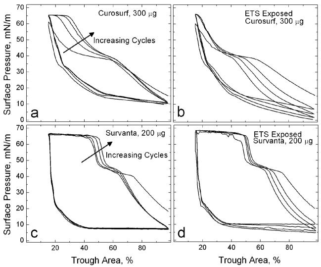
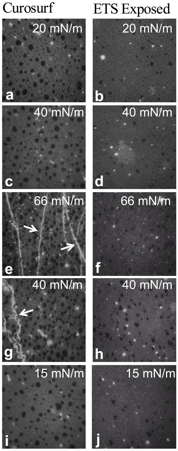
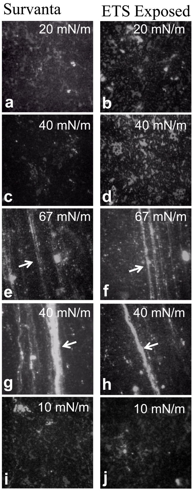
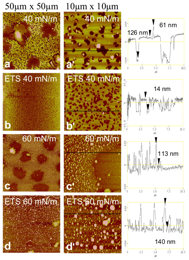
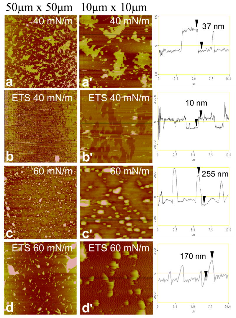
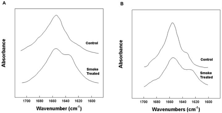
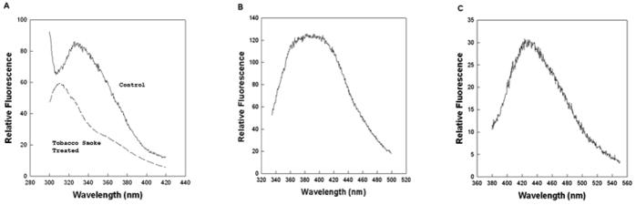
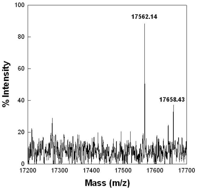
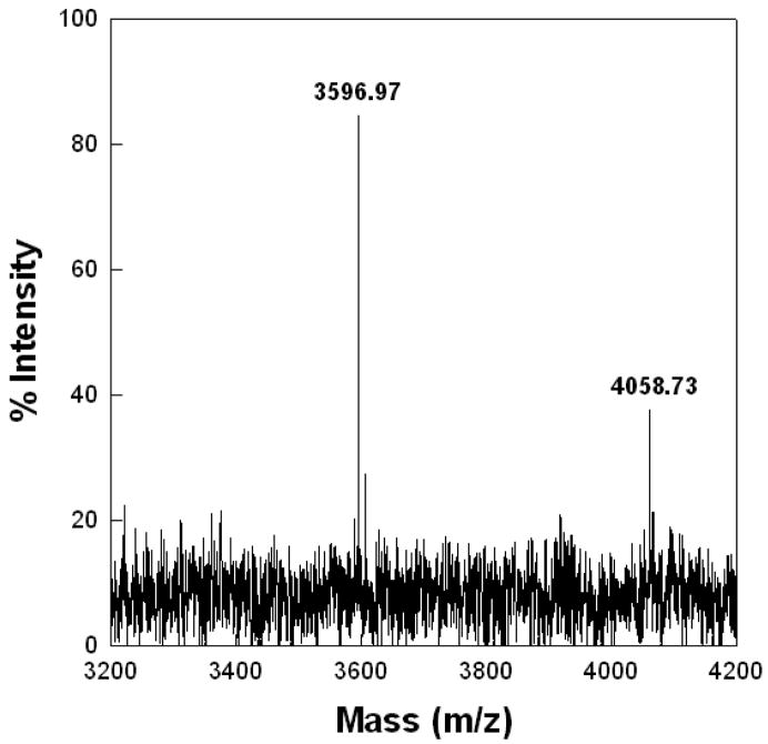
Adsorption of the clinical lung surfactants (LS) Curosurf or Survanta from aqueous suspension to the air-water interface progresses from multi-bilayer aggregates through multilayer films to a coexistence between multilayer and monolayer domains. Exposure to environmental tobacco smoke (ETS) alters this progression as shown by Langmuir isotherms, fluorescence microscopy and atomic force microscopy (AFM). After 12 h of LS exposure to ETS, AFM images of Langmuir-Blodgett deposited films show that ETS reduces the amount of material near the interface and alters how surfactant is removed from the interface during compression. For Curosurf, ETS prevents refining of the film composition during cycling; this leads to higher minimum surface tensions. ETS also changes the morphology of the Curosurf film by reducing the size of condensed phase domains from 8-12 microm to approximately 2 microm, suggesting a decrease in the line tension between the domains. The minimum surface tension and morphology of the Survanta film are less impacted by ETS exposure, although the amount of material associated with the film is reduced in a similar way to Curosurf. Fluorescence and mass spectra of Survanta dispersions containing native bovine SP-B treated with ETS indicate the oxidative degradation of protein aromatic amino acid residue side chains. Native bovine SP-C isolated from ETS exposed Survanta had changes in molecular mass consistent with deacylation of the lipoprotein. Fourier Transform Infrared Spectroscopy (FTIR) characterization of the hydrophobic proteins from ETS treated Survanta dispersions show significant changes in the conformation of SP-B and SP-C that correlate with the altered surface activity and morphology of the lipid-protein film.


Figures

Figure 1
Figure 1. Cyclic isotherms of pristine and ETS-exposed (TSP = 77 mg/m3) commercial replacement lung surfactants on a saline buffer subphase. On compression, the isotherms exhibit a characteristic shoulder at Π~40 mN/m and a collapse plateau at Πmax~66 mN/m
(a) 300 μg pristine Curosurf. The fourth cycle collapse plateau here accounts for 15% of the total trough area. (b) 300 μg ETS-exposed Curosurf. In contrast to (a), the fourth cycle collapse plateau only accounts for 2% of the total trough area. (c) 200 μg pristine Survanta (d) 200 μg ETS-exposed Survanta. In both (c) and (d), the fourth cycle collapse plateau accounts for 34% of the total trough area. Arrows indicate increasing number of compression/expansion cycles.
Figure 2
Figure 2
Fluorescence images of 300 μg pristine Curosurf and ETS-exposed (TSP = 77 mg/m3) Curosurf doped with 1% mol Texas Red-DHPE. Images are 171 μm × 171 μm and were acquired on the second cycle. Discrete circular condensed domains (dark) coexist with a continuous liquid expanded phase (light gray). (a) Curosurf and (b) ETS-exposed Curosurf on a saline buffered subphase at Π = 20 mN/m during compression. (c) Curosurf and (d) ETS-exposed Curosurf at Π = 40 mN/m during compression. (e) Curosurf and (f) ETS-exposed Curosurf at the collapse plateau, Π = 66 mN/m during compression. Arrows indicate cracks and folds in the monolayer due to collapse which are only seen in the Curosurf film. (g) Curosurf and (h) ETS-exposed Curosurf at Π = 40 mN/m during expansion. (i) Curosurf and (j) ETS-exposed Curosurf at Π = 15 mN/m during expansion. Generally, ETS exposure reduces the concentration of large condensed domains (8 – 12 μm) in favor of smaller ones (~2 μm).
Figure 3
Figure 3
Fluorescence images of 200 μg pristine Survanta and ETS-exposed (TSP = 77 mg/m3) Survanta doped with 1% mol Texas Red-DHPE. Images are 171 μm × 171 μm and were acquired on the second cycle. The darker condensed phase forms a continuous network surrounding the lighter liquid expanded phase. (a) Survanta and (b) ETS-exposed Survanta a saline buffered subphase at Π = 20 mN/m during compression. (c) Survanta and (d) ETS-exposed Survanta at Π = 40 mN/m during compression. (e) Survanta and (f) ETS-exposed Survanta at the collapse plateau, Π = 67 mN/m, during compression. Arrows indicate cracks and folds in the monolayer due to collapse. (g) Survanta and (h) ETS-exposed Survanta at Π = 40 mN/m during expansion. (i) Survanta and (j) ETS-exposed Survanta at Π = 10 mN/m during expansion. ETS exposure has less effect on the morphology of the Survanta film than on Curosurf films.
Figure 4
Figure 4
Atomic force microscopy images of pristine Curosurf and ETS-exposed Curosurf transferred onto mica substrates by conventional Langmuir-Blodgett deposition. The low resolution images (left column) are 50 × 50 μm, and the high resolution images (right column) are 10 × 10 μm sections taken from the low-resolution images. The height trace in the third column corresponds to the black line on each high-resolution image. The brightness in the image increases with the height relative to the mica substrate. Images at 40 mN/m of Curosurf at (a) low and (a) high resolution and ETS-exposed Curosurf at (b) low and (b) high resolution. Discrete dark brown circular domains up to 10–15 μm wide in Curosurf (a,a′) and 0.2–1 μm wide ETS-exposed Curosurf (b,b′) exist in a bright yellow network. The height traces indicate a greater thickness of the bright yellow liquid expanded phase in Curosurf (~126 nm) vs. ETS-exposed Curosurf (~14 nm) indicates a larger surface associated “reservoir” which is trapped underneath the monolayer during LB deposition. Images at 60 mN/m of Curosurf at (c) low and (c) high resolution and ETS-exposed Curosurf at (d) low and (d) high resolution. Both Curosurf (c) and ETS-exposed Curosurf (d) show a non-uniform liquid expanded phase with discrete raised domains (0.3–1.3 μm wide and ~110–140 nm high) where material that has been removed from the interface is held in the surface associated reservoir. The linescans indicate a greater concentration of raised domains in Curosurf (c′) demonstrating that ETS exposure (d′) alters the ways that material is removed from the interface.
Figure 5
Figure 5. Atomic force microscopy images of pristine Survanta and ETS-exposed Survanta transferred onto mica substrates by conventional Langmuir-Blodgett deposition. The low resolution images (left column) are 50 × 50 μm, and the high resolution images (right column) are 10 × 10 μm sections taken from the low-resolution images. The height trace in the third column corresponds to the black line on each high-resolution image. The brightness in the image increases with the height relative to the mica substrate
Images at 40 mN/m of Survanta at (a) low and (a) high resolution and ETS-exposed Survanta at (b) low and (b) high resolution. While Survanta shows a continuous dark brown network with discrete bright yellow fractal domains, ETS-exposed Survanta shows a greater percentage bright liquid expanded phase. The bright liquid expanded region in Survanta is ~37 nm thicker than the condensed phase versus only ~10 nm thicker in the ETS-exposed Survanta. Images at 60 mN/m of Survanta at (a) low and (a) high resolution and ETS-exposed Survanta at (b) low and (b) high resolution. Both sets of images are dominated by discrete circular raised domains, but the domains are higher and cover a greater area fraction in Survanta indicating that less material is stored near the interface in ETS-exposed Survanta.
Figure 6
Figure 6
Stack plots of FTIR spectra of the amide I spectral region of the control and Smoke treated SP-B (A) and SP-C (B). Proteins (100 μg protein) were spread as a self-film in hexafluoroisopropanol(HFIP) and allowed to air dry. The film was then overlaid with HFIP solvent and spectra accumulated and processed as described in the text.
Figure 7
Figure 7
Fluorescence emission spectra of Trp-Tyr SP-B (50 μg protein/mL) (A) and Fluorescence emission spectra of NFKyn (50 μg protein/mL)(B) and Kyn (50 μg protein/mL) (C) aromatic degradation products associated with smoke treated samples. The control emission spectra were digitally subtracted from the ETS treated sample spectra to obtain the fluorescence emission spectrum of NFKyn and Kyn in the isolated protein sample. Excitation wavelength was at 275 nm for Trp and Tyr, 325 nm for NKFyn and 365 nm for Kyn.
Figure 8
Figure 8
MALDI-TOF spectrum of Bovine SP-B dimer sample eluted from SDS gel of ETS-exposed surfactant sample. The ABI Voyager-DE STR MALDI-TOF was run in linear mode with delayed extraction and negative polarity. The instrument had an accelerating voltage of 2000 volts, an extraction delay time of 125 nsec, and a laser intensity of 1925. Treated samples had an intense mass peak at 17562.14 Da with a minor peak at 17658.43 Da (Figure 8) compared with a control sample mass of 17397.7 Da (not shown) that is close to the calculated mass of dimeric SP-B [36].
Figure 9
Figure 9
Maldi-TOF spectrum of isolated Bovine SP-C sample eluted from SDS gel of ETS-treated sample. SP-C isolated from the ETS-treated surfactant had an intense signal at 3586.97 Da indicative of completely deacylated SP-C [42]. There was also a much less intense peak at 4058.73, indicating the presence of a minor population of fully acylated SP-C.

Similar articles

Cited by

References

    1. Chan-Yeung M, Dimich-Ward H. Respiratory health effects of exposure to environmental tobacco smoke. Respirology. 2003;8:131–139. - PubMed
    1. Teague SV, Pinkerton KE, Goldsmith M, Gebremichael A, Chang S, Jenkins RA, Moneyhun JH. Sidestream cigarette-smoke generation and exposure system for environmental tobacco-smoke studies. Inhalation Toxicology. 1994;6:79–93.
    1. Adlkofer F. Lung cancer due to passive smoking - A review. International Archives of Occupational and Environmental Health. 2001;74:231–241. - PubMed
    1. Dunn A, Zeine L. California Environmental Protection Agency. Sacramento, CA: 1997.
    1. Janson C. The effect of passive smoking on respiratory health in children and adults. International Journal of Tuberculosis and Lung Disease. 2004;8:510–516. - PubMed

Publication types

MeSH terms

Substances