

Mechanisms of LPS-induced CD40 expression in human peripheral blood monocytic cells
- PMID: 19118532
- PMCID: PMC2649752
- DOI: 10.1016/j.bbrc.2008.12.082
Mechanisms of LPS-induced CD40 expression in human peripheral blood monocytic cells
Abstract
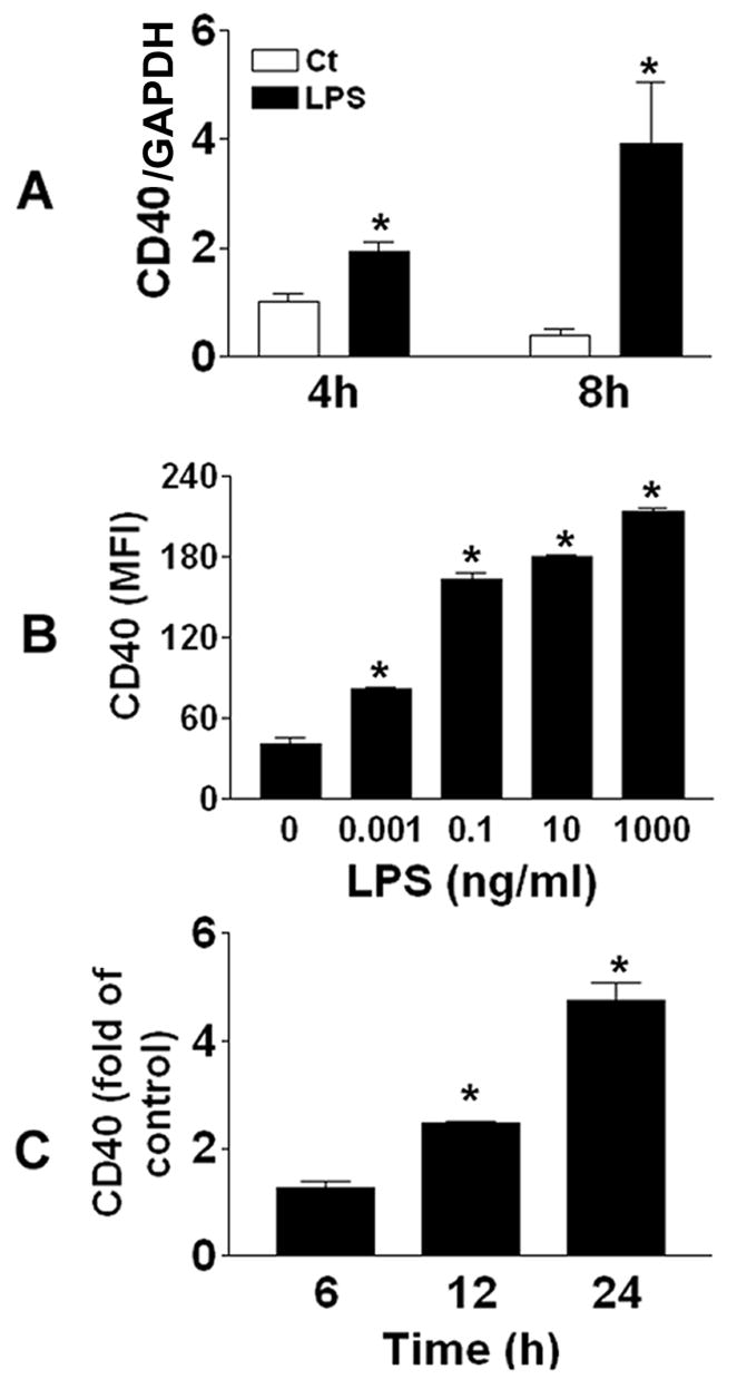
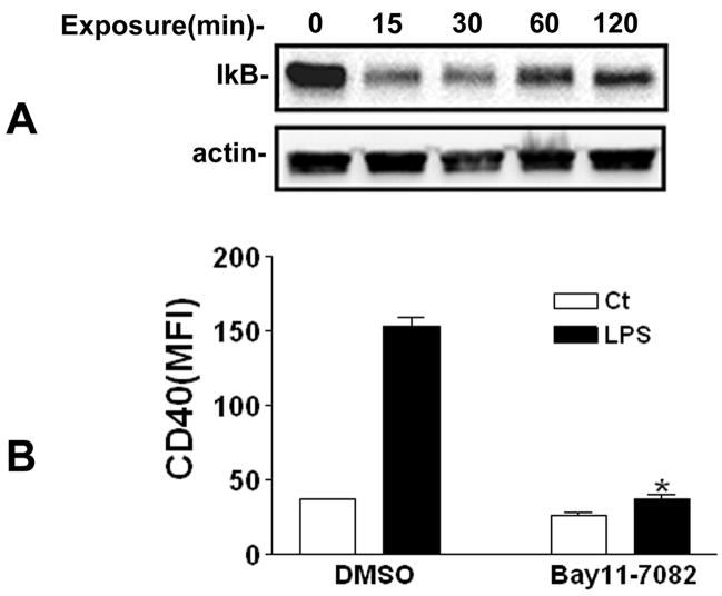
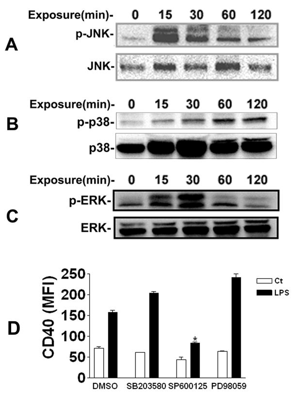
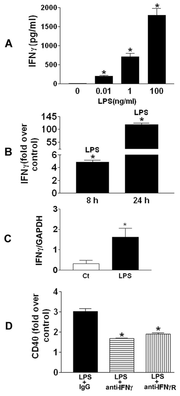
CD40 plays important roles in cell-mediated and humoral immune responses. In this study, we explored mechanisms underlying lipopolysaccharide (LPS)-induced CD40 expression in purified human peripheral blood monocytic cells (PBMCs) from healthy volunteers. Exposure to LPS induced increases in CD40 mRNA and protein expression on PBMCs. LPS stimulation caused IkappaBalpha degradation. Inhibition of NFkappaB activation abrogated LPS-induced CD40 expression. LPS stimulation also resulted in phosphorylation of mitogen-activated protein kinases, however, only Jun N-terminal kinase (JNK) was partially involved in LPS-induced CD40 expression. In addition, LPS exposure resulted in elevated interferon gamma (IFNgamma) levels in the medium of PBMCs. Neutralization of IFNgamma and IFNgamma receptor using specific antibodies blocked LPS-induced CD40 expression by 44% and 37%, respectively. In summary, LPS-induced CD40 expression on human PBMCs through activation of NFkappaB and JNK, and partially through the induction of IFNgamma production.
Figures
Similar articles
-
Involvement of mitogen-activated protein kinases and NFkappaB in LPS-induced CD40 expression on human monocytic cells.Toxicol Appl Pharmacol. 2008 Apr 15;228(2):135-43. doi: 10.1016/j.taap.2007.12.002. Epub 2007 Dec 14. Toxicol Appl Pharmacol. 2008. PMID: 18187173 Free PMC article.
-
Distinct role of p38 and c-Jun N-terminal kinases in IL-10-dependent and IL-10-independent regulation of the costimulatory molecule B7.2 in lipopolysaccharide-stimulated human monocytic cells.J Immunol. 2002 Feb 15;168(4):1759-69. doi: 10.4049/jimmunol.168.4.1759. J Immunol. 2002. PMID: 11823508
-
Regulatory role of cytosolic phospholipase A2 alpha in the induction of CD40 in microglia.J Neuroinflammation. 2017 Feb 10;14(1):33. doi: 10.1186/s12974-017-0811-z. J Neuroinflammation. 2017. PMID: 28187742 Free PMC article.
-
Intravenous immunoglobulin preparation attenuates LPS-induced production of pro-inflammatory cytokines in human monocytic cells by modulating TLR4-mediated signaling pathways.Naunyn Schmiedebergs Arch Pharmacol. 2012 Sep;385(9):891-8. doi: 10.1007/s00210-012-0765-8. Epub 2012 May 31. Naunyn Schmiedebergs Arch Pharmacol. 2012. PMID: 22644107
-
CD40 activates NF-kappa B and c-Jun N-terminal kinase and enhances chemokine secretion on activated human hepatic stellate cells.J Immunol. 2001 Jun 1;166(11):6812-9. doi: 10.4049/jimmunol.166.11.6812. J Immunol. 2001. PMID: 11359840
Cited by
-
Biological Activity of Masked Endotoxin.Sci Rep. 2017 Mar 20;7:44750. doi: 10.1038/srep44750. Sci Rep. 2017. PMID: 28317862 Free PMC article.
-
Characterization of a Broadly Reactive Anti-CD40 Agonistic Monoclonal Antibody for Potential Use as an Adjuvant.PLoS One. 2017 Jan 20;12(1):e0170504. doi: 10.1371/journal.pone.0170504. eCollection 2017. PLoS One. 2017. PMID: 28107431 Free PMC article.
-
Transcriptional splice variants of CD40 and its prognostic value in breast cancer.Turk J Biol. 2020 Apr 2;44(2):73-81. doi: 10.3906/biy-1912-21. eCollection 2020. Turk J Biol. 2020. PMID: 32256143 Free PMC article.
-
TLR7-Adjuvanted Ionizable Lipid Nanoparticles for mRNA Vaccine Delivery.AAPS J. 2025 Apr 25;27(4):80. doi: 10.1208/s12248-025-01073-2. AAPS J. 2025. PMID: 40281311
-
Human breast milk-derived exosomes attenuate lipopolysaccharide-induced activation in microglia.J Neuroinflammation. 2025 Feb 15;22(1):41. doi: 10.1186/s12974-025-03345-2. J Neuroinflammation. 2025. PMID: 39955566 Free PMC article.
References
-
- van Kooten C, Banchereau J. CD40-CD40 ligand. J Leukoc Biol. 2000;67:2–17. - PubMed
-
- Banchereau J, Bazan F, Blanchard D, Briere F, Galizzi JP, van Kooten C, Liu YJ, Rousset F, Saeland S. The CD40 antigen and its ligand. Annu Rev Immunol. 1994;12:881–922. - PubMed
-
- Benveniste EN, Nguyen VT, Wesemann DR. Molecular regulation of CD40 gene expression in macrophages and microglia. Brain Behav Immun. 2004;18:7–12. - PubMed
-
- Tone M, Tone Y, Babik JM, Lin CY, Waldmann H. The role of Sp1 and NF-kappa B in regulating CD40 gene expression. J Biol Chem. 2002;277:8890–8897. - PubMed
-
- Park JH, Chang HS, Park CS, Jang AS, Park BL, Rhim TY, Uh ST, Kim YH, Chung IY, Shin HD. Association analysis of CD40 polymorphisms with asthma and the level of serum total IgE. Am J Respir Crit Care Med. 2007;175:775–782. - PubMed
Publication types
MeSH terms
Substances
Grants and funding
LinkOut - more resources
Full Text Sources
Research Materials
Miscellaneous
