. 2009 Mar;1793(3):489-95.
doi: 10.1016/j.bbamcr.2008.11.017. Epub 2008 Dec 11.

A novel mode of translocation for cytolethal distending toxin

Affiliations

A novel mode of translocation for cytolethal distending toxin

Lina Guerra et al. Biochim Biophys Acta. 2009 Mar.

Abstract

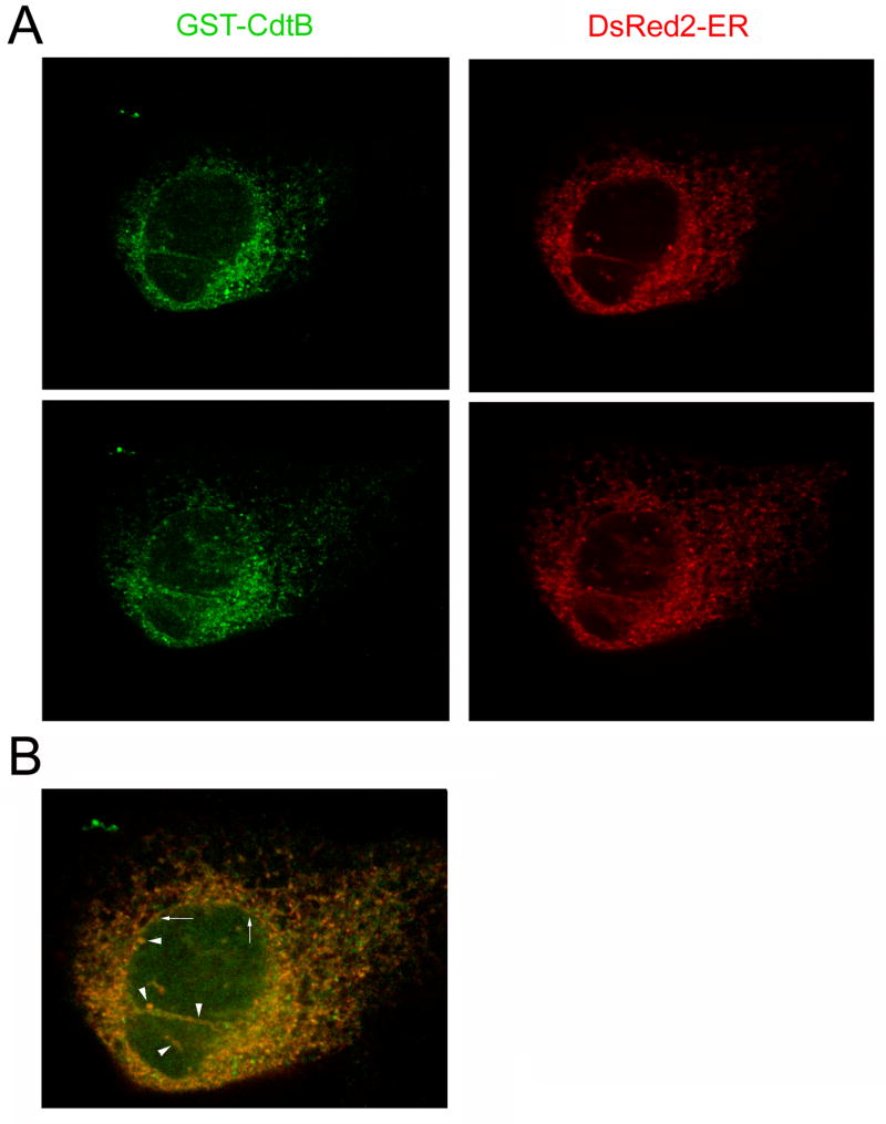
Thermal instability in the toxin catalytic subunit may be a common property of toxins that exit the endoplasmic reticulum (ER) by exploiting the mechanism of ER-associated degradation (ERAD). The Haemophilus ducreyi cytolethal distending toxin (HdCDT) does not utilize ERAD to exit the ER, so we predicted the structural properties of its catalytic subunit (HdCdtB) would differ from other ER-translocating toxins. Here, we document the heat-stable properties of HdCdtB which distinguish it from other ER-translocating toxins. Cell-based assays further suggested that HdCdtB does not unfold before exiting the ER and that it may move directly from the ER lumen to the nucleoplasm. These observations suggest a novel mode of ER exit for HdCdtB.


Figures

Figure 1
Figure 1
HdCdtB thermal stability. Temperature-induced changes to the structure of HdCdtB were monitored by fluorescence spectroscopy (A–B) and far-UV CD (C). Both measurements were conducted near-simultaneously on the same sample after equilibration at each temperature for 3 min. Three scans per spectra were collected and averaged to improve the signal-to-noise ratio. (A): The change in color from blue to red corresponds to a stepwise increase in temperature from 18°C to 60°C. Dotted lines are spectra of samples cooled to 50°C, 40°C, 37°C, 25°C, and 18°C after HdCdtB was heated to 60°C; colors indicate the same temperatures as the solid lines. (B): The maximum emission wavelengths (λmax) from panel A were plotted as a function of temperature. Data points from the cooled HdCdtB spectra are presented as open circles. (C): For far-UV CD analysis, the mean residue molar ellipticities at 220 nm ([θ]220) were plotted as a function of temperature. Data points from the cooled HdCdtB spectra are presented as open circles. The solid lines in panels B and C are best fit curves simulated as described in Ref. [11].
Figure 2
Figure 2
HdCdtB proteolysis. (A): HdCdtB or the CTA1/CTA2 heterodimer was placed in 20 mM Na-phosphate buffer (pH 7.0) containing 10 mM β-mercaptoethanol. After incubation at the indicated temperatures for 45 min, thermolysin was added for an additional 45 min at 4ºC. Proteolysis was halted by the addition of EDTA and sample buffer. The toxins were then visualized by SDS-PAGE and Coomassie staining. For both gels, the upper thermolysin band is denoted with an asterisk. (B): HdCdtB or the reduced CTA1/CTA2 heterodimer was placed in assay buffer with 100 nM of the 20S proteasome. Proteolysis was halted after 0, 4, 8, or 20 h of incubation at 37ºC. The toxins were then visualized by SDS-PAGE and Coomassie staining.
Figure 3
Figure 3
HdCDT toxicity. (A–B): HeLa cells were incubated with no toxin or with 2 μg/ml of HdCDT for 2 h in medium alone (control) or medium supplemented with 10% glycerol. As indicated, one set of cells were pretreated with 10% glycerol medium for 1 hr before a further 2 h incubation with glycerol in the absence (no toxin) or presence of HdCDT. (A): Toxin-induced phosphorylation of histone H2AX was visualized in fixed cells with a rabbit anti-phospho-H2AX antibody and a FITC-conjugated swine anti-rabbit IgG antibody. One of two experiments is shown. (B): The graph charts the average percentage (± range) of nuclei positive for H2AX foci from both experiments. (C): HeLa cells were incubated with various concentrations of ricin for 4 h in the absence or presence of 10% glycerol before protein synthesis levels were quantified. Measurements taken from unintoxicated cells incubated in the absence or presence of glycerol were used to establish the 100% value for the corresponding experimental condition. The average ± range of two independent experiments with triplicate samples is shown.
Figure 4
Figure 4
HdCdtB translocation. (A): Purified GST-HdCdtB-CVIM was incubated in the absence or presence of rabbit reticulocyte lysate before placement in cold 1% Triton X-114. Aqueous (A) and detergent (D) phases of Triton X-114 were isolated by centrifugation of the warmed samples. The distribution of GST-HdCdtB-CVIM was then determined by Western blot analysis of the separate detergent and aqueous phases. (B): For in vivo farnesylation of GST-HdCdtB-CVIM, HeLa cells were exposed for 4 h to 50μg/ml of a recombinant CDT holotoxin that contained the GST-HdCdtB-CVIM subunit. After lysis in 1% Triton X-114, aqueous (A) and detergent (D) phases were separated. GST-HdCdtB-CVIM was recovered by affinity purification and visualized by Western-blot analysis as described in Materials and methods.
Figure 5
Figure 5
Intracellular distribution of HdCdtB. Transfected HeLa cells expressing DsRed2-ER, a fluorescent marker for the ER, were exposed for 4 h to 10 μg/ml of a recombinant CDT holotoxin that contained a GST-HdCdtB subunit. The cells were then fixed and stained with a fluorescein-conjugated anti-GST antibody. (A): Labeling patterns for DsRed2-ER and GST-CdtB from two Z-sections of a single cell. (B): The merged image of DsRed2-ER and GST-CdtB distributions. Arrows denote the nuclear envelope; arrowheads denote invaginations of the nucleoplasmic reticulum.

References

    1. Thelestam M, Frisan T. Cytolethal distending toxins. In: Alouf JE, Popoff MR, editors. The Comprehensive Sourcebook of Bacterial Protein Toxins. Academic Press; San Diego: 2006. pp. 448–467.
    1. Cortes-Bratti X, Chaves-Olarte E, Lagergard T, Thelestam M. Cellular internalization of cytolethal distending toxin from Haemophilus ducreyi. Infect Immun. 2000;68:6903–6911. - PMC - PubMed
    1. Guerra L, Teter K, Lilley BN, Stenerlow B, Holmes RK, Ploegh HL, Sandvig K, Thelestam M, Frisan T. Cellular internalization of cytolethal distending toxin: a new end to a known pathway. Cell Microbiol. 2005;7:921–934. - PubMed
    1. Hazes B, Read RJ. Accumulating evidence suggests that several AB-toxins subvert the endoplasmic reticulum-associated protein degradation pathway to enter target cells. Biochemistry. 1997;36:11051–11054. - PubMed
    1. Lord JM, Roberts LM, Lencer WI. Entry of protein toxins into mammalian cells by crossing the endoplasmic reticulum membrane: co-opting basic mechanisms of endoplasmic reticulum-associated degradation. Curr Top Microbiol Immunol. 2005;300:149–168. - PubMed

Publication types

Substances

LinkOut - more resources