Comparative Study
. 2009 Jan 13;19(1):9-19.
doi: 10.1016/j.cub.2008.12.006.

Three distinct condensin complexes control C. elegans chromosome dynamics

Affiliations
Comparative Study

Three distinct condensin complexes control C. elegans chromosome dynamics

Gyorgyi Csankovszki et al. Curr Biol. .

Erratum in

  • Curr Biol. 2009 Jan 27;19(2):176. Sarkesik, Ali [corrected to Sarkeshik, Ali]

Abstract

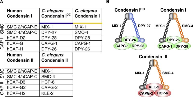
Background: Condensin complexes organize chromosome structure and facilitate chromosome segregation. Higher eukaryotes have two complexes, condensin I and condensin II, each essential for chromosome segregation. The nematode Caenorhabditis elegans was considered an exception, because it has a mitotic condensin II complex but appeared to lack mitotic condensin I. Instead, its condensin I-like complex (here called condensin I(DC)) dampens gene expression along hermaphrodite X chromosomes during dosage compensation.

Results: Here we report the discovery of a third condensin complex, condensin I, in C. elegans. We identify new condensin subunits and show that each complex has a conserved five-subunit composition. Condensin I differs from condensin I(DC) by only a single subunit. Yet condensin I binds to autosomes and X chromosomes in both sexes to promote chromosome segregation, whereas condensin I(DC) binds specifically to X chromosomes in hermaphrodites to regulate transcript levels. Both condensin I and II promote chromosome segregation, but associate with different chromosomal regions during mitosis and meiosis. Unexpectedly, condensin I also localizes to regions of cohesion between meiotic chromosomes before their segregation.

Conclusions: We demonstrate that condensin subunits in C. elegans form three complexes, one that functions in dosage compensation and two that function in mitosis and meiosis. These results highlight how the duplication and divergence of condensin subunits during evolution may facilitate their adaptation to specialized chromosomal roles and illustrate the versatility of condensins to function in both gene regulation and chromosome segregation.


Figures

Figure 1
Figure 1. The Three-Condensin Model
(A) C. elegans condensin subunits and their human homologs. We propose that C. elegans has three condensin complexes of the composition shown. Condensin I and II share the same SMC2 and SMC4 subunits but associate with either class I or class II CAP subunits, respectively. Condensin IDC differs from condensin I only by its unique SMC4 subunit, DPY-27. (B) Cartoon depicting the three proposed complexes.
Figure 2
Figure 2. Condensin Subunit Interactions Identified by Immunoprecipitation
(A) Immunoprecipitation (IP) from embryo extracts with antibodies against DPY-27, SMC-4, KLE-2, CAPG-1, and KLP-7 (control) were analyzed by MudPIT mass spectrometry. Numbers indicate the total mass spectra collected in three (KLE-2) or four (all others) IP samples corresponding to each protein indicated on the left. (B and C) IP reactions from embryo extracts with antibodies against condensin subunits were performed and analyzed on western blots probed with an array of antibodies. Preimmune IP (Pre-im) is shown as control. Selected western blots are shown in (B); summary of all IPs analyzed is shown in (C). Plus sign indicates subunit was detected in the IP; minus sign indicates subunit was not detected in the IP; blank cell indicates interaction was not scored.
Figure 3
Figure 3. Condensin I Localizes to Mitotic Chromosomes in a Pattern Distinct from Condensin IDC or Condensin II
DPY-27 antibody was used to indicate the subcellular localization of condesin IDC, anti-CAPG-1 to indicate both condensin I and condensin IDC, and anti-KLE-2 to indicate condensin II. Condensin IDC does not associate with mitotic chromosomes in early-stage hermaphrodite embryos (A) or in male embryos (D), but associates with the two hermaphrodite X chromosomes after the onset of dosage compensation, both during interphase (B) and during mitosis (C). CAPG-1 staining not shared by DPY-27 indicates that condensin I localizes to mitotic chromosomes in both hermaphrodites (E) and males (H). Once dosage compensation initiates in hermaphrodite embryos, CAPG-1 localizes to the X chromosomes in interphase, as part of condensin IDC (F). During mitosis, CAPG-1 localizes to two bright X foci (as part of condensin IDC) and less intensely to other chromosomes (as part of condensin I) (G). Condensin II shows no distinct pattern during interphase (J), and a centromere-like pattern on mitotic chromosomes in both hermaphrodites (I, K) and males (L). Antibody in green, DNA in red, merge in yellow.
Figure 4
Figure 4. Condensin I Localizes to Meiotic Chromosomes in a Pattern Distinct from Condensin IDC or Condensin II
(A) DPY-27 antibody was used to indicate the localization of condesin IDC, anti-CAPG-1 to indicate condensin I and condensin IDC, and anti-KLE-2 to indicate condensin II. DPY-27 was detected in the nucleoplasm of mature oocytes, but not at other stages of meiosis. CAPG-1 staining indicates that condensin I surrounds chromosomes in prophase then localizes to the interface between homologs (meiosis I) or sister chromatids (meiosis II). In contrast, KLE-2 staining indicates that condensin II localizes to the core of each sister chromatid. Antibody in green, DNA in red, merge in yellow. (B) MIX-1 localization is a hybrid of condensin I and condensin II patterns. MIX-1 (green) partially colocalizes with CAPG-1 (red, representing condensin I) on sperm chromosomes (blue). MIX-1 and CAPG-1 colocalize surrounding chromosomes in meiosis prophase I, between homologs in metaphase I, and between sisters at metaphase II (filled arrows). In addition, MIX-1 localizes to the chromosome core at each stage (open arrows), a pattern resembling condensin II and not shared by CAPG-1.
Figure 5
Figure 5. CAPG-1 Functions in Dosage Compensation
(A) CAPG-1 (green) localizes to X chromosomes in wild-type hermphrodites, but not in animals carrying mutated subunits of condensin IDC. (B) Condensin IDC subunits (green) localize to hermaphrodite X chromosomes in control vector RNAi animals, but not in animals fed RNAi food targeting capg-1. Shown are intestinal nuclei (red) of adult hermaphrodites. (C) Genetic assays testing dosage compensation function. Feeding RNAi targeting condensin IDC subunits was minimally lethal in wild-type embryos, and enhanced lethality in the sex-1 mutant background. By contrast, xol-1 sex-1 males die because of inappropriate activation of dosage compensation, but can be rescued by RNAi depletion of a condensin IDC subunit. In both assays, capg-1 RNAi yields results similar to RNAi of other condensin IDC subunits. Percent lethality was calculated as dead progeny/total progeny × 100. Male rescue was calculated as number of males/expected number of males × 100. At least 200 animals were scored for RNAi of each gene.
Figure 6
Figure 6. Depleting Each Condensin II Subunit Results in Common Chromosomal and Developmental Defects
(A) Images from time-lapse movies of control or capg-2 RNAi-depleted worms carrying a GFP::histone H2B transgene. At the first embryonic mitosis, capg-2-depleted chromosomes fail to condense into distinct rod shapes during prophase (left). At anaphase, capg-2-depleted chromosomes fail to separate completely and abnormal DNA connections are observed (middle). Separating chromatids during meiosis anaphase II also show abnormal DNA connections in capg-2-depleted animals (right). (B) Wild-type or homozygous condensin II subunit mutant adult hermaphrodites, stained with DNA dye. Gut nuclei (top row) are separated in wild-type, but often connected in condensin II subunit mutants. Germline nuclei (bottom row) are uniformly sized and evenly spaced in wild-type. Condensin II subunit mutants have fewer germline nuclei, which are abnormally sized and unevenly distributed.
Figure 7
Figure 7. Condensin I Is Required for Mitotic and Meiotic Chromosome Segregation
(A) Gut nuclei from adults raised since hatching on bacteria expressing control vector or dsRNA against the subunit indicated. Gut nuclei (stained for DNA) are normally separated in control or dpy-27 RNAi-treated animals, but abnormally connected after depleting condensin I subunits dpy-28, capg-1, and dpy-26. (B) Quantification of defects shown in (A), scored in hermaphrodites (magenta), males (blue), and a mutant situation with half XO hermaphrodites and half XX hermaphrodites (tra-2(e2531), purple). Percentage calculated as the number of gut nuclei with an obvious connection/total gut nuclei × 100. Standard deviation from three experiments is shown. In comparison to control vector by Fisher's exact test, dpy-27 p > 0.2 in each sex (not significant); for dpy-28, capg-1, and dpy-26 each p < 0.0001 (significant). (C) Still images from time-lapse movies of the first mitosis in F2 embryos after two generations of RNAi feeding in a GFP:histone H2B strain. Chromosome segregation errors characterized by DNA strands between separating sets of chromosomes were observed with dpy-28, capg-1, and dpy-26 RNAi but not with control or dpy-27 RNAi depletion. (D) FISH analysis of nuclei from mid-stage (about 100-cell) embryos (top) or adult nerve cord cells (bottom) with a 5S rDNA probe. After two generations of RNAi feeding, progeny of animals fed control vector contain diploid nuclei with two signals. Only 3 of 15 vector-treated embryos contained nuclei with more than two spots, and in each case, it was limited to a single nucleus within the embryo. By contrast, 14 of 16 capg-1 RNAi-fed progeny contained aneuploid nuclei with more than two signals (arrow). In adults, nuclei in control worms had two signals, indicating diploidy. Only 1 of 8 control-treated animals had a nucleus with more than two signals. By contrast, 12 of 13 capg-1 RNAi-treated worms had nuclei with multiple signals, indicating aneuploidy (arrow). (E) Meiosis II after two generations of RNAi feeding in a GFP:histone H2B strain. The two meiotic divisions produce two small polar bodies and a separated oocyte pronucleus in control-treated animals. When condensin I subunit DPY-26 is depleted, the second polar body often fails to segregate from the oocyte pronucleus.

Similar articles

Cited by

References

    1. Belmont AS. Mitotic chromosome structure and condensation. Curr. Opin. Cell Biol. 2006;18:632–638. - PubMed
    1. Hirano T. Condensins: organizing and segregating the genome. Curr. Biol. 2005;15:R265–R275. - PubMed
    1. Hirota T, Gerlich D, Koch B, Ellenberg J, Peters JM. Distinct functions of condensin I and II in mitotic chromosome assembly. J. Cell Sci. 2004;117:6435–6445. - PubMed
    1. Ono T, Fang Y, Spector DL, Hirano T. Spatial and temporal regulation of Condensins I and II in mitotic chromosome assembly in human cells. Mol. Biol. Cell. 2004;15:3296–3308. - PMC - PubMed
    1. Ono T, Losada A, Hirano M, Myers MP, Neuwald AF, Hirano T. Differential contributions of condensin I and condensin II to mitotic chromosome architecture in vertebrate cells. Cell. 2003;115:109–121. - PubMed

Publication types

MeSH terms