. 2009 Jan 16;30(1):92-107.
doi: 10.1016/j.immuni.2008.11.005.

Late developmental plasticity in the T helper 17 lineage

Affiliations

Late developmental plasticity in the T helper 17 lineage

Yun Kyung Lee et al. Immunity. .

Abstract

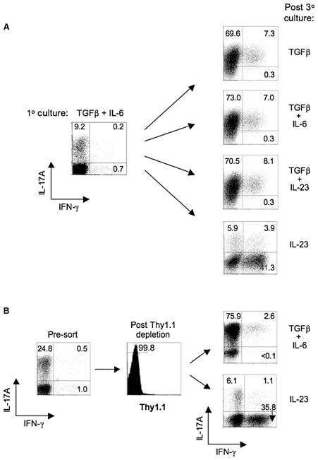
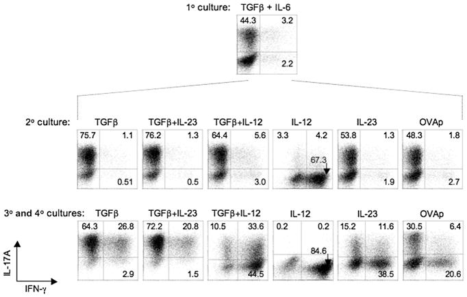
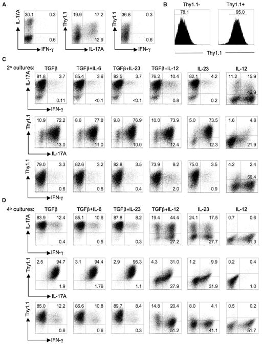
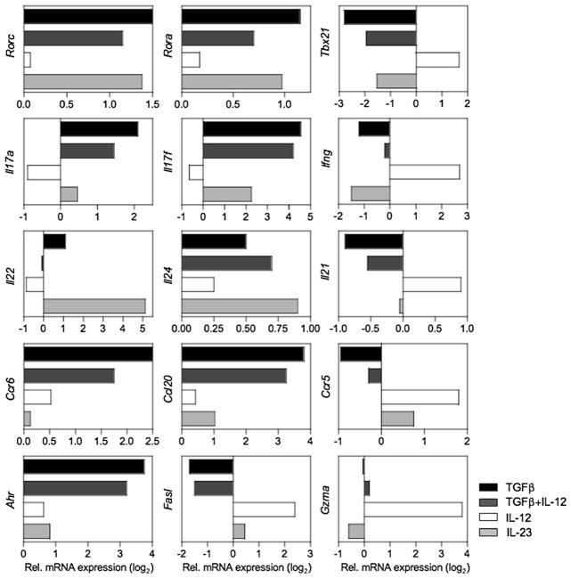
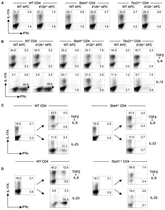
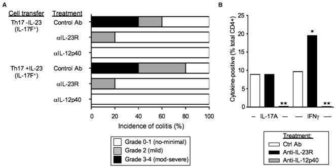
Development of T helper (Th) 17 cells requires transforming growth factor (TGF)-beta and interleukin (IL)-6 and is independent of the Th1 pathway. Although T cells that produce interferon (IFN)-gamma are a recognized feature of Th17 cell responses, mice deficient for STAT4 and T-bet-two prototypical Th1 transcription factors-are protected from autoimmunity associated with Th17 pathogenesis. To examine the fate and pathogenic potential of Th17 cells and origin of IFN-gamma-producing T cells that emerge during Th17 immunity, we developed IL-17F reporter mice that identify cells committed to expression of IL-17F and IL-17A. Th17 cells required TGF-beta for sustained expression of IL-17F and IL-17A. In the absence of TGF-beta, both IL-23 and IL-12 acted to suppress IL-17 and enhance IFN-gamma production in a STAT4- and T-bet-dependent manner, albeit with distinct efficiencies. These results support a model of late Th17 developmental plasticity with implications for autoimmunity and host defense.


Figures

Figure 1
Figure 1. Requirement for TGF-β in the Maintenance of IL-17 Expression by Th17-Polarized Cells
(A) FACS-sorted naive CD4+ T cells from OT-II TCR transgenic mice were cultured with irradiated Il12b−/− (IL-12p40-deficient) splenic feeder cells and 5 μg/ml OVAp for 7 days under Th17-polarizing conditions (TGF-β, 5 ng/ml; IL-6, 20 ng/ml; anti-IFN-γ, 10 μg/ml; anti-IL-4, 10 μg/ml), then stained intracellularly for IL-17A and IFN-γ after PMA/ionomycin activation for 5 hr in the presence of monensin (1° culture; left panel). Cells were harvested and restimulated with irradiated Il12b−/− splenic feeder cells in the presence of indicated cytokine(s) at the same doses or, in the case of IL-23 and IL-12, at 1 ng/ml, with anti-IFN-γ, anti-IL-4, and OVAp for an additional two rounds (7 days each) and stained intracelluarly for IL-17A and IFN-γ after PMA-ionomycin activation (post 3° culture; right panels). (B) IfngThy1.1/Thy1.1 OT-II naive CD4+ T cells were cultured with irradiated Il12b−/− splenic feeder cells under Th17-polarizing conditions for 6 days and a fraction of the recovered cells were stained intracellularly for IL-17A and IFN-γ after PMA-ionomycin activation (left panel). A second fraction of recovered CD4+ T cells were activated for 5 hr with OVAp and Il12b−/− feeder cells, and Thy1.1+ (IFN-γ+) cells were depleted by magnetic sorting. Thy1.1 depletion was confirmed by surface staining for Thy1.1 (middle panel), and the isolated Thy1.1- cells were cultured with Il12b−/− feeder cells and OVAp and either TGF-β plus IL-6 or IL-23 in the presence of anti-IFNγ plus anti-IL-4 for three additional rounds (right panels), then stained intracellulary for IL-17A and IFN-γ after PMA-ionomycin activation. Numbers in each quadrant indicate percentages of total CD4+ T cells. All data are representative of at least three independent experiments.
Figure 2
Figure 2. Th17 Cells Respond to IL-12 to Rapidly Upregulate IFN-γ and Extinguish IL-17
OT-II CD4+ T cells were cultured with irradiated Il12b−/− splenic feeder cells under Th17-polarizing condition for 7 days (1° culture, top panel), and a fraction of recovered T cells were restimulated and analyzed for intracellular IL-17A and IFN-γ as in Figure 1. The remaining T cells were cultured again with irradiated Il12b−/− splenic feeder cells in the presence of indicated cytokine(s), anti-IFNγ, anti-IL-4, and OVAp as in Figure 1 for 7 days (2° culture, middle panel), and a fraction was reanalyzed for intracellular cytokine expression. The remaining recovered cells from secondary cultures were recultured under identical cytokine conditions for a third and fourth round (3° and 4° cultures, bottom panel), then analyzed for intracellular IL-17A and IFN-γ after PMA-ionomycin activation as before. Numbers in quadrants indicate percentage of total CD4+ cells in each. Data are representative of at least three experiments.
Figure 3
Figure 3. IFN-γ-Producing Effectors Arise from IL-17-Producing Precursors
(A) FACS-sorted naive CD4+ T cells from Il17fThy1.1/Thy1.1 reporter mice were cultured with irradiated Il12b−/− splenic feeder cells under Th17-polarizing conditions for 6 days as in Figure 1. A fraction of recovered cells were analyzed for expression of CD4, Thy1.1 (IL-17F), and intracellular cytokine IL-17A and IFN-γ after PMA-ionomycin stimulation for 5 hr in the presence of monensin. The flow-cytometric plots are gated on CD4+ cells. (B) Cells from (A) were collected and Thy1.1+ (IL-17F+) cells isolated by magnetic sorting. The purity of sorted Thy1.1+ cells was confirmed by surface staining of Thy1.1. Numbers in the histograms are the fraction of CD4+ T cells in the indicated gates. (C and D) Thy1.1+ cells were restimulated with irradiated Il12b−/− splenic feeder cells in the presence of indicated cytokine(s), anti-IFNγ, anti-IL-4, and anti-CD3 as in Figure 1 for a second (C) and fourth round (D) and were stained for CD4, Thy1.1 (IL-17F), and intracellular IL-17A and IFN-γ after PMA-ionomycin activation for 5 hr. Numbers in each quadrant indicate the percentage of total CD4+ T cells. Data are representative of at least three independent experiments.
Figure 4
Figure 4. Divergence of Transcriptomes of Th17 Progeny Restimulated under Different Cytokine Conditions
FACS-sorted naive CD4+ T cells from Il17fThy1.1/Thy1.1 mice were cultured with irradiated Il12b−/− splenic feeder cells under Th17-polarizing conditions for 6 days. Cells were collected and Thy1.1+ (IL-17F+) cells were isolated by magnetic sorting as in Figure 3. Isolated Thy1.1+ cells were restimulated with anti-CD3 and anti-CD28 in the presence of indicated cytokine(s) and processed for mRNA quantification of the indicated genes at 6 hr (Fasl and Gzma) or 18 hr after stimulation by real-time PCR. All data were normalized to 18S RNA and are expressed as fold differences (log2) relative to anti-CD3- and anti-CD28-stimulated cells. Data are representative of two or three experiments.
Figure 5
Figure 5. Transition of Th17 Precursors into IFN-γ-Producing Cells Is Stat4 and T-bet Dependent
(A) CD4+ T cells from WT (BALB/c), Stat4−/−, or Tbx21−/− mice were cultured with either WT or Il12b−/− splenic feeder cells and anti-CD3 under Th17-polarizing conditions for 7 days, and then a fraction was analyzed for CD4 and intracellular IL-17A and IFN-γ after PMA-ionomycin stimulation for 5 hr. (B) Recovered CD4+ T cells were restimulated with either irradiated WT or Il12b−/− splenic feeder cells in the presence of indicated cytokine(s), anti-IFNγ, anti-IL-4, and anti-CD3 for an additional 5 days and analyzed as in (A). (C and D) Naive CD4+ T cells from BALB or B6 WT, BALB.Stat4−/−, or B6.Tbx21−/− mice were cultured as in (A) for one round, a fraction was analyzed for CD4 and intracellular cytokines (IL-17A and IFN-γ) as in (A), and the remainder was recultured for three additional rounds under the indicated cytokine conditions as in (B). After the third round of culture, recovered BALB.WT and BALB.Stat4−/− CD4+ T cells ([C], left and right panels) or B6.WT and B6.Tbx21−/− ([D], left and right panels) were activated with PMA-ionomycin and analyzed for CD4 and intracellular IL-17A and IFN-γ. Numbers in quadrants indicate percentages of total CD4+ T cells in each. Data are representative of at least two independent experiments.
Figure 6
Figure 6. A Subset of Th17 Cells Extinguish IL-17 and Transition to Single-Positive IFN-γ Cells in Transfer Model of Colitis
(A and B) FACS-sorted CD4+CD45RBhi T cells (4 ×10⁵) or CD4+CD45RBlo (2 ×10⁵) T cells isolated from pooled spleen and LNs of Il17fThy1.1/Thy1.1 mice were transferred into congenic Rag1−/− mice as indicated. Th17-polarized T cells were generated from naive Il17fThy1.1/Thy1.1 CD4+ T cells with Il12b−/− APCs and anti-CD3 (2.5 μg/ml) as in Figure 1, in the absence (Th17 − IL-23) or presence (Th17 + IL-23) of 10 ng/ml IL-23. Thy1.1 (IL-17F ) and Thy1.1+ (IL-17F+) cells were isolated by FACS sorting (see Figure S8), and 3 ×10⁵ cells were transferred as indicated. As shown in (A), 8 weeks after transfer, mice were sacrificed and intestinal tissues were collected for histological processing and analysis. Shown are representative sections of colon from indicated experimental groups; all magnifications represent 40×. (B) shows the incidence and severity of colitis 4 (upper panel) or 8 (lower panel) weeks after transfer of the indicated cell populations. n represents the number of recipients in each group. (C and D) IL-17A, Thy1.1 (IL-17F), and IFN-γ expression of CD4+ T cells isolated from MLNs of indicated recipient mice 4 weeks (upper two panels) or 8 weeks (lower panel) after cell transfer. Recovered cells were stimulated ex vivo with PMA-ionomycin and analyzed for CD4, Thy1.1, and intracellular IL-17A and IFN-γ. Numbers in each quadrant indicate percentages of total CD4+ T cells (C). Cumulative data for frequencies of cytokine-positive CD4+ T cells, as analyzed as in (C), are shown in (D). Data are the means ± SEM for tissues and groups indicated. All data are representative of results from five similar experiments.
Figure 7
Figure 7. Effects of IL-23 and IL-12 Blockade on Th17 Precursors in Transfer Model of Colitis
(A) Th17-polarized T cells were generated from naive Il17fThy1.1/Thy1.1 CD4+ T cells with Il12b−/− APCs and anti-CD3 (5 μg/ml) as in Figure 1, in the absence (Th17 -IL-23) or presence (Th17 +IL-23) of 5 ng/ml IL-23. Thy1.1+ (IL-17F+) cells were isolated by FACS sorting and 3 × 10⁵ cells were transferred into congenic Rag1−/− mice as indicated. Recipients were administered 100 μg of anti-IL-23R, anti-IL-12p40, or isotype control via i.p. injection in saline 1 day prior and 1 day after T cell transfers and once weekly thereafter (five total doses). Mice were sacrificed 4 weeks after transfer and analyzed histologically and scored for development of colitis. (B) Mice that received Th17 -IL-23 (IL-17F+) T cells and treated as in (A) were sacrificed 4 weeks after transfer, and cells recovered from MLNs were stimulated ex vivo with PMA ionomycin and analyzed for CD4 and intracellular IL-17A and IFN-γ by FACS. Data are the means from five mice in each treatment group. *p < 0.05 and **p < 0.01 versus control treatment group. Data are representative of two similar experiments.

Comment in

References

    1. Aggarwal S, Ghilardi N, Xie MH, de Sauvage FJ, Gurney AL. Interleukin-23 promotes a distinct CD4 T cell activation state characterized by the production of interleukin-17. J Biol Chem. 2003;278:1910–1914. - PubMed
    1. Annunziato F, Cosmi L, Santarlasci V, Maggi L, Liotta F, Mazzinghi B, Parente E, Fili L, Ferri S, Frosali F, et al. Phenotypic and functional features of human Th17 cells. J Exp Med. 2007;204:1849–1861. - PMC - PubMed
    1. Batten M, Li J, Yi S, Kljavin NM, Danilenko DM, Lucas S, Lee J, de Sauvage FJ, Ghilardi N. Interleukin 27 limits autoimmune encephalomyelitis by suppressing the development of interleukin 17-producing T cells. Nat Immunol. 2006;7:929–936. - PubMed
    1. Becher B, Durell BG, Noelle RJ. Experimental autoimmune encephalitis and inflammation in the absence of interleukin-12. J Clin Invest. 2002;110:493–497. - PMC - PubMed
    1. Bettelli E, Carrier Y, Gao W, Korn T, Strom TB, Oukka M, Weiner HL, Kuchroo VK. Reciprocal developmental pathways for the generation of pathogenic effector TH17 and regulatory T cells. Nature. 2006;441:235–238. - PubMed

Publication types

Associated data