. 2009 Feb 27;284(9):5807-18.
doi: 10.1074/jbc.M807593200. Epub 2008 Dec 31.

A DNA polymerase-{alpha}{middle dot}primase cofactor with homology to replication protein A-32 regulates DNA replication in mammalian cells

Affiliations

A DNA polymerase-{alpha}{middle dot}primase cofactor with homology to replication protein A-32 regulates DNA replication in mammalian cells

Darren E Casteel et al. J Biol Chem. .

Abstract

alpha-Accessory factor (AAF) stimulates the activity of DNA polymerase-alpha.primase, the only enzyme known to initiate DNA replication in eukaryotic cells ( Goulian, M., Heard, C. J., and Grimm, S. L. (1990) J. Biol. Chem. 265, 13221-13230 ). We purified the AAF heterodimer composed of 44- and 132-kDa subunits from cultured cells and identified full-length cDNA clones using amino acid sequences from internal peptides. AAF-132 demonstrated no homologies to known proteins; AAF-44, however, is evolutionarily related to the 32-kDa subunit of replication protein A (RPA-32) and contains an oligonucleotide/oligosaccharide-binding (OB) fold domain similar to the OB fold domains of RPA involved in single-stranded DNA binding. Epitope-tagged versions of AAF-44 and -132 formed a complex in intact cells, and purified recombinant AAF-44 bound to single-stranded DNA and stimulated DNA primase activity only in the presence of AAF-132. Mutations in conserved residues within the OB fold of AAF-44 reduced DNA binding activity of the AAF-44.AAF-132 complex. Immunofluorescence staining of AAF-44 and AAF-132 in S phase-enriched HeLa cells demonstrated punctate nuclear staining, and AAF co-localized with proliferating cell nuclear antigen, a marker for replication foci containing DNA polymerase-alpha.primase and RPA. Small interfering RNA-mediated depletion of AAF-44 in tumor cell lines inhibited [methyl-(3)H]thymidine uptake into DNA but did not affect cell viability. We conclude that AAF shares structural and functional similarities with RPA-32 and regulates DNA replication, consistent with its ability to increase polymerase-alpha.primase template affinity and stimulate both DNA primase and polymerase-alpha activities in vitro.


Figures
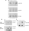
FIGURE 1.
FIGURE 1.
AAF-44 and AAF-132 mRNA expression in murine tissues and expression of Myc epitope-tagged AAF-44 and AAF-132 proteins. A, a Northern blot prepared with 2 μg of poly(A)+ RNA/lane was hybridized to radioactively labeled cDNA probes encoding murine AAF-132 (top panel), AAF-44 (middle panel), or human β-actin (lower panel). RNA molecular weight markers (in kb) are shown on the left. Lanes 1-8 show RNA isolated from adult murine heart (H), brain (B), spleen (Sp), lung (Lu), liver (Li), skeletal muscle (Sk), kidney (K), and testis (T). B, cDNA sequences encoding AAF-44 (lanes 2 and 4) and/or AAF-132 (lanes 3 and 4) with an N-terminal Myc epitope tag were subjected to in vitro transcription and translation in a coupled T7 RNA polymerase/reticulocyte lysate system using [35S]methionine. Lane 1 shows a control reaction with “empty vector” template. Aliquots of the reaction product were analyzed by SDS-PAGE/autoradiography (upper panel) and Western blotting using a Myc epitope-specific antibody (lower panel). C, 293T cells were transfected with equal amounts of expression vectors encoding Myc-tagged AAF-44 (lanes 1 and 3), AAF-132 (lanes 2 and 3), or empty vector (lane 4), and cell lysates were analyzed by SDS-PAGE/Western blotting using an anti-Myc antibody. AAF-132 appears to form high molecular weight aggregates that do not enter the gel properly (marked with an asterisk).
FIGURE 2.
FIGURE 2.
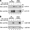
Association of AAF-44 with AAF-132 in intact cells. A, 293T cells were transfected with expression vectors encoding HA epitope-tagged AAF-44 and/or Myc epitope-tagged AAF-132 as indicated, and cell lysates were subjected to immunoprecipitation (IP) with anti-Myc antibody (lanes 1-3). Proteins present in the immunoprecipitates were analyzed by SDS-PAGE/Western blotting using an anti-HA epitope antibody (upper panel) or an anti-Myc epitope antibody (lower panel). Ten percent of total cell lysate input was analyzed in parallel (lanes 4-6). B, the experiment was similar to that described in A, but cell lysates were subjected to immunoprecipitation with anti-HA antibody (lanes 1-3).
FIGURE 3.
FIGURE 3.
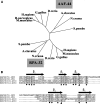
Phylogenetic relationship of AAF-44 and RPA-32 and alignment of AAF and RPA OB fold domains. A, AAF-44 sequences were aligned with RPA-32 sequences of human, rat, mouse, chicken, zebrafish, and fungal origin using the ClustalW program (25). An unrooted phylogenetic tree was drawnby the neighbor-joining method using the program Phylodendron. GenBank accession numbers for AAF-44 are as follows: Homo sapiens, NP_079204; Rattus norvegicus, NP_001011943; Mus musculus, NP_780569; Gallus gallus, XP_421742; Danio rerio, NP_956683; Neurospora crassa, XP_960343; Aspergillus clavatus, XP_001275592; and S. pombe, CAL44730. GenBank accession numbers for RPA-32 are as follows: H. sapiens, NP_002937; R. norvegicus, NP_067593; M. musculus, NP_035414; G. gallus, NP_001026063; D. rerio, NP_571786; N. crassa, XP_961967; A. clavatus, XP_001268210; and S. pombe, NP_588227. B, human AAF-44 was aligned with the sequences of the OB fold domains of human RPA-70 and RPA-32 using ClustalW with slight manual modifications; RPA-70 and RPA-32 were aligned according to published crystal structures, and regions of β-strands conserved among the OB folds are shown above as black arrows (28, 30). Numbers in parentheses refer to amino acid positions in the human sequences, and residues that are identical or conserved between AAF-44 and RPA are shaded in gray. Conserved hydrophobic residues in alternating amino acid positions within the β-sheets are indicated by black dots, and conserved glycine and aspartate residues are marked by their bold letter symbols. Two arrowheads indicate aromatic amino acid residues that are thought to be important for ssDNA binding of RPA-32 and are conserved in members of the S1 family of OB fold proteins (30).
FIGURE 4.
FIGURE 4.
ssDNA binding of AAF and effects of mutations in the putative OB fold domain of AAF-44. A, 293T cells were transfected with equal amounts of expression vectors encoding Myc epitope-tagged AAF-44 (lanes 1-3), Myc-tagged AAF-132 (lanes 7-9), or both (lanes 4-6), and 20, 60, or 200 μg of cell lysate protein (designated by the triangle above) were incubated with 10 pmol of biotinylated oligo(dC)30 as described under “Experimental Procedures.” Biotinylated oligo(dC)30 was isolated using streptavidin-agarose beads, and proteins bound to the washed beads were analyzed by SDS-PAGE/Western blotting with anti-Myc antibody. The arrows indicate migration of full-length AAF-132 and -44. B, cells were transfected with equal amounts of Myc-tagged AAF-44 and -132 (left panel) or with each subunit separately (right panels), and cell lysates (20 and 100 μg of protein in the left or right panels, respectively) were incubated with 30 pmol of biotinylated oligo(dC)30 in the absence (lane 1) and presence (lanes 2-5) of increasing amounts of non-biotinylated competitor (Comp.) oligo(dC) (90, 300, 900, and 3000 pmol) as indicated. Protein·DNA complexes were isolated on streptavidin-agarose beads and analyzed as described in A. C, cells were co-transfected with vectors expressing Myc-tagged AAF-132 and FLAG-tagged wild type AAF-44 (wt; lanes 1-3) or mutant AAF-44 containing alanine substitutions for Trp-96 and Phe-151 (mut; lanes 4-6). Binding to oligo(dC)30 was assessed as described in A except that bound proteins were detected by immunoblotting with anti-Myc plus anti-FLAG antibodies (upper panel). The lower panel shows a Western blot of cell lysates demonstrating similar amounts of wild type and mutant AAF-44 present in the lysates used for the DNA pulldown assay. D, three independent experiments were performed as described in C, and relative amounts of wild type versus mutant AAF-44 bound to oligo(dC)30 were quantified by densitometry scanning. Shown are the mean ± S.D.; asterisk, p < 0.05 for the comparison between wild type and mutant AAF-44.
FIGURE 5.
FIGURE 5.
DNA binding of AAF requires both subunits. A, 293T cells were co-transfected with FLAG-tagged AAF-44 and Myc-tagged AAF-132 (lanes 1-5) or received vector encoding AAF-44 plus empty vector (lanes 6-9). Cell lysate proteins were passed over an anti-FLAG antibody affinity matrix, and AAF-44 was eluted with FLAG peptide as described under “Experimental Procedures.” Analysis of eluates by SDS-PAGE/silver staining and Western blotting is shown in supplemental Fig. 2. Increasing amounts of the eluates containing purified FLAG-AAF-44 with (lanes 1-5) or without (lanes 6-9) co-purified Myc-AAF-132 were incubated with 30 pmol of biotinylated oligo(dC)30 probe as indicated by +. Protein·DNA complexes were isolated on streptavidin-agarose, and bound proteins were detected by Western blotting with anti-FLAG plus anti-Myc antibodies as described under “Experimental Procedures.” In lane 5, the eluate was incubated with streptavidin-agarose in the absence of biotinylated probe. The arrows indicate migration of full-length AAF-132 and -44. B, purified FLAG-tagged AAF-44 was incubated with cell lysates from 293T cells transfected with either empty vector (lane 1) or Myc-tagged AAF-132 (lane 3). Lane 2 shows the cell lysate containing AAF-132 in the absence of purified AAF-44. Proteins bound to biotinylated oligo(dC)30 were analyzed as described in A. C, FLAG-tagged AAF-44 and Myc-tagged AAF-132 were co-purified as described in A and incubated with increasing amounts of biotinylated oligo(dC)30 probe as indicated. Protein·DNA complexes were analyzed as described in A. D, the relative amounts of DNA-bound AAF-44 were determined by densitometry scanning of two independent experiments performed as described in C. A double reciprocal plot of 1/[bound AAF-44] versus 1/[probe] is shown (with the probe concentration in μm).
FIGURE 6.
FIGURE 6.
Recombinant AAF stimulates DNA primase activity. Immunoaffinity-purified FLAG-AAF-44 with (gray bars) or without (black bars) co-purified Myc-AAF-132 was incubated with 0.1 unit of purified pol-α·primase complex, and primase activity was determined as described under “Experimental Procedures.” Primase activity measured in the presence of an equal volume of FLAG peptide-containing elution buffer was assigned a relative value of 1. Asterisk, p < 0.05 for the comparison between AAF-44 and the AAF-44·AAF-132 complex. conc., concentrated; dil, diluted. Shown are the means ± S.D. of three experiments.
FIGURE 7.
FIGURE 7.
Subcellular localization of AAF subunits and co-localization with PCNA. A, HeLa cells were co-transfected with expression vectors encoding HA epitope-tagged AAF-44 and Myc epitope-tagged AAF-132 and analyzed by double immunofluorescence staining as described under “Experimental Procedures.” B, HeLa cells were co-transfected with Myc epitope-tagged AAF-44 and AAF-132, serum-starved, exposed to aphidicolin in full growth media, and released from aphidicolin blockade to allow semisynchronous progression through S phase. Two and 6 h later, cells were fixed and stained with antibodies specific for PCNA (green) and the Myc-epitope (red) as indicated; yellow fluorescence indicates co-localization in the merged images. Cells were sectioned at 2-μm intervals and midnuclear sections are shown; images were obtained using a Delta Vision deconvolution microscope.
FIGURE 8.
FIGURE 8.
Effect of AAF-44 siRNAs on DNA replication. A, MDA-MB-231 cells were transfected with siRNAs targeting two different sequences in AAF-44 or with control siRNAs (one control siRNA targeting GFP and the other not targeting any known mRNA). Forty-eight hours later, AAF-44 mRNA levels were quantified by real time reverse transcription-PCR. Expression levels were normalized to glyceraldehyde-3-phosphate dehydrogenase mRNA levels, and the relative AAF-44 mRNA level in cells transfected with GFP siRNA was assigned a value of 100%. B, MDA-MB-231 cells were transfected as described in A and grown in medium containing either 0.1% (gray bars) or 10% (black bars) dialyzed FBS. Forty-eight hours after transfection, cells were incubated with [methyl-3H]thymidine for 4 h, and thymidine uptake into DNA was measured as described under “Experimental Procedures”; the uptake observed in GFP siRNA-transfected cells was assigned a value of 100%. For GFP siRNA-transfected cells grown in 0.1 or 10% FBS, [methyl-3H]thymidine uptake into DNA was 42,040 ± 14,545 and 149,175 ± 48,030 cpm/105 cells/h, respectively. C, MCF-7 and PC-3 cells were transfected with siRNAs targeting GFP or AAF-44, and AAF-44 mRNA levels were quantified as described in A. D, MCF-7 and PC-3 cells were transfected as in C and transferred to medium containing 0.1% FBS, and [methyl-3H]thymidine uptake into DNA was determined as in B. Asterisk, p < 0.05 for the comparison between cells transfected with AAF siRNA versus GFP siRNA. Shown are the means ± S.D. of at least three independent experiments.

References

    1. Bell, S. P., and Dutta, A. (2002) Annu. Rev. Biochem. 71333 -374 - PubMed
    1. Garg, P., and Burgers, P. M. (2005) Crit. Rev. Biochem. Mol. Biol. 40115 -128 - PubMed
    1. Fanning, E., Klimovich, V., and Nager, A. R. (2006) Nucleic Acids Res. 344126 -4137 - PMC - PubMed
    1. Bochkareva, E., Frappier, L., Edwards, A. M., and Bochkarev, A. (1998) J. Biol. Chem. 2733932 -3936 - PubMed
    1. Frick, D. N., and Richardson, C. C. (2001) Annu. Rev. Biochem. 7039 -80 - PubMed

Publication types

MeSH terms