. 2009 Jan;5(1):e1000326.
doi: 10.1371/journal.pgen.1000326. Epub 2009 Jan 2.

A novel role for Mc1r in the parallel evolution of depigmentation in independent populations of the cavefish Astyanax mexicanus

Affiliations

A novel role for Mc1r in the parallel evolution of depigmentation in independent populations of the cavefish Astyanax mexicanus

Joshua B Gross et al. PLoS Genet. 2009 Jan.

Abstract

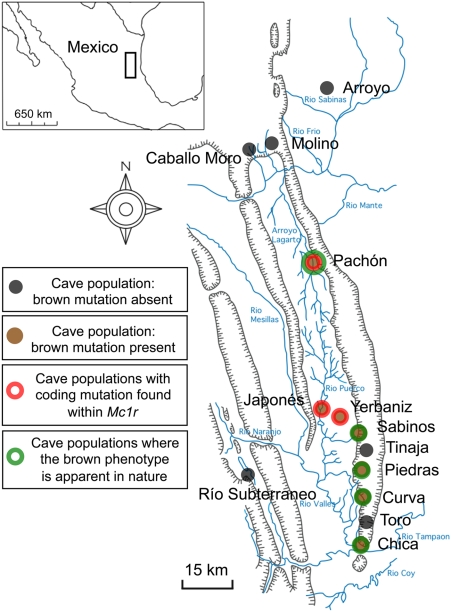
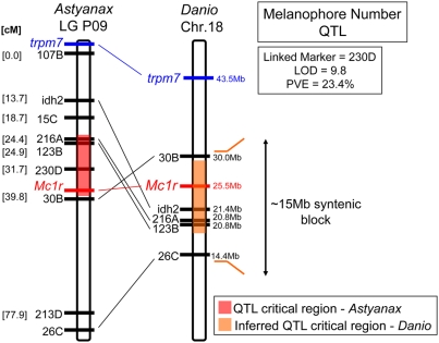
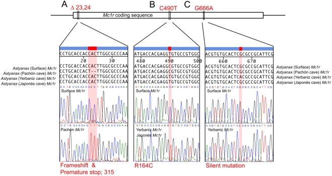
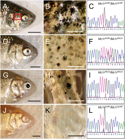
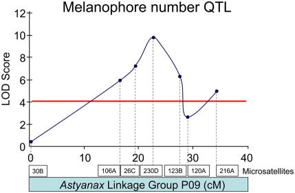
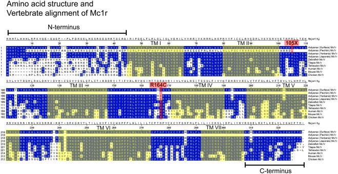
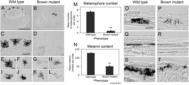
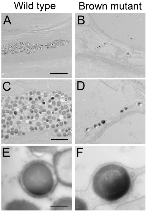
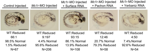
The evolution of degenerate characteristics remains a poorly understood phenomenon. Only recently has the identification of mutations underlying regressive phenotypes become accessible through the use of genetic analyses. Focusing on the Mexican cave tetra Astyanax mexicanus, we describe, here, an analysis of the brown mutation, which was first described in the literature nearly 40 years ago. This phenotype causes reduced melanin content, decreased melanophore number, and brownish eyes in convergent cave forms of A. mexicanus. Crosses demonstrate non-complementation of the brown phenotype in F(2) individuals derived from two independent cave populations: Pachón and the linked Yerbaniz and Japonés caves, indicating the same locus is responsible for reduced pigmentation in these fish. While the brown mutant phenotype arose prior to the fixation of albinism in Pachón cave individuals, it is unclear whether the brown mutation arose before or after the fixation of albinism in the linked Yerbaniz/Japonés caves. Using a QTL approach combined with sequence and functional analyses, we have discovered that two distinct genetic alterations in the coding sequence of the gene Mc1r cause reduced pigmentation associated with the brown mutant phenotype in these caves. Our analysis identifies a novel role for Mc1r in the evolution of degenerative phenotypes in blind Mexican cavefish. Further, the brown phenotype has arisen independently in geographically separate caves, mediated through different mutations of the same gene. This example of parallelism indicates that certain genes are frequent targets of mutation in the repeated evolution of regressive phenotypes in cave-adapted species.


Conflict of interest statement

The authors have declared that no competing interests exist.

Figures

Figure 1
Figure 1. Schematic map and localities of cave and Surface populations of the Mexican tetra, Astyanax mexicanus.
Several cave and Surface populations have been described throughout the Sierra de el Abra region of northeastern Mexico (inset). Cave populations in which the brown mutation is either absent (Molino cave) or has not been described (Tinaja cave) are depicted by a black dot. Cave populations in which the brown mutation has been described are denoted by a brown dot, while populations housing the brown mutation and carrying coding mutations in the gene Mc1r are depicted by a red/brown dot. Cave localities in which the brown phenotype is apparent in nature are labeled in green. Most localities do not harbor albinism (e.g., Chica, Curva, Piedras and Sabinos) and therefore, the phenotypic expression of the brown mutation in nature is inferred in these caves. Caves in which albinism has been reported include the Molino, Pachón, Yerbaniz and Japonés populations. Interestingly, of these caves only the Molino cave does not also harbor the brown mutation. While brown individuals have been reported in the Pachón cave , the same has not been reported in the literature in fish derived from the albino Yerbaniz/Japonés cave populations. Surface populations of Astyanax are found widely present throughout the rivers and streams of the region. Reports of the brown mutation and map of localities adapted from , , , –.
Figure 2
Figure 2. Astyanax linkage group P09 anchors strongly to Danio chromosome 18.
Genomic sequences flanking microsatellites on Astyanax linkage group P09 are localized to within a ∼15 Mb stretch of Danio chromosome (Chr.) 18. Two candidate genes, Mc1r and trpm7, are present on chromosome 18 in Danio, however only Mc1r co-localizes to the critical region (red rectangle) of the melanophore number QTL on Astyanax linkage group P09. Legend: QTL critical region on Astyanax linkage group P09 (red rectangle), inferred QTL critical region on Danio chromosome 18 (orange triangle).
Figure 3
Figure 3. Sequence analyses of Mc1r open reading frame in Surface, Pachón, Yerbaniz and Japonés cavefish of Astyanax mexicanus reveal three coding mutations.
A schematic representation of the Mc1r coding sequence is shown with each of three mutations identified in our sequence analyses. (A–C) Each of the three mutations is magnified to show nucleotide alignments for the Surface, Pachón, Yerbaniz and Japonés alleles of Mc1r. Below each alignment is the representative chromatogram sequence data for the Surface (wild type) compared with variant sequences. Three coding alterations were observed in three of a total nine caves completely sequenced for Mc1r in this study. (A) A 2-bp deletion at positions 23,24 was discovered in members of the Pachón cave population of Astyanax. This 2-bp deletion (red) causes a frame-shift and the introduction of a premature stop codon at nucleotide position 315. (B) We discovered an additional mutation at position 490 that causes an arginine to cysteine modification at amino acid position 164 (red). The identical mutation was discovered through analyses of both the Yerbaniz and Japonés populations, however these caves are likely members of the same cave population, possibly joined via a subterranean interconnection. (C) An additional silent mutation (G666A; red) was observed only in Yerbaniz individuals.
Figure 4
Figure 4. Mc1r genotype-phenotype correlation in representative members of an F2 pedigree derived from a Surface×Pachón cavefish cross.
(A,D,G,J) We crossed two F1 hybrid individuals to generate a small pedigree of individuals demonstrating a range of pigmentation phenotypes. (A) Each fish was photographed from the right side to visualize the entire head or a region just posterior to the orbit of the eye (red rectangle). Each individual was also genotyped for the polymorphic region of Mc1r ORF housing the fixed 2-bp deletion found in members of the Pachón cave. (C,F,I,L) The resulting chromatogram sequence data is depicted to the right of each individual image. (A–C) We found that the darkest individual, with the most melanophores, carries two copies of the Surface form of Mc1r. (D–F) The heterozygous individual demonstrates roughly the same number of melanophores, however each cell appears to produce less eumelanin. (G–I) A non-albino F2 individual with two copies of the Pachón allele of Mc1r demonstrates the least amount of pigmentation compare to other pigmented genotypes, however this individual clearly differs from (J) albino phenotypes. (J–L) An albino individual carrying two mutant copies of the gene Oca2 who carries two Surface copies of Mc1r cannot produce pigment, demonstrating the epistatic nature of the albino mutation. Scale bars: A,D,G,J = 3 mm; B,E,H,K = 500 µm.
Figure 5
Figure 5. A melanophore number QTL resides on Astyanax linkage group P09.
The genetic architecture of the melanophore number trait in our cross yields a single strong QTL consistent with the predictions of breeding experiments, as previously described . The x-axis depicts the relative positions of microsatellite markers on linkage group P09, LOD score values (y-axis) across markers are depicted in dark blue. A LOD score significance value of 4.0 is depicted by a horizontal red line.
Figure 6
Figure 6. Vertebrate alignment of the amino acid sequence of Mc1r.
Mc1r protein sequences of Surface (wild type) and three populations of cave Astyanax, three teleost fish species (zebrafish, Tilapia, Tetraodon), and three amniotes (human, mouse, chicken) were aligned using MegAlign 6.1. Conserved protein sequences are depicted in blue, unconserved regions are shown in white. The seven (I–VII) transmembrane domains (TM) are shaded in yellow. The positions of the premature stop codon introduced at position 105 in the Pachón form of Mc1r, and the arginine mutation at position 164 found in individuals derived from the Yerbaniz and Japonés caves are depicted in red. Transmembrane domains are predicted from .
Figure 7
Figure 7. Variation in melanophore number and melanin content in scales derived from Surface (wild type) and brown mutant fish.
Individual scales were collected from (A) wild type and (B) brown mutant individuals and assessed for (M) total number of melanophores. (B,D,M) Scales drawn from individuals demonstrating the brown phenotype (carrying two copies of the Pachón Δ23,24 allele) had significantly less numbers of melanophores per scale. (A,C) In wild type scales, melanophores were frequently localized to the periphery of the scale, while (B,D) brown mutant melanophores were rarely at the periphery. Multiple melanophores observed in both (E,F,I,J) wild type and (G,H,K,L) brown mutant scales demonstrated a variety of morphologies, demonstrated by representatives from both phenotypes. (N) The amount of melanin is significantly lower in brown mutant melanophores. Skin tissues derived from the identical region of the dorsal flank demonstrated qualitatively lower amounts of melanin per melanophore in (P,R,T) brown mutants (carrying two copies of the Pachón Δ23,24 allele) compared to (O,Q,S) wild type individuals. Three representative regions were analyzed in each individual. Overall, less melanophores were observed in brown mutant individuals, demonstrated by the absence of pigment in R. Scale bars: A,B = 500 µm; C,D = 100 µm; E–L = 50 µm; O–T = 30 µm.
Figure 8
Figure 8. Brown mutant melanophores contain a lower number of melanin granules compared to wild type.
Corresponding ultra-thin (95 nm) sections through tail tissue of a representative (A,C,E) Surface and (B,D,F) brown mutant individual (carrying two copies of the Pachón Δ23,24 allele) were processed and imaged using high-resolution electron microscopy. Wild type tissue sections routinely demonstrated densely packed granules of melanin, while sections through brown mutant tissue contained far fewer melanin granules. Overall, melanin granules identified in wild type and brown mutants were the same size and contain comparable amounts of melanin. Scale bars: A,B = 10 µm; C,D = 2.5 µm; E,F = 250 nm.
Figure 9
Figure 9. The brown mutation is recapitulated in Mc1r-MO knockdown experiments and is rescued by the Surface form of Mc1r in the zebrafish.
Single-celled zebrafish embryos were injected with 0.2 µM of a MO-oligonucleotide directed against the first 25 base pairs of the Danio rerio form of Mc1r (see Methods). Compared with embryos receiving a mock injection of 2× 1 nl of Danieaux's solution, embryos receiving MO injections demonstrate significantly reduced pigmentation. This reduced pigmentation phenotype was highly penetrant, occurring in 95.6% of individuals (n = 206) receiving the injection. Rescue experiments were performed in which individuals receiving Mc1r-MO injection were co-injected with in vitro transcribed RNA from Surface, Pachón, or Yerbaniz constructs. Individuals co-injected with Surface Mc1r RNA were rescued from the reduced pigmentation phenotype (86.1%, n = 108). Individuals co-injected with RNA derived from Pachón or Yerbaniz allele failed to rescue the diminished pigmentation phenotype (20.7% in 58 individuals; 7.4% in 54 individuals, respectively). Scale bar: 500 µm.

References

    1. Protas ME, Hersey C, Kochanek D, Zhou Y, Wilkens H, et al. Genetic analysis of cavefish reveals molecular convergence in the evolution of albinism. Nat Genet. 2006;38:107–111. - PubMed
    1. Protas M, Tabansky I, Conrad M, Gross JB, Vidal O, et al. Multi-trait evolution in a cave fish, Astyanax mexicanus. Evol Dev. 2008;10:196–209. - PubMed
    1. Bensouilah M, Denizot J-P. Taste buds and neuromasts of Astyanax jordani: Distribution and immunochemical demonstration of co-localized substance P and enkephalins. Eur J Neurosci. 1991;3:407–414. - PubMed
    1. Wilkens H. Evolution and genetics of epigean and cave Astyanax fasciatus (Characidae, Pisces): Support for the neutral mutation theory. In: Hecht MK, Wallace B, editors. Evolutionary biology. New York: Plenum Publishing Corporation; 1988. pp. 271–367.
    1. Jeffery WR. Cavefish as a model system in evolutionary developmental biology. Dev Biol. 2001;231:1–12. - PubMed

Publication types