

Clear cell "sugar" tumor of the lung: a well-enhanced mass with an early washout pattern on dynamic contrast-enhanced computed tomography
- PMID: 19119463
- PMCID: PMC2610653
- DOI: 10.3346/jkms.2008.23.6.1121
Clear cell "sugar" tumor of the lung: a well-enhanced mass with an early washout pattern on dynamic contrast-enhanced computed tomography
Abstract
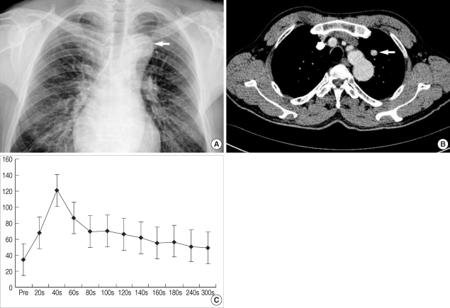
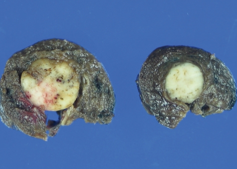
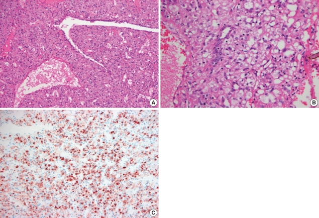
Clear cell tumor of the lung is a rare and very unusual benign pulmonary tumor. As clear cell tumor of the lung contains abundant cytoplasmic glycogen, this tumor is called "sugar tumor". We report a case of sugar tumor in a 64-yr-old man presenting as a round pulmonary nodule. On dynamic computed tomography (CT) scans, the solitary pulmonary nodule showed early wash-in enhancement with an early washout pattern like a lung malignancy. The patient underwent wedge resection for the tumor. Pathologic examination, including immunohistochemical studies, revealed that the nodule was a benign clear cell tumor, so-called "sugar tumor". Because only a small number of cases have been reported previously, clinical aspects, radiological characteristics on dynamic contrast-enhanced CT, and differential diagnosis of the tumor are not well established. Herein we present a clear cell tumor of the lung and discuss the clinical, radiological, and pathological features of the tumor.
Keywords: Clear Cell Tumor; Dynamic Contrast-Enhanced CT; Lung; Sugar Tumor; Washout Pattern.
Figures
References
-
- Shimosato Y, Miller RR. Biopsy interpretation of the lung. New York: Raven Press; 1995.
-
- Andrion A, Mazzucco G, Gugliotta P, Monga G. Benign clear cell (sugar) tumor of the lung: a light microscopic, histochemical, and ultrastructural study with a review of the literature. Cancer. 1985;56:2657–2663. - PubMed
-
- Ozdemir IA, Zaman NU, Rullis I, Webb WR. Benign clear cell tumor of lung. J Thorac Cardiovasc Surg. 1974;68:131–133. - PubMed
Publication types
MeSH terms
Substances
LinkOut - more resources
Full Text Sources
Medical
Research Materials
