

Rabbit Model for in vivo Study of Intervertebral Disc Degeneration and Regeneration
- PMID: 19119470
- PMCID: PMC2612571
- DOI: 10.3340/jkns.2008.44.5.327
Rabbit Model for in vivo Study of Intervertebral Disc Degeneration and Regeneration
Abstract
Objective: The purpose of this study is to verify the usefulness of the rabbit model for disc degeneration study.
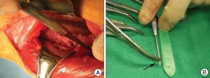
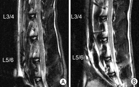
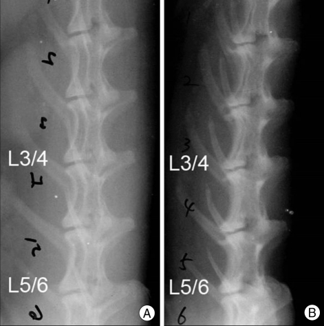
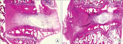
Materials: The L1-L2, L2-L3, L3-L4, or L4-L5 lumbar intervertebral disc (IVD) of 9 mature male New Zealand White rabbits were injured by inserting a 16-gauge needle to a depth of 5 mm in the left anterolateral annulus fibrosus while leaving L5-L6 IVD uninjured. Three other rabbits also received intradiscal injections of rabbit disc cells transfected with adenovirus and bone morphogenetic protein-2 (ad-BMP-2) at L4-L5 in addition to injury by 16-gauge needle at the L1-L2 level. Using digitized radiographs, measurements of IVD height were made and analyzed by using the disc height index (DHI). Magnetic resonance imaging (MRI) scans of the injured discs, injected discs, and uninjured L5-L6 discs were performed at 15 weeks post surgery and compared with preoperative MRI scans.
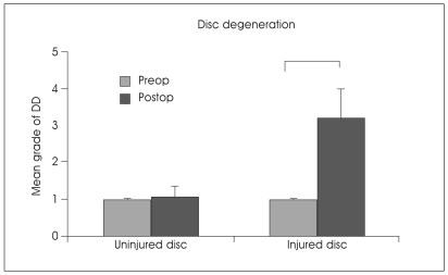
Results: All twelve rabbits showed consistent results of disc degeneration within 15 weeks following annular puncture. DHIs of injured discs were significantly lower than that of the uninjured L5-L6 discs (p<0.05). The mean value of disc degeneration grade of injured discs was significantly higher than that of uninjured discs (p<0.05). The injection of disc cell transfected with ad-BMP-2 did not induce disc regeneration at 15 weeks after injection.
Conclusion: This study showed that the injured disc had a significant change in DHI on simple lateral radiograph and disc degeneration grade on MRI scans within 15 weeks in all rabbits. Rabbit annular puncture model can be useful as a disc degeneration model in vivo.
Keywords: Animal model; Disc degeneration; Intervertebral disc; Rabbit; in vivo.
Figures

Similar articles
-
Intradiscal injection of monosodium iodoacetate induces intervertebral disc degeneration in an experimental rabbit model.Arthritis Res Ther. 2021 Dec 8;23(1):297. doi: 10.1186/s13075-021-02686-6. Arthritis Res Ther. 2021. PMID: 34876212 Free PMC article.
-
A slowly progressive and reproducible animal model of intervertebral disc degeneration characterized by MRI, X-ray, and histology.Spine (Phila Pa 1976). 2005 Jan 1;30(1):15-24. doi: 10.1097/01.brs.0000148048.15348.9b. Spine (Phila Pa 1976). 2005. PMID: 15626975
-
Intradiscal injections of osteogenic protein-1 restore the viscoelastic properties of degenerated intervertebral discs.Spine J. 2006 Nov-Dec;6(6):692-703. doi: 10.1016/j.spinee.2006.04.014. Epub 2006 Oct 10. Spine J. 2006. PMID: 17088200
-
A percutaneous, minimally invasive annulus fibrosus needle puncture model of intervertebral disc degeneration in rabbits.J Orthop Surg (Hong Kong). 2018 May-Aug;26(3):2309499018792715. doi: 10.1177/2309499018792715. J Orthop Surg (Hong Kong). 2018. PMID: 30114959
-
Revealing the presence of lymphatic vessels within intervertebral discs: Novel insights into disc degeneration.Innovation (Camb). 2025 Mar 3;6(6):100865. doi: 10.1016/j.xinn.2025.100865. eCollection 2025 Jun 2. Innovation (Camb). 2025. PMID: 40528887 Free PMC article. Review. No abstract available.
Cited by
-
Needle puncture in rabbit functional spinal units alters rotational biomechanics.J Spinal Disord Tech. 2015 Apr;28(3):E146-53. doi: 10.1097/BSD.0000000000000196. J Spinal Disord Tech. 2015. PMID: 25370985 Free PMC article.
-
Link protein N-terminal peptide and fullerol promote matrix production and decrease degradation enzymes in rabbit annulus cells.Connect Tissue Res. 2018 Mar;59(2):191-200. doi: 10.1080/03008207.2017.1330333. Epub 2017 Jun 8. Connect Tissue Res. 2018. PMID: 28509587 Free PMC article.
-
Animal Models of Intervertebral Disc Diseases: Advantages, Limitations, and Future Directions.Neurol Int. 2024 Dec 9;16(6):1788-1818. doi: 10.3390/neurolint16060129. Neurol Int. 2024. PMID: 39728755 Free PMC article. Review.
-
Animal models of regenerative medicine for biological treatment approaches of degenerative disc diseases.Exp Biol Med (Maywood). 2021 Feb;246(4):483-512. doi: 10.1177/1535370220969123. Epub 2020 Nov 11. Exp Biol Med (Maywood). 2021. PMID: 33175609 Free PMC article. Review.
-
BMP-2 and TGF-β3 do not prevent spontaneous degeneration in rabbit disc explants but induce ossification of the annulus fibrosus.Eur Spine J. 2012 Sep;21(9):1724-33. doi: 10.1007/s00586-012-2371-3. Epub 2012 May 26. Eur Spine J. 2012. PMID: 22639297 Free PMC article.
References
-
- An HS, Takegami K, Kamada H, Nguyen CM, Thonar EJ, Singh K, et al. Intradiscal administration of osteogenic protein-1 increases intervertebral disc height and proteoglycan content in the nucleus pulposus in normal adolescent rabbits. Spine. 2005;30:25–31. discussion 31-32. - PubMed
-
- An HS, Thonar EJ, Masuda K. Biological repair of intervertebral disc. Spine. 2003;28(15 Suppl):S86–S92. - PubMed
-
- Boos N, Weissbach S, Rohrbach H, Weiler C, Spratt KF, Nerlich AG. Classification of age-related changes in lumbar intervertebral discs. Spine. 2002;27:2631–2644. - PubMed
-
- Chiba K, Andersson GBJ, Masuda K, Momohara S, Williams JM, Thonar EJ. A new culture system to study the metabolism of the intervertebral disc in vitro. Spine. 1998;23:1821–1827. - PubMed
LinkOut - more resources
Full Text Sources
