Intraventricular cavernous malformation radiologically mimicking meningioma
- PMID: 19119474
- PMCID: PMC2612575
- DOI: 10.3340/jkns.2008.44.5.345
Intraventricular cavernous malformation radiologically mimicking meningioma
Abstract
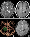
We report a case of trigonal cavernous malformation (CM) radiologically mimicking meningioma. The computed tomographic (CT) head angiography and magnetic resonance imaging (MRI) showed a partially calcified lesion with slight contrast enhancement located in the area of the left atrium of lateral ventricle. The lesion was completely removed using microsurgery with a parieto-occipital transcortical approach. The resected mass was histologically confirmed as CM. CM should be considered as differential diagnosis in case of the atrial mass lesion due to lack of hemosiderin ring characteristically seen other seated CM.
Keywords: Atrium; Cavernous malformation; Meningioma; Trigone.
Figures


References
-
- Anderson RC, Connolly ES, Jr, Ozduman K, Laurans MS, Gunel M, Khandji A, et al. Clinicopathologic review : giant intraventricular cavernous malformation. Neurosurgery. 2003;53:374–378. discussion 378-379. - PubMed
-
- Del Curling O, Jr, Kelly DL, Jr, Elster AD, Craven TE. An analysis of the natural history of cavernous angioma. J Neurosurg. 1991;75:702–708. - PubMed
-
- Fagundes-Pereyra WJ, Marques JA, Sousa LD, Carvalho GT, Sousa AA. Carvernoma of the lateral ventricle : case report. Arq Neuropsiquiatr. 2000;58:958–964. - PubMed
-
- Ishikawa M, Handa H, Moritake K, Mori K, Nakano Y, Aii H. Computed tomography of cerebral cavernous hemangiomas. J Comput Assist Tomogr. 1980;4:587–591. - PubMed
-
- Jellinger K. Vascular malformations of the central nervous system : a morphological overview. Neurosurg Rev. 1986;9:177–216. - PubMed
Publication types
LinkOut - more resources
Full Text Sources

