Review
. 2009 May;11(5):1059-81.
doi: 10.1089/ars.2008.2291.

Mechanistic and kinetic details of catalysis of thiol-disulfide exchange by glutaredoxins and potential mechanisms of regulation

Affiliations
Review

Mechanistic and kinetic details of catalysis of thiol-disulfide exchange by glutaredoxins and potential mechanisms of regulation

Molly M Gallogly et al. Antioxid Redox Signal. 2009 May.

Abstract

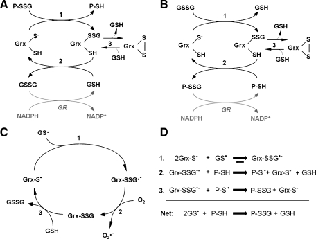
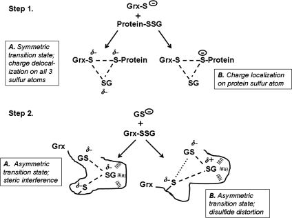
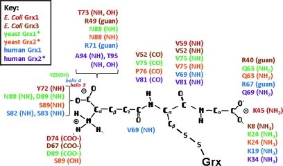
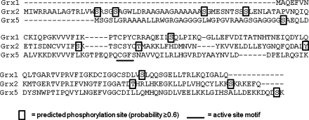
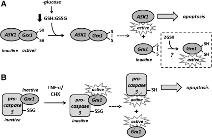
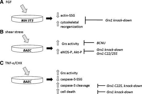
Glutaredoxins are small, heat-stable proteins that exhibit a characteristic thioredoxin fold and a CXXC/S active-site motif. A variety of glutathione (GSH)-dependent catalytic activities have been attributed to the glutaredoxins, including reduction of ribonucleotide reductase, arsenate, and dehydroascorbate; assembly of iron sulfur cluster complexes; and protein glutathionylation and deglutathionylation. Catalysis of reversible protein glutathionylation by glutaredoxins has been implicated in regulation of redox signal transduction and sulfhydryl homeostasis in numerous contexts in health and disease. This forum review is presented in two parts. Part I is focused primarily on the mechanism of the deglutathionylation reaction catalyzed by prototypical dithiol glutaredoxins, especially human Grx1 and Grx2. Grx-catalyzed protein deglutathionylation proceeds by a nucleophilic, double-displacement mechanism in which rate enhancement is attributed to special reactivity of the low pK(a) cysteine at its active site, and to increased nucleophilicity of the second substrate, GSH. Glutaredoxins (and Grx domains) have been identified in most organisms, and many exhibit deglutathionylation or other activities or both. Further characterization according to glutathionyl selectivity, physiological substrates, and intracellular roles may lead to subclassification of this family of enzymes. Part II presents potential mechanisms for in vivo regulation of Grx activity, providing avenues for future studies.


Figures

FIG. 1.
FIG. 1.
Sequential thiol-disulfide exchange between hydroxyethyl disulfide (HEDS) and GSH. In the first step, the GSH thiolate attacks one sulfur of HEDS, forming hydroxyethyl-SSG (βME-SSG) and β-mercaptoethanol (βME). In the second step, a second GSH attacks the βME-SSG mixed disulfide, forming GSSG and a second molecule of β-mercaptoethanol. When HEDS is used as a pro-substrate for Grx, the first step of this reaction creates the glutathionyl mixed-disulfide substrate that can be deglutathionylated by the enzyme (81).
FIG. 2.
FIG. 2.
Grx catalysis of reversible thiol-disulfide interchange—approach to equilibrium. Reaction mixtures were 2.2 ml at 30°C, containing 0.091 M K phosphate, pH 7.5. Lower lines: initial reaction mixtures also contained 0.91 mM HEDS and 1.82 mM GSH ± Grx1 from human red blood cells (0.01 units). Upper lines: initial reaction mixtures contained 0.91 mM GSSG and 1.82 mM β-mercaptoethanol (β-ME) ± Grx1 (0.01 units). Solid symbols, +Grx; open symbols, -Grx. Separate stock solutions of the reactants were prewarmed to 30°C, and each reaction was initiated by adding the reduced substrate (GSH or β-ME). At time points indicated, 0.2 ml of each reaction mixture was withdrawn and added to 0.8 ml of 0.25 mM NADPH in 0.125 mM K phosphate, pH 7.5; after mixing and reading an initial A340nm value, 2 units of GSSG reductase was added, and the decrease in A340nm was monitored until it reached a plateau (ca. 2 min). Total GSSG was calculated from the values of A340nm (1 nmol/0.005 ΔA340nm), determined from a separate standard curve for authentic GSSG). All data points represent the mean ± standard error of two separate experiments. Reprinted with permission from (83).
FIG. 3.
FIG. 3.
(A) Catalytic mechanism of deglutathionylation by human glutaredoxins (38, 47, 122). In the first step, the thiolate of the Grx N-terminal active-site cysteine attacks the glutathionyl sulfur of the protein-glutathione mixed disulfide (P-SSG), forming the Grx-SSG intermediate and releasing reduced protein-SH (P-SH). In the second step, free GSH attacks the glutathionyl sulfur of the Grx-SSG intermediate, releasing reduced Grx and GSSG. GSSG is then reduced to 2GSH by GSSG reductase (GR) and NADPH. Step 3 represents a side reaction in which the Grx C-terminal active-site cysteine competes with GSH for reduction of Grx-SSG, forming a Grx active-site disulfide and releasing GSH. The Grx-S2 side product is reduced by GSH and recruited back into the catalytic cycle. (B) Grx can use GSSG as an oxidized substrate (38, 83, 124). This scheme is analogous to Scheme 1A, except that the first substrate for Grx is glutathionylated glutathione (i.e., GSSG), and the second substrate is protein-SH. This reaction occurs under oxidizing conditions (i.e., low GSH/GSSG ratio) until the protein-SH/protein-SSG ratio reaches equilibrium. (C) Proposed mechanism of glutathione thiyl radical (GS) scavenging by Grx (124). In the first step, the N-terminal active-site cysteine of Grx attacks GS, forming a Grx disulfide anion radical intermediate. This radical then reacts with O2 in Step 2, forming superoxide (O2•−) and the typical Grx-SSG intermediate. In Step 3, the Grx-SSG intermediate is reduced by GSH, forming GSSG and reduced enzyme. (D) Proposed mechanism of glutathionyl transfer by Grx (124). In the first step of the reaction, the Grx catalytic cysteine thiolate attacks GS, forming the Grx-SSG•− disulfide anion radical intermediate. This intermediate can proceed to react with protein-SH (P-SH, Step 2), forming protein thiyl radical (P-S). In Step 3, another Grx-SSG•− molecule reacts with P-S (Step 3), quenching the radical reaction and forming protein-SSG (P-SSG). The net reaction yields protein-SSG from two GS and two P-SH molecules.
FIG. 4.
FIG. 4.
Complete commitment to catalysis vs. encounter-type catalytic mechanisms. (A) In the case of high commitment to catalysis, a reversible binding step exists between enzyme and substrate, followed by a chemistry step (k2) that is very fast compared with the rate of enzyme–substrate dissociation (k-1). Thus, essentially every substrate molecule that binds to enzyme undergoes a nucleophilic displacement reaction. (B) In the case of an encounter-type mechanism, enzyme and substrate react on association, but without formation of a reversible complex. This latter model is supported by two-substrate kinetic analysis of Grx1 (122) and Grx2 (38), which predicts “true” KMint values approaching infinity for both substrates.
FIG. 5.
FIG. 5.
Potential transition states in catalysis of deglutathionylation by Grx. Step 1, the Grx thiolate attacks the protein-SSG mixed-disulfide bond. (A) A symmetric transition state is depicted in which the negative charge of the attacking Grx thiolate is shared equally among all three sulfur atoms involved in the thiol-disulfide exchange reaction. (B) An asymmetric charge distribution is shown in which the negative charge is localized mainly to the protein thiolate, the exclusive leaving group in the reaction. Step 2 shows the glutathionyl thiolate attacking the Grx-SSG intermediate. (A) Steric interference is pictured, preventing interaction between the attacking glutathionyl sulfur and the catalytic cysteinyl sulfur of Grx, resulting in an asymmetric transition state. (B) An asymmetric transition state is depicted in which distortion of the mixed disulfide bond between Grx and the adducted glutathionyl moiety polarizes the disulfide bond, creating a more electrophilic site for the attack by the GS-thiolate.
FIG. 6.
FIG. 6.
Grx residues reported to interact with a covalently bound GS-moiety in the Grx-SSG mixed disulfide. Residues identified as making ion-pair or H bond contacts or both to the bound GS moiety are indicated next to the chemical group with which they interact. The functionality of the interacting amino acid is indicated parentheses (guan, guanidino group). Colors indicate the species from which the Grx-SSG structure was determined [red, E. coli Grx1 (15); brown, E. coli Grx3 (28, 91); green, yeast Grx1 (48, 145); orange, yeast Grx2 (24), blue, human Grx1 (143)]. For human Grx2 [represented in purple (60)], the depicted interactions correspond to those identified in a co-crystallized complex of reduced hGrx2 and GSH rather than the Grx2-SSG mixed disulfide. *A modified orientation of the γ-glutamyl group of the associated glutathionyl moiety, in comparison to the schematic representation [adapted from Nikkola et al. (89)]. (For interpretation of the references to color in this figure legend, the reader is referred to the web version of this article at www.liebertonline.com/ars).
FIG. 7.
FIG. 7.
Predicted phosphorylation sites for human Grx isoforms. General [NetPhos 2.0 (12)] and kinase-specific [NetPhosK 1.0 (13)] phosphorylation sites were predicted using FASTA sequences of human Grx1, Grx2, and Grx5. All sites with probability scores >0.8 are boxed. The CXXC active-site sequence is underlined. The sequence alignment was adapted from Johansson et al. (60).
FIG. 8.
FIG. 8.
Proposed interactions between Grx1 and cytosolic enzymes. (A) Reduced Grx1 is shown bound to ASK1 in MCF-7/ADR (multidrug-resistant breast cancer) cells under resting conditions (120). On glucose deprivation, the GSH/GSSG ratio decreases, resulting in oxidation of thiols in the Grx1 active site and disruption of its association with ASK1. Dissociation of Grx1 and ASK1 leads to ASK1 activation and increased apoptosis. (B) Grx1 and caspase-3 are shown associated under nonstressed conditions in BAECs. TNF-α–induced activation of Grx leads to pro-caspase-3 deglutathionylation, release from the complex, and cleavage by caspase-8 to active caspase-3, triggering apoptosis (94).
FIG. 9.
FIG. 9.
Examples of acute regulation of Grx1. (A) Treatment of NIH 3T3 fibroblasts with FGF resulted in robust deglutathionylation of actin within 15 min (139). Actin deglutathionylation and associated cytoskeletal changes were blocked by knockdown of Grx1. (B) Bovine aortic endothelial cells (BAECs) exposed to shear stress in culture exhibited increased Grx activity within the first 5–10 min of treatment (138). Increased Grx activity was correlated to increases in phosphorylation of eNOS and Akt. Changes in Grx activity were blocked by treatment with BCNU, whereas the changes in downstream events were blocked by Grx1 knockdown or transfection with catalytically inactive mutant enzyme. (C) Treatment of BAECs with TNF-α and cycloheximide was correlated with increased Grx activity within 3–6 h, deglutathionylation of pro-caspase-3, increased caspase-3 activity, and increased apoptosis (94). Events downstream of caspase-3 deglutathionylation were blocked by Grx1 knockdown or transfection of a catalytically inactive mutant.

References

    1. Adachi T. Pimentel DR. Heibeck T. Hou X. Lee YJ. Jiang B. Ido Y. Cohen RA. S-glutathiolation of Ras mediates redox-sensitive signaling by angiotensin II in vascular smooth muscle cells. J Biol Chem. 2004;279:29857–29862. - PubMed
    1. Adachi T. Weisbrod RM. Pimentel DR. Ying J. Sharov VS. Schoneich C. Cohen RA. S-Glutathiolation by peroxynitrite activates SERCA during arterial relaxation by nitric oxide. Nat Med. 2004;10:1200–1207. - PubMed
    1. Askelof P. Axelsson K. Eriksson S. Mannervik B. Mechanism of action of enzymes catalyzing thiol-disulfide interchange: thioltransferases rather than transhydrogenases. FEBS Lett. 1974;38:263–267. - PubMed
    1. Aslund F. Ehn B. Miranda-Vizuete A. Pueyo C. Holmgren A. Two additional glutaredoxins exist in Escherichia coli: glutaredoxin 3 is a hydrogen donor for ribonucleotide reductase in a thioredoxin/glutaredoxin 1 double mutant. Proc Natl Acad Sci U S A. 1994;91:9813–9817. - PMC - PubMed
    1. Aykac-Toker G. Bulgurcuoglu S. Kocak-Toker N. Effect of peroxynitrite on glutaredoxin. Hum Exp Toxicol. 2001;20:373–376. - PubMed

Publication types

LinkOut - more resources