. 2009 Aug;13(8B):2304-2316.
doi: 10.1111/j.1582-4934.2008.00608.x.

Small-molecule peptides inhibit Z alpha1-antitrypsin polymerization

Affiliations

Small-molecule peptides inhibit Z alpha1-antitrypsin polymerization

Yi-Pin Chang et al. J Cell Mol Med. 2009 Aug.

Abstract

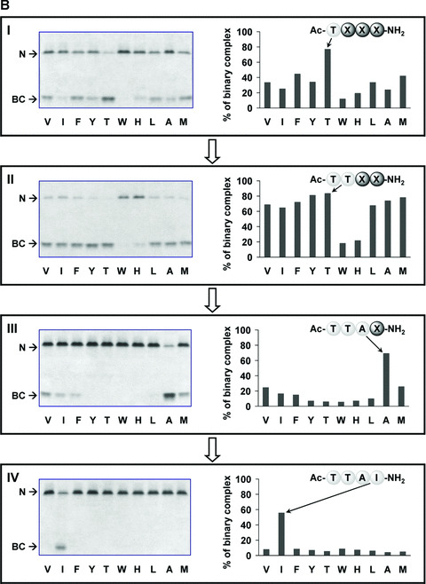
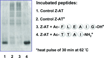
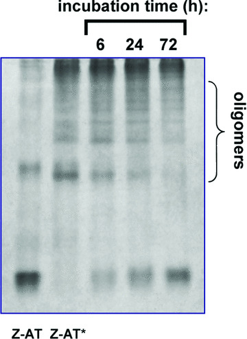
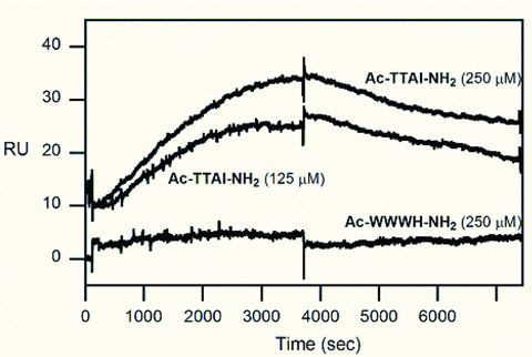
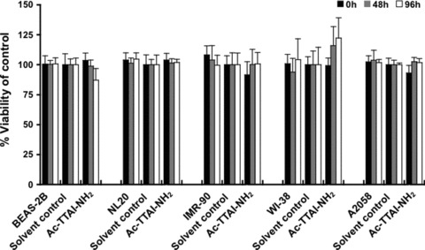
The Z variant of 1-antitrypsin (AT) polymerizes within the liver and gives rise to liver cirrhosis and the associated plasma deficiency leads to emphysema. In this work, a combinatorial approach based on the inhibitory mechanism of (alpha1)-AT was developed to arrest its pathogenic polymerization. One peptide, Ac-TTAI-NH(2), emerged as the most tight-binding ligand for Z (alpha1)-AT. Characterization of this tetrapeptide by gel electrophoresis and biosensor analysis revealed its markedly improved binding specificity and affinity compared with all previously reported peptide inhibitors. In addition, the peptide is not cytotoxic to lung cell lines. A model of the peptide-protein complex suggests that the peptide interacts with nearby residues by hydrogen bonds, hydrophobic interactions, and cavity-filling stabilization. The combinatorially selected peptide not only effectively blocks the polymerization but also promotes dissociation of the oligomerized (alpha1)-AT. These results are a significant step towards the potential treatment of Z (alpha1)-AT related diseases.


Figures

Figure 1
Figure 1
Iterative deconvolution of β‐strand‐directed tetrapeptide library binding to M and Z‐AT. The potency of each sub‐library was determined by the ability to form binary complex (BC) with native M‐ (A) and Z‐AT (B) as screened by 8 M urea gels (left side), and further quantitatively measured to give the percentage of BC formation by densitometric analysis (right side). The unbound M‐ and Z‐AT were designated as N in the figures. Urea was an additive and served as a chaotropic reagent to distinguish the native AT and its peptide‐inserted BC. More anodal migration of the BC most likely was due to the acquisition of additional stability of a fully antiparallel β‐sheet A from the incorporated peptide, leading to greater resistance to unfolding and hence faster migration in electrophoresis. The letters below each graph were standard amino acid symbols. The most reactive sub‐library in each screening cycle was indicated on the right side (X represented the randomized position), which defined the synthesis of next round sub‐libraries for subsequent screening. Panels I, II, III and IV showed the screening results of rounds 1, 2, 3 and 4, respectively. All screening were performed with a calculated 10‐fold molar excess of each individual peptide in each sub‐library compared with AT, apart from panel V which was performed with a 100‐fold molar excess of peptide compared with AT. Incubations were performed at 37°C for 2 hrs, apart from panels IV and V which were 1‐hr incubations.
Figure 1
Figure 1
Iterative deconvolution of β‐strand‐directed tetrapeptide library binding to M and Z‐AT. The potency of each sub‐library was determined by the ability to form binary complex (BC) with native M‐ (A) and Z‐AT (B) as screened by 8 M urea gels (left side), and further quantitatively measured to give the percentage of BC formation by densitometric analysis (right side). The unbound M‐ and Z‐AT were designated as N in the figures. Urea was an additive and served as a chaotropic reagent to distinguish the native AT and its peptide‐inserted BC. More anodal migration of the BC most likely was due to the acquisition of additional stability of a fully antiparallel β‐sheet A from the incorporated peptide, leading to greater resistance to unfolding and hence faster migration in electrophoresis. The letters below each graph were standard amino acid symbols. The most reactive sub‐library in each screening cycle was indicated on the right side (X represented the randomized position), which defined the synthesis of next round sub‐libraries for subsequent screening. Panels I, II, III and IV showed the screening results of rounds 1, 2, 3 and 4, respectively. All screening were performed with a calculated 10‐fold molar excess of each individual peptide in each sub‐library compared with AT, apart from panel V which was performed with a 100‐fold molar excess of peptide compared with AT. Incubations were performed at 37°C for 2 hrs, apart from panels IV and V which were 1‐hr incubations.
Figure 2
Figure 2
The comparison of the binding of the reported peptides Ac‐FLEAIG, WMDF‐NH2, Ac‐FLAA‐NH2 and the combinatorially selected Ac‐TTAI‐NH2 to AT by 8 M urea gels. (A) M‐ (lanes 1–3) and Z‐AT (lanes 4–6) were incubated with the peptides Ac‐FLEAIG (lanes 2 and 5; AT/peptide = 1:100) and Ac‐TTAI‐NH2 (lanes 3 and 6; AT/peptide = 1:10) at 37°C for 2 hrs. Each lane contained 2.5 μg of protein. The stronger affinity of the peptide Ac‐TTAI‐NH2 with Z‐AT was clearly distinguished by its lower molar ratio and short incubation required to form BC, whereas as predicted, no BC was formed with Ac‐FLEAIG. In addition, the specificity of the peptide Ac‐TTAI‐NH2 was also demonstrated as no BC was formed with M‐AT. (B) Z‐AT (lane 1) was incubated with the peptides WMDF‐NH2 (lane 2; AT/peptide = 1:100), Ac‐FLAA‐NH2 (lane 3; AT/peptide = 1:50) and Ac‐TTAI‐NH2 (lane 4; AT/peptide = 1:10) at 37°C for 2 hrs. As anticipated, no BC was formed with WMDF‐NH2 and Ac‐FLAA‐NH2 under this strict condition. The potency of the peptide Ac‐TTAI‐NH2 was demonstrated by the fact that a low molar ratio and short incubation time were required to form BC. Each lane contained 2.5 μg of protein.
Figure 3
Figure 3
The 4‐mer peptide Ac‐TTAI‐NH2 effectively blocked the polymerization of Z‐AT. Z‐AT was incubated with the previous identified peptide Ac‐FLEAIG (lane 3; 100‐fold molar excess) and the combinatorially selected peptide Ac‐TTAI‐NH2 (lane 4; 20‐fold molar excess) at 37°C for 4 hrs, and then heated to 62°C for 30 min. to intentionally accelerate the polymerization process before assay. Lane 2 showed the effect of heat pulse on Z‐AT without peptide. While Ac‐FLEAIG was able to inhibit polymerization under physiological conditions [25], it was unable to prevent polymerization under such harsh conditions. In contrast, polymerization of Z‐AT was completely inhibited by the peptide Ac‐TTAI‐NH2 at a low molar ratio. Each lane contained 2.5 μg of protein.
Figure 4
Figure 4
Reversal of Z‐AT oligomers by the identified peptide Ac‐TTAI‐NH2. Oligomers of Z‐AT were prepared by heating Z‐AT (0.5 mg/ml) at 58°C for 1 hr and labelled as Z‐AT* in the figure. The oligomerized Z‐AT was then incubated with the peptide Ac‐TTAI‐NH2 (100‐fold molar excess) for 6, 24 and 72 hrs at 37°C. After 6 hrs of incubation, BC was observable and increased with time, and as indicated there was the gradual resolution of oligomers of Z‐AT. Note that the native protein and BC bands appeared at almost the same position on a native PAGE. Each lane contained 3.5 μg of protein.
Figure 5
Figure 5
Binding of the combinatorially selected peptide Ac‐TTAI‐NH2 to a Z‐AT chip by SPR. Overlaid plot demonstrated the specific binding of the peptide Ac‐TTAI‐NH2 (125 and 250 μM), and the control peptide Ac‐WWWH‐NH2 (250 μM) was not recognized by the immobilized Z‐AT. The sensorgrams of the peptide Ac‐TTAI‐NH2 were shifted (10 resonance units) for clarity. The resonance unit was converted from the refractive index changes (measured by resonance angle shift in reflected light) upon binding on the sensor chip surface. The peptides and injection concentrations were as indicated in the figure.
Figure 6
Figure 6
MTT assay of various human cell lines treated with the Ac‐TTAI‐NH2 peptide. Five human cell lines including BEAS‐2B and NL20 normal lung epithelial cell lines, WI‐38 and IMR‐90 normal lung fibroblast cell lines, and A2058 melanoma cell line were treated for 0, 48 or 96 hrs with DMSO solvent control or 10 μM of the synthetic Ac‐TTAI‐NH2 peptide. The percentage of viable cells, relative to the solvent control‐treated cells, was measured by MTT cell proliferation assay. The results showed that no obvious cytotoxicity was detected for the Ac‐TTAI‐NH2 tetramer peptide. All experiments were performed in octuplicate in 96‐well microplates as described in the Materials and Methods section. Data are presented as mean ± S.D.
Figure 7
Figure 7
Proposed structure of the identified peptide Ac‐TTAI‐NH2 binding to AT. The peptide inserted into the lower part of A‐sheet (labelled in blue) of AT and interacted with its nearby residues. The NH and CO groups from the backbone of the incorporated peptide were hydrogen bonded to the backbones of adjacent strands 3 and 5 (residues in yellow) of AT (square panel on the left) and rendered the A‐sheet into a six‐stranded antiparallel β‐sheet. The circle on the upper right revealed the hydrogen bond (light green dashed line) between the N‐terminal Thr (P8) of peptide Ac‐TTAI‐NH2 and Ser56. Additional hydrogen bonds derived from the acetyl group of peptide with the side chain of His334 (light green dashed line) and the backbone NH group of Lys335 (light green dashed line) were also illustrated. The circle on the lower right showed the hydrophobic side chain of Ile occupies and interacted with a pocket surrounding by residues of Val173, Glu175, Leu176, Ala183 and Lys331. The complex orientations in the two circle panels on the right were changed to better present the peptide‐protein interactions. Carbon, nitrogen and oxygen atoms were shown in white, light blue and red, respectively.

Similar articles

Cited by

References

    1. Brantly M, Nukiwa T, Crystal RG. Molecular basis of alpha‐1‐antitrypsin deficiency. Am J Med. 1988; 84: 13–31. - PubMed
    1. Mahadeva R, Lomas DA. Alpha1‐antitrypsin deficiency, cirrhosis and emphysema. Thorax. 1998; 53: 501–5. - PMC - PubMed
    1. Elliott PR, Lomas DA, Carrell RW, et al . Inhibitory conformation of the reactive loop of α1‐antitrypsin. Nat Struct Biol. 1996; 3: 676–81. - PubMed
    1. Loebermann H, Tokuoka R, Deisenhofer J, et al . Human α1‐proteinase inhibitor: crystal structure analysis of two crystal modifications, molecular model, and preliminary analysis of the implications for function. J Mol Biol. 1984; 177: 531–57. - PubMed
    1. Stratikos E, Gettins PWG. Major proteinase movement upon stable serpin‐pro teinase complex formation. Proc Natl Acad Sci USA. 1997; 94: 453–8. - PMC - PubMed

Publication types