

A distributed neural system for top-down face processing
- PMID: 19121364
- PMCID: PMC2634849
- DOI: 10.1016/j.neulet.2008.12.039
A distributed neural system for top-down face processing
Abstract
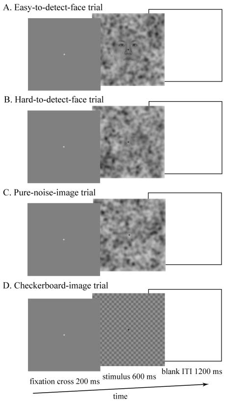
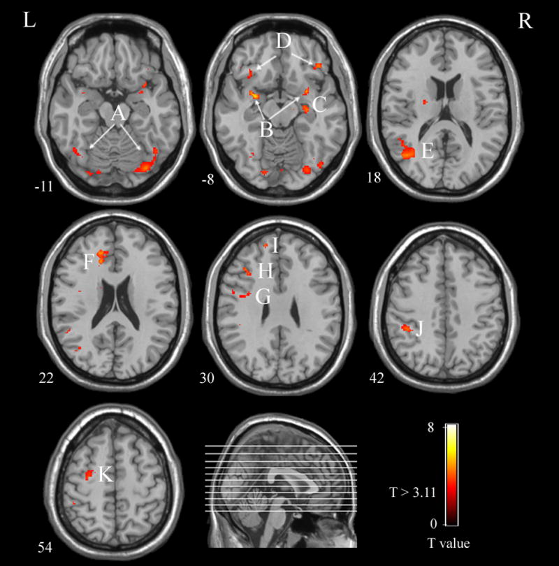
Evidence suggests that the neural system associated with face processing is a distributed cortical network containing both bottom-up and top-down mechanisms. While bottom-up face processing has been the focus of many studies, the neural areas involved in the top-down face processing have not been extensively investigated due to difficulty in isolating top-down influences from the bottom-up response engendered by presentation of a face. In the present study, we used a novel experimental method to induce illusory face-detection. This method allowed for directly examining the neural systems involved in top-down face processing while minimizing the influence of bottom-up perceptual input. A distributed cortical network of top-down face processing was identified by analyzing the functional connectivity patterns of the right fusiform face area (FFA). This distributed cortical network model for face processing includes both "core" and "extended" face processing areas. It also includes left anterior cingulate cortex (ACC), bilateral orbitofrontal cortex (OFC), left dorsolateral prefrontal cortex (DLPFC), left premotor cortex, and left inferior parietal cortex. These findings suggest that top-down face processing contains not only regions for analyzing the visual appearance of faces, but also those involved in processing low spatial frequency (LSF) information, decision-making, and working memory.
Figures
References
-
- Bar M. A cortical mechanism for triggering top-down facilitation in visual object recognition. J Cogn Neurosci. 2003;15:600–609. - PubMed
-
- Barth H, La Mont K, Lipton J, Dehaene S, Kanwisher N, Spelke E. Non-symbolic arithmetic in adults and young children. Cognition. 2006;98:199–222. - PubMed
-
- Breiter HC, Etcoff NL, Whalen PJ, Kennedy WA, Rauch SL, Buckner RL, Strauss MM, Hyman SE, Rosen BR. Response and habituation of the human amygdala during visual processing of facial expression. Neuron. 1996;17:875–887. - PubMed
Publication types
MeSH terms
Grants and funding
LinkOut - more resources
Full Text Sources
