. 2009 Feb 15;207(1-2):45-56.
doi: 10.1016/j.jneuroim.2008.11.009. Epub 2009 Jan 3.

Depression is an early disease manifestation in lupus-prone MRL/lpr mice

Affiliations

Depression is an early disease manifestation in lupus-prone MRL/lpr mice

Hua-Xin Gao et al. J Neuroimmunol. .

Abstract

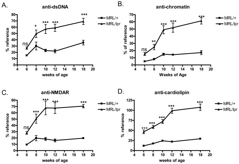
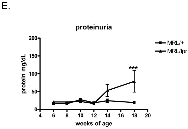
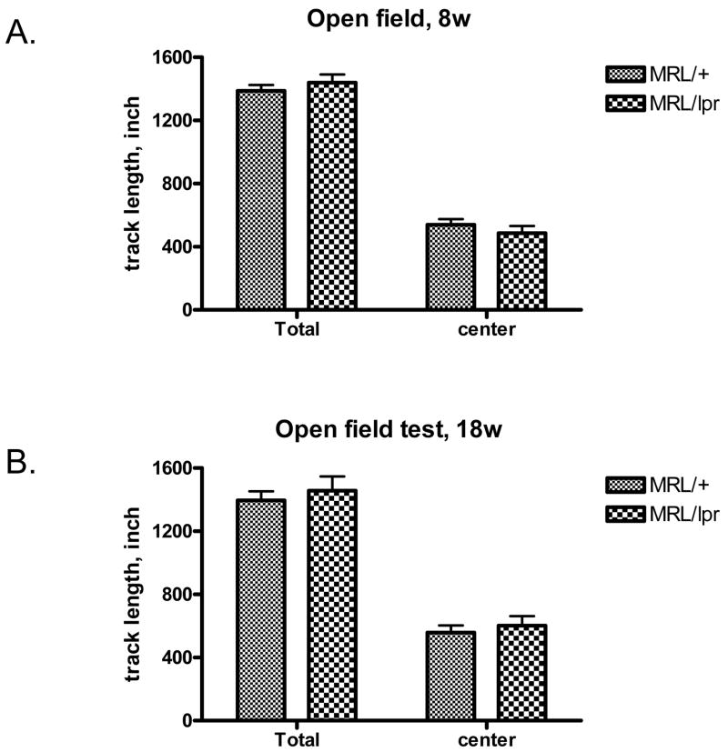
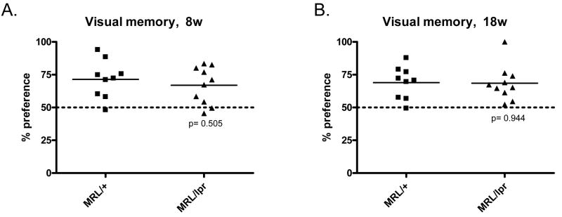
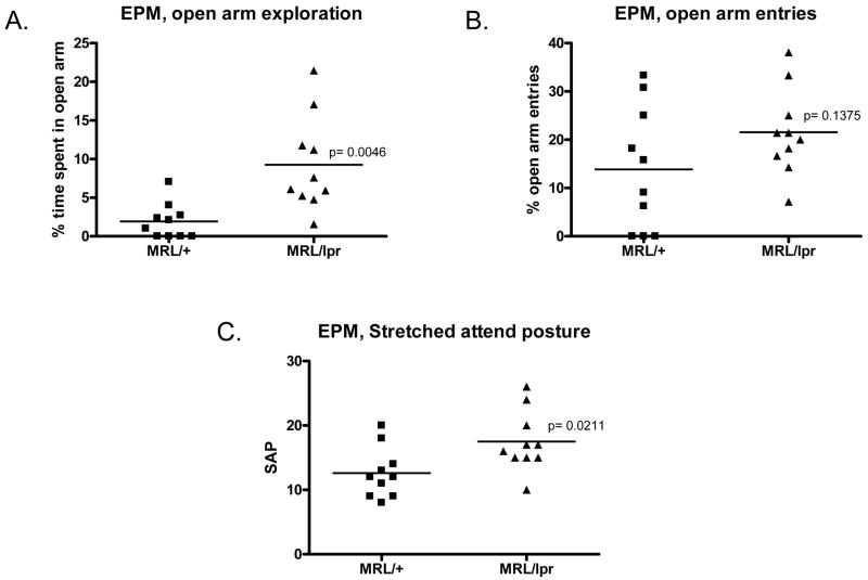
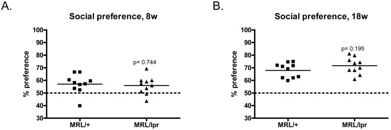
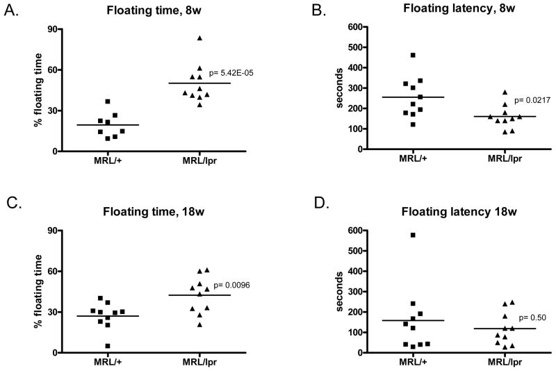
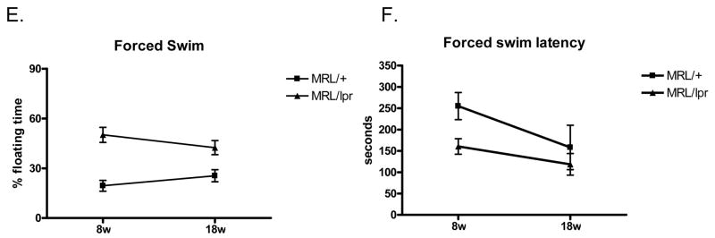
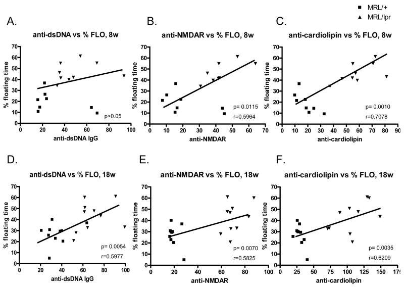
Many lupus patients develop neuropsychiatric manifestations, including cognitive dysfunction, depression, and anxiety. However, it is not clear if neuropsychiatric lupus is a primary disease manifestation, or is secondary to non-CNS disease. We found that MRL/lpr lupus-prone mice exhibited significant depression-like behavior already at 8 weeks of age, despite normal visual working memory, locomotor coordination and social preference. Moreover, depression was significantly correlated with titers of autoantibodies against DNA, NMDA receptors and cardiolipin. Our results indicate that lupus mice develop depression and CNS dysfunction very early in the course of disease, in the absence of substantial pathology involving other target organs.


Figures

Fig. 1
Fig. 1
Proteinuria and autoantibody levels in MRL/lpr and MRL/+ mice. A–D, Mouse serum autoantibody titers examined by ELISA, from age 6w to 18w. A. IgG anti-dsDNA titers. B. IgG anti-chromatin titers. C. IgG anti-NMDAR titers. D. IgG anti-cardiolipin titers. E. Urine protein concentrations in MRL/+ and MRL/lpr mice at 6–18 weeks. N=10 in each of the mice groups in this Figure. *, p<0.05; **, p<0.01; ***, p<0.001
Fig. 1
Fig. 1
Proteinuria and autoantibody levels in MRL/lpr and MRL/+ mice. A–D, Mouse serum autoantibody titers examined by ELISA, from age 6w to 18w. A. IgG anti-dsDNA titers. B. IgG anti-chromatin titers. C. IgG anti-NMDAR titers. D. IgG anti-cardiolipin titers. E. Urine protein concentrations in MRL/+ and MRL/lpr mice at 6–18 weeks. N=10 in each of the mice groups in this Figure. *, p<0.05; **, p<0.01; ***, p<0.001
Fig. 2
Fig. 2
Lupus-prone MRL/lpr mice behave normally in the open field test. The total track lengths and center track lengths in the open field of 8w (A) and 18w (B) MRL/+ and MRL/lpr mice (n=10) were measured. Total time in the field: 15 minutes.
Fig. 3
Fig. 3
Lupus-prone MRL/lpr mice display normal behavior in the balance beam walking test. 8w old MRL/+ and MRL/lpr mice were tested in beam walking for locomotor coordination. A. Number of slips recorded during beam crossing. B. Total time each group spent crossing the beam.
Fig. 4
Fig. 4
Cognition is preserved in 8w and 18w MRL/lpr mice. Object recognition (visual memory) tests on 8w (A) and 18w (B) mice were performed. A 45 minute delay time was used between the first and second object exposures. Mice having normal visual memory are expected to have >50% preference for a novel object (above the dotted line).
Fig. 5
Fig. 5
Elevated plus maze test detects less anxiety in lupus mice than control mice. Anxiety levels were tested in an EPM test. A. Time spent in the open arms was recorded and converted to a percentage of the total time in the EPM (5 minutes). The percent time spent on exploration is shown in the graph. B. Open arm entries were recorded. C. stretched attend postures counted during EPM tests.
Fig. 6
Fig. 6
MRL/lpr mice had similar social preference as MRL/+ controls. 8w (A) and 18w (B) MRL/lpr mice were tested for social activity in a Y maze. Preference was calculated based on the social target contacting time over total exploring time. Mice having normal social activities are expected to have >50% preference to a social target (above the dotted line).
Fig. 7
Fig. 7
MRL/lpr mice display depression-like behavior at 8 weeks of age. The forced swim test was used to assess depression-like behavior in mice. % floating time: total floating time converted to percentage of total test time (A, test on 8 weeks of age. C, test on 18 weeks of age). B and D, floating latency is the delay time until mice give up struggling in the water and start to show the ‘still’ floating behavior. Time was recorded in seconds. B, testing 8 week old mice. D, testing 18 week old mice. E, F. Two way ANOVA analysis of the effects of age and genotype on total floating time (E) and floating latency (F) in the tested mice.
Fig. 7
Fig. 7
MRL/lpr mice display depression-like behavior at 8 weeks of age. The forced swim test was used to assess depression-like behavior in mice. % floating time: total floating time converted to percentage of total test time (A, test on 8 weeks of age. C, test on 18 weeks of age). B and D, floating latency is the delay time until mice give up struggling in the water and start to show the ‘still’ floating behavior. Time was recorded in seconds. B, testing 8 week old mice. D, testing 18 week old mice. E, F. Two way ANOVA analysis of the effects of age and genotype on total floating time (E) and floating latency (F) in the tested mice.
Fig. 8
Fig. 8
Lupus autoantibody levels correlate with depression behavior. Linear regression correlation of % floating time in forced swim test with serum anti-dsDNA titers (A, 8w; D, 18w), anti-NMDAR antibody titers (B, 8w; E, 18w), or anti-cardiolipin antibody titers (C, 8w; F, 18w) were shown. MRL/+: ■, MRL/lpr: ▼. P values and Pearson’s R values for each correlation are also shown in the graphs.
Fig. 9
Fig. 9
MRI/MRSI analyses show differences in regional brain metabolism between control and lupus mice. The figure illustrates the type of spectra obtained from the hippocampus (top) and thalamus (bottom), with a corresponding anatomical image of a lupus mouse brain. Data from the comparative analysis of lupus and control mouse brains is provided in Table 2.

References

    1. Bessa JM, Oliveira M, Cerqueira JJ, Almeida OF, Sousa N. Age-related qualitative shift in emotional behaviour: paradoxical findings after re-exposure of rats in the elevated-plus maze. Behav Brain Res. 2005;162:135–142. - PubMed
    1. Bluestein HG. The central nervous system in systemic lupus erythematosus. In: Lahita RG, editor. Systemic lupus erythematosus. Churchill Livingstone; New York: 1992. pp. 639–655.
    1. Bonfa E, Marshak-Rothstein A, Weissbach H, Brot N, Elkon K. Frequency and epitope recognition of anti-ribosome P antibodies from humans with systemic lupus erythematosus and MRL/lpr mice are similar. J Immunol. 1988;140:3434–3437. - PubMed
    1. Brey RL, Abbott RD, Curb JD, Sharp DS, Ross GW, Stallworth CL, Kittner SJ. beta(2)-Glycoprotein 1-dependent anticardiolipin antibodies and risk of ischemic stroke and myocardial infarction: the honolulu heart program. Stroke. 2001;32:1701–1706. - PubMed
    1. Brooks WM, Sabet A, Sibbitt WL, Jr, Barker PB, van Zijl PC, Duyn JH, Moonen CT. Neurochemistry of brain lesions determined by spectroscopic imaging in systemic lupus erythematosus. J Rheumatol. 1997;24:2323–2329. - PubMed

MeSH terms