

Architectural analysis and intraoperative measurements demonstrate the unique design of the multifidus muscle for lumbar spine stability
- PMID: 19122093
- PMCID: PMC2663324
- DOI: 10.2106/JBJS.G.01311
Architectural analysis and intraoperative measurements demonstrate the unique design of the multifidus muscle for lumbar spine stability
Abstract
Background: Muscular instability is an important risk factor for lumbar spine injury and chronic low-back pain. Although the lumbar multifidus muscle is considered an important paraspinal muscle, its design features are not completely understood. The purpose of the present study was to determine the architectural properties, in vivo sarcomere length operating range, and passive mechanical properties of the human multifidus muscle. We hypothesized that its architecture would be characterized by short fibers and a large physiological cross-sectional area and that it would operate over a relatively wide range of sarcomere lengths but would have very stiff passive material properties.
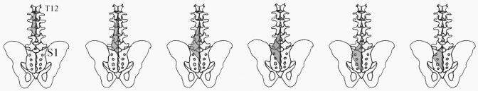
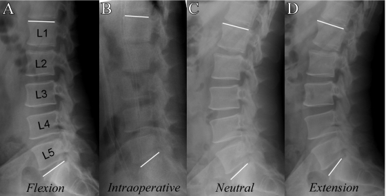
Methods: The lumbar spines of eight cadaver specimens were excised en bloc from T12 to the sacrum. Multifidus muscles were isolated from each vertebral level, permitting the architectural measurements of mass, sarcomere length, normalized fiber length, physiological cross-sectional area, and fiber length-to-muscle length ratio. To determine the sarcomere length operating range of the muscle, sarcomere lengths were measured from intraoperative biopsy specimens that were obtained with the spine in the flexed and extended positions. The material properties of single muscle fibers were obtained from passive stress-strain tests of excised biopsy specimens.
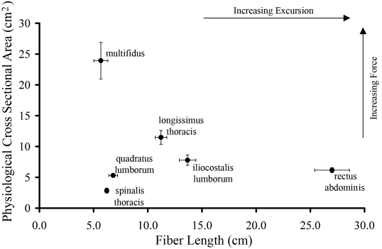
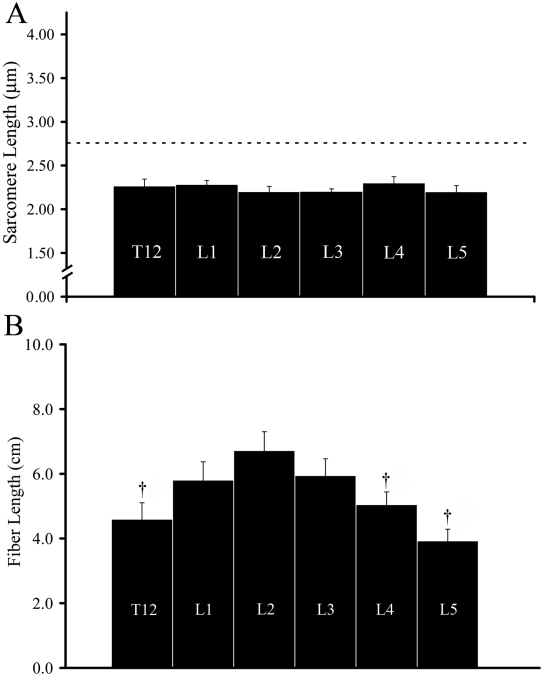
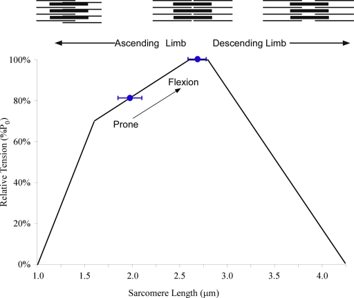
Results: The average muscle mass (and standard error) was 146 +/- 8.7 g, and the average sarcomere length was 2.27 +/- 0.06 microm, yielding an average normalized fiber length of 5.66 +/- 0.65 cm, an average physiological cross-sectional area of 23.9 +/- 3.0 cm(2), and an average fiber length-to-muscle length ratio of 0.21 +/- 0.03. Intraoperative sarcomere length measurements revealed that the muscle operates from 1.98 +/- 0.15 microm in extension to 2.70 +/- 0.11 microm in flexion. Passive mechanical data suggested that the material properties of the muscle are comparable with those of muscles of the arm or leg.
Conclusions: The architectural design (a high cross-sectional area and a low fiber length-to-muscle length ratio) demonstrates that the multifidus muscle is uniquely designed as a stabilizer to produce large forces. Furthermore, multifidus sarcomeres are positioned on the ascending portion of the length-tension curve, allowing the muscle to become stronger as the spine assumes a forward-leaning posture.
Figures
References
-
- Bogduk N, Macintosh JE, Pearcy MJ. A universal model of the lumbar back muscles in the upright position. Spine. 1992;17:897-913. - PubMed
-
- Macintosh JE, Bogduk N. The biomechanics of the lumbar multifidus. Clin Biomech (Bristol, Avon). 1986;1:205-13. - PubMed
-
- Macintosh JE, Pearcy MJ, Bogduk N. The axial torque of the lumbar back muscles: torsion strength of the back muscles. Aust N Z J Surg. 1993;63:205-12. - PubMed
-
- Delp SL, Suryanarayanan S, Murray WM, Uhlir J, Triolo RJ. Architecture of the rectus abdominis, quadratus lumborum, and erector spinae. J Biomech. 2001;34:371-5. - PubMed
-
- Stokes IA, Gardner-Morse M. Quantitative anatomy of the lumbar musculature. J Biomech. 1999;32:11-6. - PubMed
Publication types
MeSH terms
Grants and funding
LinkOut - more resources
Full Text Sources
Other Literature Sources
Research Materials
