. 2009 Jan;21(1):197-215.
doi: 10.1105/tpc.108.061317. Epub 2009 Jan 2.

A polypyrimidine tract binding protein, pumpkin RBP50, forms the basis of a phloem-mobile ribonucleoprotein complex

Affiliations

A polypyrimidine tract binding protein, pumpkin RBP50, forms the basis of a phloem-mobile ribonucleoprotein complex

Byung-Kook Ham et al. Plant Cell. 2009 Jan.

Abstract

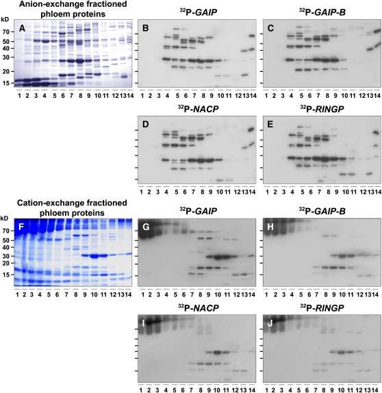
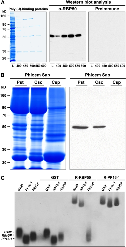
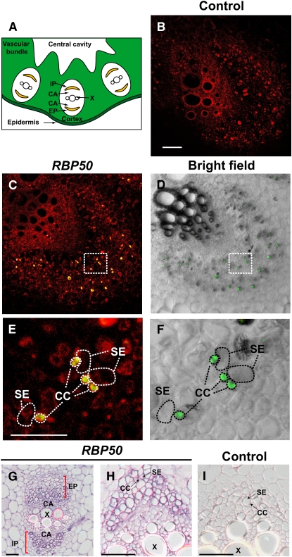
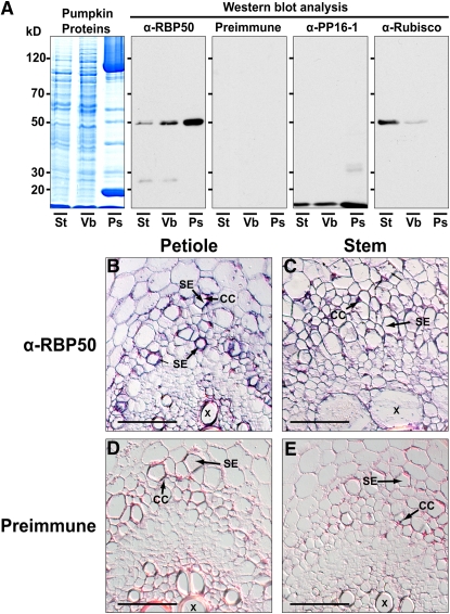
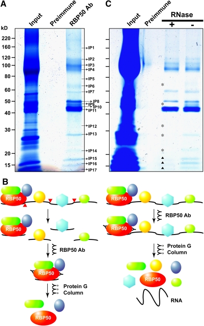
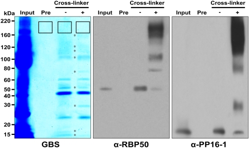
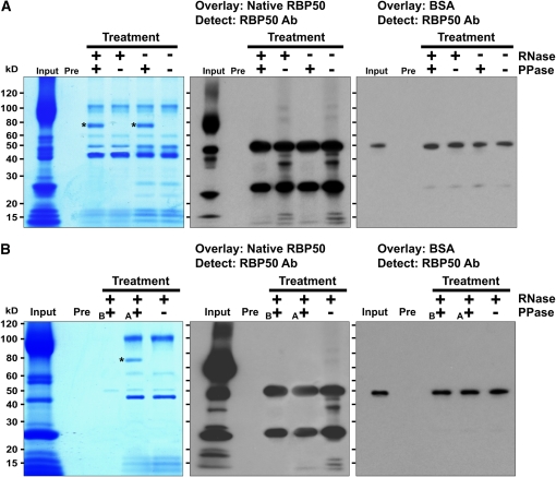
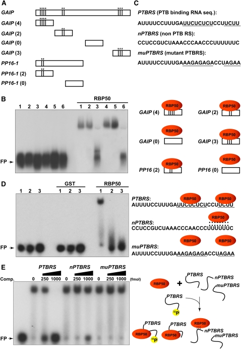
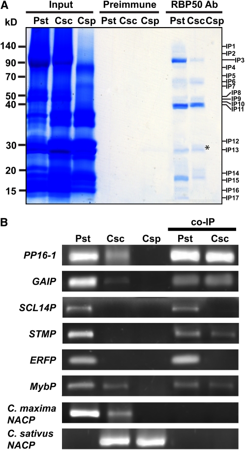
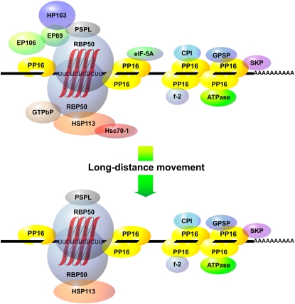
RNA binding proteins (RBPs) are integral components of ribonucleoprotein (RNP) complexes and play a central role in RNA processing. In plants, some RBPs function in a non-cell-autonomous manner. The angiosperm phloem translocation stream contains a unique population of RBPs, but little is known regarding the nature of the proteins and mRNA species that constitute phloem-mobile RNP complexes. Here, we identified and characterized a 50-kD pumpkin (Cucurbita maxima cv Big Max) phloem RNA binding protein (RBP50) that is evolutionarily related to animal polypyrimidine tract binding proteins. In situ hybridization studies indicated a high level of RBP50 transcripts in companion cells, while immunolocalization experiments detected RBP50 in both companion cells and sieve elements. A comparison of the levels of RBP50 present in vascular bundles and phloem sap indicated that this protein is highly enriched in the phloem sap. Heterografting experiments confirmed that RBP50 is translocated from source to sink tissues. Collectively, these findings established that RBP50 functions as a non-cell-autonomous RBP. Protein overlay, coimmunoprecipitation, and cross-linking experiments identified the phloem proteins and mRNA species that constitute RBP50-based RNP complexes. Gel mobility-shift assays demonstrated that specificity, with respect to the bound mRNA, is established by the polypyrimidine tract binding motifs within such transcripts. We present a model for RBP50-based RNP complexes within the pumpkin phloem translocation stream.


Figures

Figure 1.
Figure 1.
Pumpkin Phloem Sap Contains a Spectrum of RNA Binding Proteins. (A) Pumpkin phloem sap proteins separated by anion-exchange FPLC. Proteins were separated on a 13% SDS-PAGE gel and then stained with GBS reagent. Numbers represent the elution fractions from anion-exchange FPLC. (B) to (E) RNA overlay–protein blot assays performed on FPLC-fractionated proteins from (A) using the following riboprobes specific for phloem-mobile transcripts: GAIP (B), GAIP-B (C), NACP (D), and RINGP (E). (F) Pumpkin phloem sap proteins separated by cation-exchange FPLC. Proteins were separated on a 13% SDS-PAGE gel and then stained with GBS reagent. Numbers represent the elution fractions from cation-exchange FPLC. (G) to (J) RNA overlay–protein blot assays performed on FPLC-fractionated proteins from (F) using the following riboprobes specific for phloem-mobile transcripts: GAIP (G), GAIP-B (H), NACP (I), and RINGP (J).
Figure 2.
Figure 2.
RBP50, a 50-kD Poly(U) Binding Protein, Moves through the Translocation Stream and Has the Capacity to Bind, in a Sequence-Specific Manner, to Phloem-Mobile Transcripts. (A) A poly(U)-affinity column was used to purify pumpkin phloem proteins with poly(U) binding capacity. Candidate proteins were eluted using the following step concentrations of NaCl: 400, 450, 500, 550, and 600 mM. Protein profiles (L; loading sample) were separated on a 12% SDS-PAGE gel (volumes of 20 μL were applied for each fraction) and then visualized with GBS reagent. Purified anti-RBP50 antibody (α-RBP50) specifically detected the 50-kD protein (middle panel), whereas preimmune sera did not cross-react with any protein, including those in the loading sample. (B) Long-distance movement of RBP50 is confirmed by heterografting studies. Phloem sap was collected from pumpkin stock (Pst), cucumber scions grafted onto pumpkin stocks (Csc), and wild-type cucumber plant (Csp). Phloem sap proteins were resolved by SDS-PAGE (left panel) and subjected to protein gel blot analysis with anti-RBP50 antibody (right panel). Detection of RBP50 in phloem sap collected from cucumber scions establishes that this protein is phloem-mobile. (C) Gel mobility-shift assays reveal that recombinant (R) RBP50 binds to phloem-mobile transcripts in a sequence-specific manner. RNA binding capacities of RBP50, PP16-1, and GST were tested against 32P-labeled in vitro–transcribed riboprobes for GAIP, PP16-1, and RINGP. Note that GST did not bind to these transcripts, RBP50 bound to GAIP and PP16-1 but not RINGP, and PP16-1 bound all three probes.
Figure 3.
Figure 3.
In Situ RT-PCR– and RNA in Situ Hybridization–Based Detection of RBP50 Transcripts in Companion Cells. (A) Schematic transverse section of a portion of the pumpkin petiole/stem. Vascular bundles are composed of internal and external phloem (IP and EP, respectively) and xylem (X), with an intervening cambium (CA). (B) to (F) Transverse sections from pumpkin stem tissue analyzed by in situ RT-PCR using RBP50 gene-specific primers. Images were collected with a confocal laser scanning microscope; positive signal (green) represents incorporation of Alexa Fluor–labeled nucleotides. (B) Negative control in which primers were omitted from the RT-PCR mixture. Red fluorescence represents tissue autofluorescence. Bar = 500 μm (common for [B] to [D]). (C) Transverse section of a vascular bundle demonstrating the presence of RBP50 in phloem cells. Note that the green signal from the Alexa Fluor–labeled nucleotides appears yellow due to the red autofluorescence background. (D) Bright-field image of (C) used to illustrate the cellular architecture of the vascular bundle. RBP50 transcripts (green fluorescent signal) accumulated in companion cells. (E) Higher magnification of the boxed area shown in (C). CC, companion cell; SE, sieve element. Bar = 100 μm (common for [E] and [F]). (F) Higher magnification of the boxed area shown in (D). Images presented are representative of those obtained from at least three replicate experiments. (G) to (I) Pumpkin stem sections analyzed by in situ hybridization. Transverse sections were hybridized with an in vitro–transcribed antisense RNA probe to RBP50 ([G] and [H]); purple signal represents the presence of RBP50 transcripts in the small companion cells. Control in situ hybridization was performed with an in vitro–transcribed sense RNA probe to RBP50 (I). Bars = 500 μm.
Figure 4.
Figure 4.
RBP50 Is Synthesized in Companion Cells and Accumulates in Sieve Elements. (A) RBP50 accumulates in the phloem. Proteins extracted from pumpkin stem (St), vascular bundles (Vb), and phloem sap (Ps) were resolved on a 12% SDS-PAGE gel and visualized with GBS reagent. Proteins were transferred to nitrocellulose membranes, and blotting was performed with anti-RBP50 antibody, preimmune serum, anti-PP16-1 antibody, or anti-Rubisco antibody. Protein gel blot analysis indicated that RBP50 and PP16-1 accumulate in sieve elements. Preimmune serum failed to detect proteins extracted from these pumpkin tissues. Anti-Rubisco antibody, which served as a control against phloem sap contamination by proteins from surrounding tissue, did not detect the presence of Rubisco in the phloem sap. Results are based on three independent replicate experiments. (B) Immunohistochemical detection of RBP50 within companion cells (CC) and sieve elements (SE) of pumpkin petiole vascular bundles. RBP50 was detected using a combination of anti-RBP50 primary antibody followed by anti-rabbit IgG as a secondary antibody. (C) Immunohistochemical detection of RBP50 within companion cells and sieve elements of pumpkin stem vascular bundles. (D) and (E) Immunohistochemical controls performed on petiole and stem sections with preimmune sera. Note the absence of signal within the phloem tissues. Bars = 500 μm for (B) to (E).
Figure 5.
Figure 5.
Identification of the Pumpkin Phloem Proteins Coimmunoprecipitated with RBP50. (A) Total phloem proteins (input protein) were coimmunoprecipitated using either preimmune serum or purified anti-RBP50 antibody (Ab). Proteins were visualized by GBS reagent. The co-IP complex(es) included interacting proteins IP1 to IP17; note that IP8 was confirmed to be RBP50 by LC-MS/MS analysis. (B) Schematic illustration of phloem proteins that potentially could be bound to a phloem-mobile transcript contained within a RBP50–RNP complex. Note that transcripts bound by RBP50 may also interact with additional RNA binding proteins. RNase treatment was employed to separate a bound transcript (red darts) into individual RNP fragments, thereby allowing identification of proteins contained within the RBP50 complex. (C) Co-IP of the RBP50 RNP complex was performed with or without RNase treatment. Asterisks indicate phloem proteins likely not directly contained within the RBP50 complex; darts indicate proteins likely bound to both the RBP50 and other protein complexes located on the same/different RNA molecule(s).
Figure 6.
Figure 6.
Chemical Cross-Linking Identifies Proteins of the Core RBP50–RNP Complex. Phloem proteins (Input) were coimmunoprecipitated using either preimmune serum (Pre) or purified RBP50 antibody. Isolated RBP50–RNP complexes were then treated with the chemical cross-linker, bis(sulfosuccinimidyl) suberate, and subjected to SDS-PAGE. Protein profiles were visualized using GBS reagent (left panel). To confirm the cross-linkage of RBP50 with its interacting proteins, protein gel blot assays were performed using purified anti-RBP50 antibody (middle panel) or purified anti-PP16-1 antibody (right panel). Asterisks (left panel) indicate protein bands that underwent changes in intensity after cross-linker treatment. The boxed regions on the SDS-PAGE gel were excised for protein analysis using LC-MS/MS.
Figure 7.
Figure 7.
Protein Phosphorylation Is Required for RBP50-Based RNP Complex Formation. (A) Total phloem sap (containing proteins and mRNA) was treated with or without RNase, followed by co-IP. Purified RBP50-based RNP complexes were then treated with or without CIP (PPase), the proteins were separated by SDS-PAGE, and their profiles were visualized by GBS reagent (left panel). Proteins were then blotted onto nitrocellulose membranes and overlaid with either native phloem-purified RBP50 or BSA, and interacting proteins were detected by anti-RBP50 antibody (Ab; middle and right panels, respectively). Asterisks indicate the bands for CIP. (B) Total phloem sap was pretreated with RNase and CIP (PPase) before (B) the co-IP with anti-RBP50 antibodies. A second aliquot of total phloem sap was given a CIP treatment after (A) RNase pretreatment and co-IP. Proteins were separated by SDS-PAGE, and their profiles were visualized by GBS reagent (left panel). After blotting onto nitrocellulose membranes, proteins were overlaid with either native phloem-purified RBP50 or BSA, and interacting proteins were detected with anti-RBP50 antibody (middle and right panels, respectively). The asterisk indicates the band for CIP.
Figure 8.
Figure 8.
Selectivity of RBP50 RNA Binding Is Conferred through PTB Binding Motifs. (A) Schematic illustration of the deletion mutant series of GAIP and PP16-1 RNA employed in gel mobility-shift assays. Asterisks indicate the locations of PTB motifs on wild-type and mutant RNAs. Values in parentheses indicate the number of PTB motifs. (B) Gel mobility-shift assays establish that RBP50 binding to RNA is dependent on the presence of PTB motifs. Experiments were performed using recombinant RBP50 and 10 nM aliquots of the indicated 32P-labeled riboprobe. Lane 1, GAIP (4); lane 2, GAIP (2); lane 3, GAIP (0); lane 4, GAIP (3); lane 5, PP16-1 (2); lane 6, PP16-1 (0). FP, free riboprobe. (C) Engineered 27-nucleotide PTB motif variants; PTB motifs are underlined. The two PTB motifs present in the PTBRS RNA sequence were derived from the first two motifs located in the 5′ region of the GAIP mRNA. The nPTBRS RNA represents a sequence located in the 3′ region of the GAIP mRNA devoid of PTB motifs. The muPTBRS represents PTBRS in which the two PTB motifs have been mutated. (D) Synthetic 27-nucleotide riboprobes confirm the sequence requirement for RBP50 binding. Gel mobility-shift assays were performed using 27-nucleotide RNA probes (10 nM) and GST or recombinant RBP50. Lane 1, PTBRS; lane 2, nPTBRS; lane 3, muPTBRS. Dashed lines (right panel) indicate a weak interaction between RBP50 and nPTBRS RNA. (E) Competition assay performed by preincubating RBP50 (250 ng) with increasing concentrations of unlabeled PTBRS, nPTBRS, or muPTBRS followed by competition with 32P-labeled synthesized PTBRS (10 nM).
Figure 9.
Figure 9.
RBP50-Based RNP Complexes Move into the Cucumber Scion. (A) Phloem sap collected from pumpkin stock (Pst), cucumber scion (Csc), and control (ungrafted) cucumber plants (Csp) was subjected to co-IP against RBP50 or purified preimmune serum. Proteins were visualized by GBS staining. Note the similarity between the protein profiles for co-IP performed with phloem sap collected from pumpkin stock and cucumber scions. The absence of proteins in the co-IP experiment performed with phloem sap collected from control cucumber plants indicates the specificity of the interaction between RBP50 and the purified anti-RBP50 antibody preparation. The asterisk indicates the presence of a 28-kD unknown cucumber phloem protein that copurified with the pumpkin RBP50-based RNP complexes. (B) RT-PCR–based detection of phloem transcripts contained within the pumpkin stock (Pst), cucumber scion (Csc), and ungrafted cucumber stock (Csp) and RBP50 RNP complexes isolated from pumpkin stock or cucumber scion phloem sap. As controls, Cm NACP and Cs NACP transcripts were amplified using specific primer pairs. Results represent three independent RT-PCR experiments.
Figure 10.
Figure 10.
Schematic Illustration of a Phloem RBP50-Based Ribonucleoprotein Complex. RBP50 binds to PTB motifs (UUCUCUCUCUU) present within a subclass of phloem-mobile, polyadenylated transcripts, and this interaction leads to the binding of additional RBP50 (shown as a homodimer). PP16-1/-2 interact with both the RBP50 and the target mRNA, thereby forming the core of the RNP complex. RBP50 interacts directly with a GTPbP–HSP113–Hsc70-1 complex that may chaperone the RNP complex to and through the companion cell–sieve element plasmodesmata. Another set of four proteins, composed of the 89-kD expressed protein (EP89), the 103-kD hypothetical protein (HP103), the 106-kD expressed protein, and the PSPL are shown interacting with the RBP50. eIF-5A is a component of the RBP50 core and binds directly to PP16-1/-2. Regions outside the PTB motifs are bound by PP16-1/-2 along with five additional proteins: CPI, the Csf-2 related protein (Cmf-2), the 44-kD putative ATP binding protein (ATPase), the glutathione-regulated potassium-efflux system protein (GPSP), and the shikimate kinase precursor (SKP). The bottom image shows the composition of the phloem-mobile RBP50-based RNP complex based on co-IP results obtained using cucumber scion phloem sap.

Similar articles

Cited by

References

    1. Aoki, K., Kragler, F., Xoconostle-Cázares, B., and Lucas, W.J. (2002). A subclass of plant heat shock cognate 70 chaperones carries a motif that facilitates trafficking through plasmodesmata. Proc. Natl. Acad. Sci. USA 99 16342–16347. - PMC - PubMed
    1. Aoki, K., Suzui, N., Fujimaki, S., Dohmae, N., Yonekura-Sakakibara, K., Fujiwara, T., Hayashi, H., Yamaya, T., and Sakakibara, H. (2005). Destination-selective long-distance movement of phloem proteins. Plant Cell 17 1801–1814. - PMC - PubMed
    1. Aung, K., Lin, S.I., Wu, C.C., Huang, Y.T., Su, C.L., and Chiou, T.J. (2006). pho2, a phosphate overaccumulator, is caused by a nonsense mutation in a microRNA399 target gene. Plant Physiol. 141 1000–1011. - PMC - PubMed
    1. Auweter, S.D., and Allain, F.H.-T. (2008). Structure-function relationships of the polypyrimidine tract binding protein. Cell. Mol. Life Sci. 65 516–527. - PMC - PubMed
    1. Bari, R., Datt Pant, B., Stitt, M., and Scheible, W.R. (2006). PHO2, microRNA399, and PHR1 define a phosphate-signaling pathway in plants. Plant Physiol. 141 988–999. - PMC - PubMed

MeSH terms

LinkOut - more resources