. 2009 Feb 27;104(4):455-65.
doi: 10.1161/CIRCRESAHA.108.182568. Epub 2009 Jan 2.

Free cholesterol accumulation in macrophage membranes activates Toll-like receptors and p38 mitogen-activated protein kinase and induces cathepsin K

Affiliations

Free cholesterol accumulation in macrophage membranes activates Toll-like receptors and p38 mitogen-activated protein kinase and induces cathepsin K

Yu Sun et al. Circ Res. .

Abstract

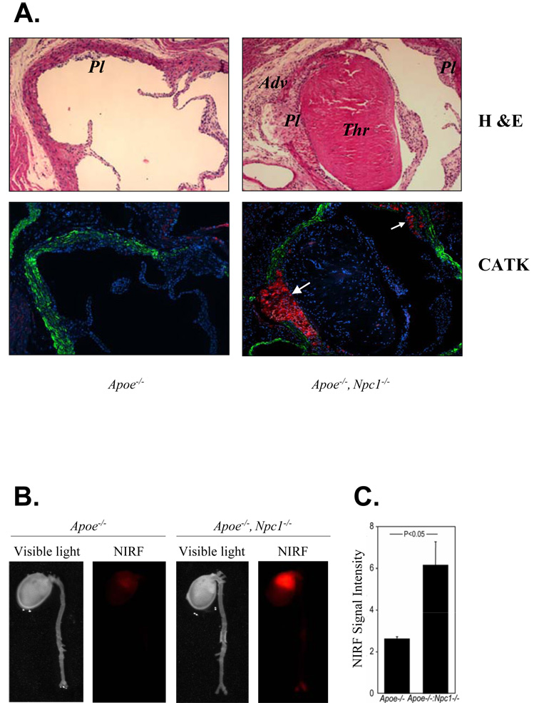
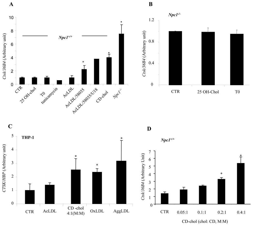
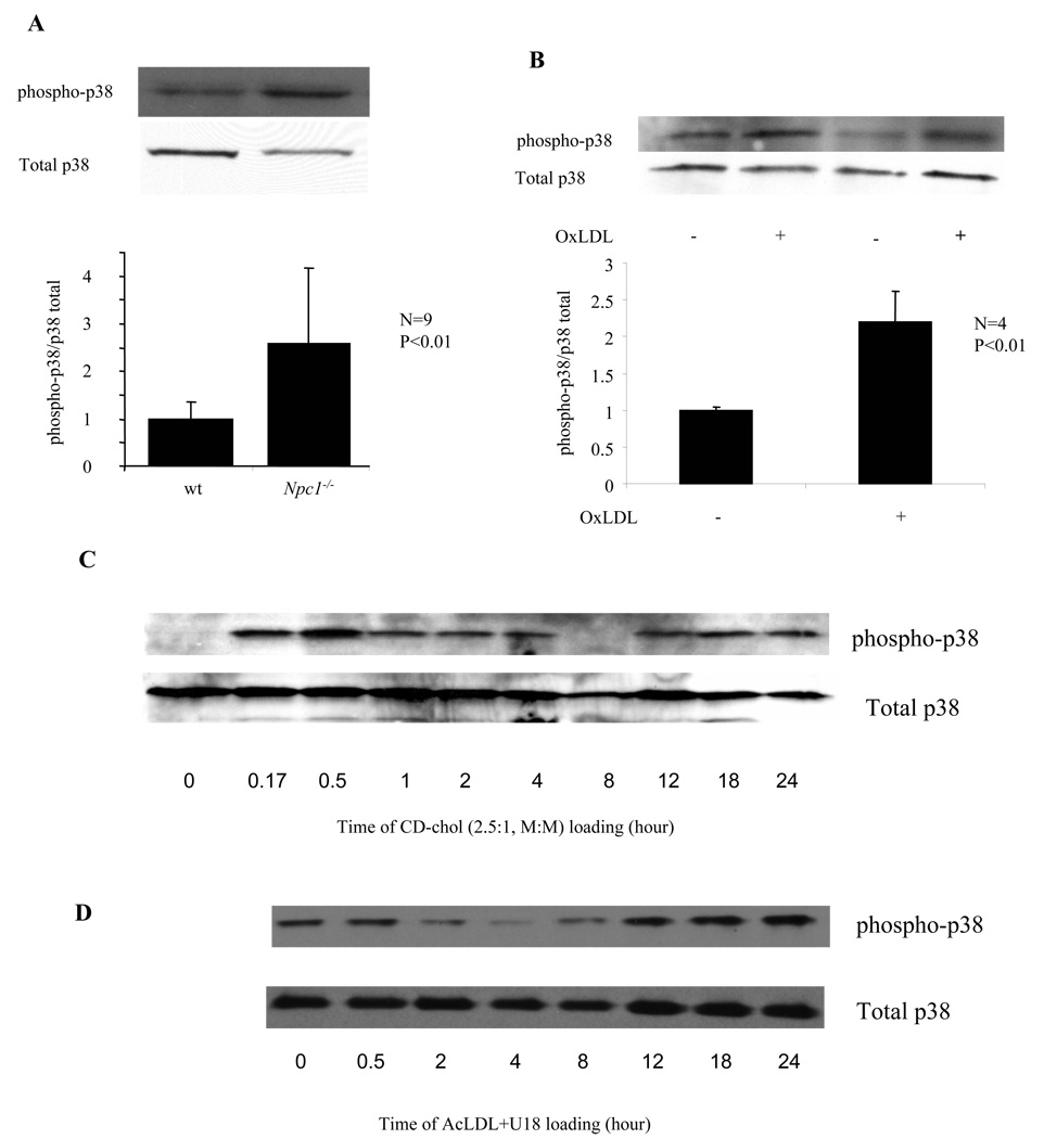
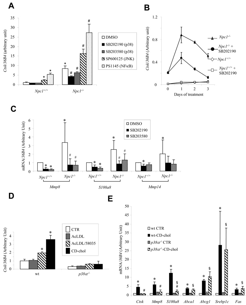
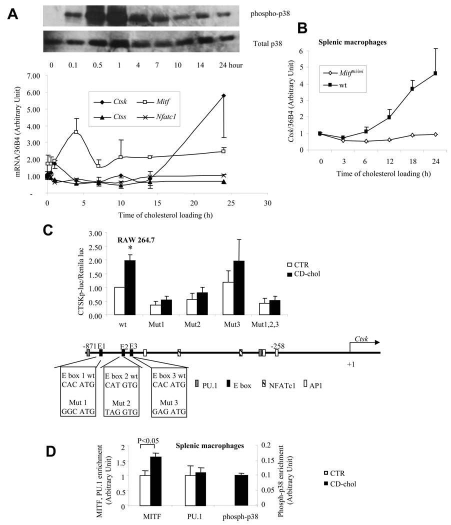
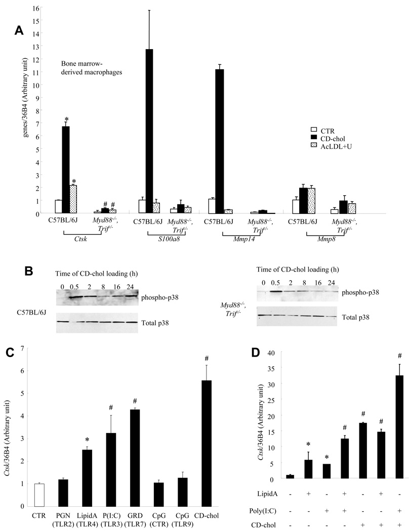
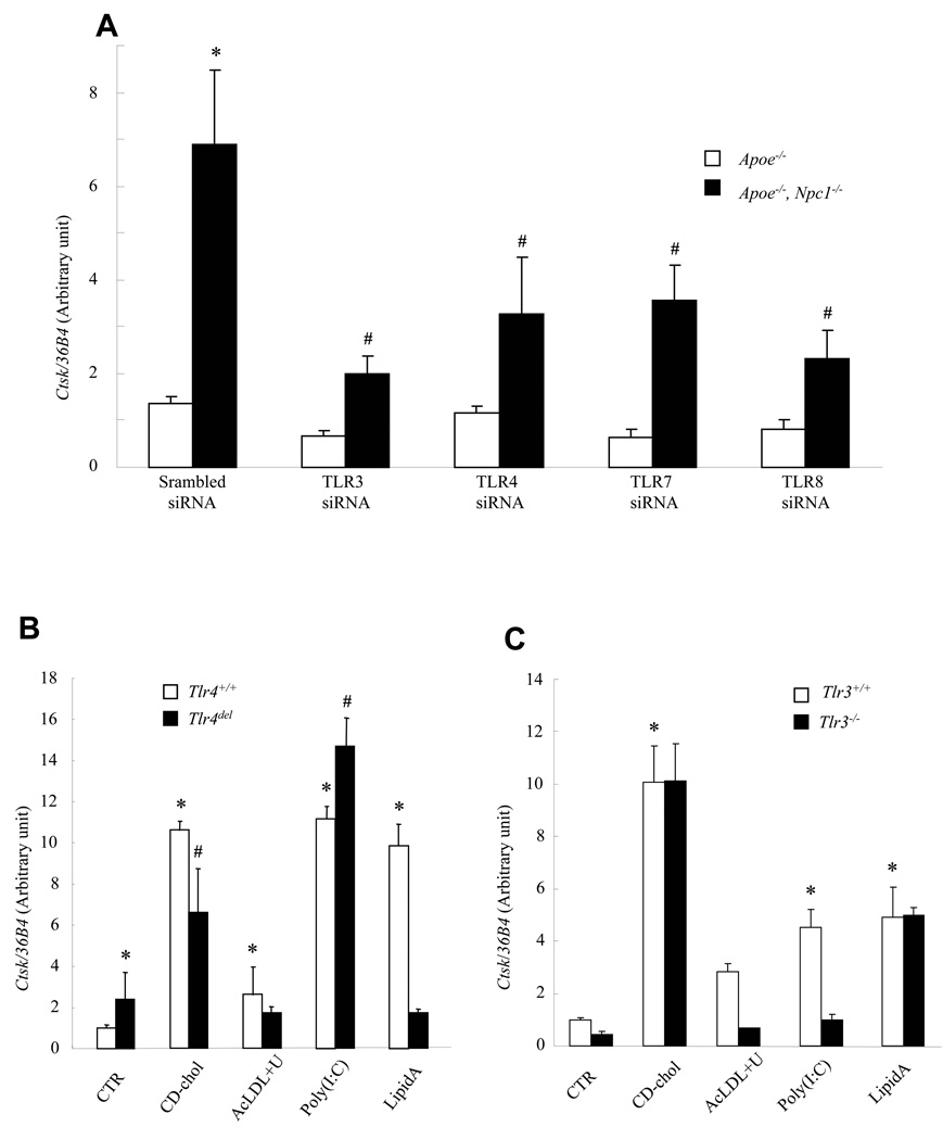
The molecular events linking lipid accumulation in atherosclerotic plaques to complications such as aneurysm formation and plaque disruption are poorly understood. BALB/c-Apoe(-/-) mice bearing a null mutation in the Npc1 gene display prominent medial erosion and atherothrombosis, whereas their macrophages accumulate free cholesterol in late endosomes and show increased cathepsin K (Ctsk) expression. We now show increased cathepsin K immunostaining and increased cysteinyl proteinase activity using near infrared fluorescence imaging over proximal aortas of Apoe(-/-), Npc1(-/-) mice. In mechanistic studies, cholesterol loading of macrophage plasma membranes (cyclodextrin-cholesterol) or endosomal system (AcLDL+U18666A or Npc1 null mutation) activated Toll-like receptor (TLR) signaling, leading to sustained phosphorylation of p38 mitogen-activated protein kinase and induction of p38 targets, including Ctsk, S100a8, Mmp8, and Mmp14. Studies in macrophages from knockout mice showed major roles for TLR4, following plasma membrane cholesterol loading, and for TLR3, after late endosomal loading. TLR signaling via p38 led to phosphorylation and activation of the transcription factor Microphthalmia transcription factor, acting at E-box elements in the Ctsk promoter. These studies suggest that free cholesterol enrichment of either plasma or endosomal membranes in macrophages leads to activation of signaling via various TLRs, prolonged p38 mitogen-activated protein kinase activation, and induction of Mmps, Ctsk, and S100a8, potentially contributing to plaque complications.


Figures

Fig. 1
Fig. 1. CATK immunostaining and Cathespin activity are induced in atherosclerotic lesion-containing aortas of Apoe−/−, Npc1−/− mice compared to Apoe−/− controls
A. Immunofluorescent staining of aortic cross-sections using specific antibodies against CATK (red, white arrows) and α-actin (green). Blue is hoechts staining. M, media; Pl, plaque; Thr, thrombus. 10X magnification. Mice were chow-fed and 10-wk-old. B. Ex vivo fluorescence reflectance imaging of mouse aortas using a probe that reports on cysteinyl protease activity. A visible light image indicates formation of early atherosclerotic changes in the aortas of Apoe−/− mice. A NIRF channel detected no substantial red signal in control. Npc1 mutation increased NIRF signal intensity in the aortas, particularly at the level of aortic root. C. Quantification of NIRF signal intensities (the target-to-background ratio, n=3).
Fig. 2
Fig. 2. Induction of Ctsk mRNA by cellular cholesterol loading
Ctsk mRNA levels measured by quantitative PCR and normalized to ribosomal 36B4 (mouse) or HBP (human). A., B. ConA-elicited mouse peritoneal macrophages with different treatments: 10 uM 25-OH-chol, 3 uM T0, 2 ug/ml tunicamycin, 50 ug/ml AcLDL, or 5 mM CD-chol (2.5:1, M:M) for 24 hrs; 50 ug/ml AcLDL+10 ug/ml ACAT inhibitor 58035±200 nMU18666A for 18 hrs. C. Differentiated human THP-1 cells treated with 100 ug/ml AcLDL, 100 ug/ml OxLDL, or 75 ug/ml AggLDL for 3 days; 5 mM CD-chol (4:1, M:M) for 24 hrs; or untreated. D. ConA-elicited mouse peritoneal macrophages treated with 5 mM CD-chol at increasing cholesterol:CD ratio for 24 hrs. CTR, control. *p<0.05 compared to CTR.
Fig. 3
Fig. 3. Npc1 mutation or cellular cholesterol loading induces p38 MAP kinase activation
Detection of total and phosphorylated p38 in protein lysates using specific antibodies. A. ConA-elicited peritoneal macrophages. B. Differentiated THP-1 cells incubated with or without 100 ug/ml OxLDL for 3 days. C, D. ConA-elicited wild-type macrophages treated with 5 mM CD-chol (2.5:1, M:M) (C) or 50 ug/ml AcLDL+400 nM U18666A (D) for indicated time.
Fig. 4
Fig. 4. Npc1 mutation- or cholesterol-mediated Ctsk induction is inhibited by chemical p38 inhibition or genetic p38 deletion
mRNA levels measured in ConA-elicited peritoneal macrophages by quantitative PCR and normalized to ribosomal 36B4. A, C. Cells treated with different inhibitors (10 uM) or DMSO for 24 hrs. *P<0.05 vs. wt/DMSO, #P<0.05 vs. Npc1−/−/DMSO. B. Cells treated with 10 uM SB202190 for indicated time. D, E. wt or p38α−/− cells treated with 50 ug/ml AcLDL±10 ug/ml ACAT inhibitor 58035 for 18 hrs, 5 mM CD-chol (2.5:1, M:M) for 24 hrs, or untreated (CTR). *P<0.05 vs. wt CTR, #P<0.05 vs. wt-CD-chol, §P<0.05 vs. p38α−/− CTR.
Fig. 5
Fig. 5. Cholesterol-mediated Ctsk induction requires the transcription factor MITF
A. CD-chol induces the expression of Mitf and Ctsk, as well as p38 MAP kinase activation. ConA-elicited macrophages were treated with 5 mM CD-cho (2.5:1, M:M) for indicated time. mRNA levels were measured by Taqman real-time PCR. Phospho- and total p38 protein were detected with specific antibodies. B. Inducation of Ctsk by CD-chol was abolished by Mitf dominant negative mutation. Splenic macrophages from wt or Mitfmi/mi mice were treated with 5 mM CD-chol (2.5:1, M:M) for indicated time. Ctsk mRNA level in untreated condition was set as 1. C. Disruption of three upstream E boxes on human CTSK promoter blunted the induction by CD-chol loading. RAW cells were transfected with indicated promoter-luciferase constructs and TK-Renilla luciferase. Transfection media was changed to DMEM/10%FBS (control) or 5 mM CD-chol (2.5:1, M:M) in DMEM/10%FBS (CD-chol) 6 hours after transfection. Cells were collected for luciferase assay 28 hours later. A schematic representation of human Ctsk promoter was shown at the bottom. The sequences of wild type and mutated E boxes used were shown in the boxes. D. CD-chol loading enriched MITF and phospho-p38 on Ctsk promoter. Splenic macrophages from wt mice were loaded with 5 mM CD-chol (2.5:1, M:M) for 18 hours. ChIP assay was performed in duplicates to determine the MITF, PU.1 and phospho-p38 on Ctsk promoter. N=2.
Fig. 6
Fig. 6. TLR signaling mediates cholesterol-induced Ctsk expression
A. Reduced expression of Ctsk and other genes in basal and CD-chol-loaded bone marrow derived macrophages from Myd88−/−, Trif−/− mice. B. Inhibition of late-phase p38 phosphorylation in Myd88−/−, Trif−/− cells compared to wild-type (C57BL/6J) controls. C. Induction of Ctsk expression by TLR3, 4 and 7 ligands in wild-type peritoneal macrophages. D. Synergistic effect of Poly (I:C) and CD-chol, but not LipidA, on Ctsk expression. *P<0.05 vs CTR, #P<0.004 vs C57BL/6J, same treatment. All treatments were for 24 hrs.
Fig. 7
Fig. 7. Ctsk induction by cholesterol loading is regulated by various TLRs
Ctsk mRNA levels measured in ConA-elicited mouse peritoneal macrophages by quantitative PCR and normalized to ribosomal 36B4. A. Knockdown of TLR 3 (66%), 4 (62%), 7(79%) or 8 (64%) by siRNA reduced Ctsk expression in Apoe−/−, Npc1−/− macrophages. *P<0.0001 vs CTR/Apoe−/−, #P<0.0001 vs CTR/Apoe−/−, Npc1−/−. B. Ctsk expression induced by cholesterol loading was blunted in Tlr4−/− macrophages. C. Ctsk expression induced by endosomal cholesterol loading was blocked in Tlr3−/− macrophages. Cells were treated for 24 hrs. *P<0.05 vs. CTR/Tlr4+/+ or Tlr3+/+, # P<0.005 vs. CTR/Tlr4del.

References

    1. Virmani R, Burke AP, Farb A, Kolodgie FD. Pathology of the vulnerable plaque. J Am Coll Cardiol. 2006;47:C13–C18. - PubMed
    1. Libby P, Simon DI. Inflammation and thrombosis: the clot thickens. Circulation. 2001;103:1718–1720. - PubMed
    1. Rader DJ, Daugherty A. Translating molecular discoveries into new therapies for atherosclerosis. Nature. 2008;451:904–913. - PubMed
    1. Gough PJ, Gomez IG, Wille PT, Raines EW. Macrophage expression of active MMP-9 induces acute plaque disruption in apoE-deficient mice. J Clin Invest. 2006;116:59–69. - PMC - PubMed
    1. Welch CL, Sun Y, Arey BJ, Lemaitre V, Sharma N, Ishibashi M, Sayers S, Li R, Gorelik A, Pleskac N, Collins-Fletcher K, Yasuda Y, Bromme D, D'Armiento JM, Ogletree ML, Tall AR. Spontaneous atherothrombosis and medial degradation in Apoe−/−, Npc1−/− mice. Circulation. 2007;116:2444–2452. - PubMed

Publication types

MeSH terms

Substances